

Leukodystrophies
- PMID: 35938662
- PMCID: PMC11320896
- DOI: 10.1212/CON.0000000000001130
Leukodystrophies
Abstract
Purpose of review: This article reviews the most common leukodystrophies and is focused on diagnosis, clinical features, and emerging therapeutic options.
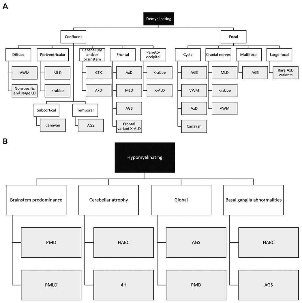
Recent findings: In the past decade, the recognition of leukodystrophies has exponentially increased, and now this class includes more than 30 distinct disorders. Classically recognized as progressive and fatal disorders affecting young children, it is now understood that leukodystrophies are associated with an increasing spectrum of neurologic trajectories and can affect all ages. Next-generation sequencing and newborn screening allow the opportunity for the recognition of presymptomatic and atypical cases. These new testing opportunities, in combination with growing numbers of natural history studies and clinical consensus guidelines, have helped improve diagnosis and clinical care. Additionally, a more granular understanding of disease outcomes informs clinical trial design and has led to several recent therapeutic advances. This review summarizes the current understanding of the clinical manifestations of disease and treatment options for the most common leukodystrophies.
Summary: As early testing becomes more readily available through next-generation sequencing and newborn screening, neurologists will better understand the true incidence of the leukodystrophies and be able to diagnose children within the therapeutic window. As targeted therapies are developed, it becomes increasingly imperative that this broad spectrum of disorders is recognized and diagnosed. This work summarizes key advances in the leukodystrophy field.
Copyright © 2022 American Academy of Neurology.
Figures
Similar articles
-
Recent Advancements in the Diagnosis and Treatment of Leukodystrophies.Semin Pediatr Neurol. 2021 Apr;37:100876. doi: 10.1016/j.spen.2021.100876. Epub 2021 Feb 10. Semin Pediatr Neurol. 2021. PMID: 33892849 Review.
-
Leukodystrophies.Continuum (Minneap Minn). 2018 Feb;24(1, Child Neurology):130-149. doi: 10.1212/CON.0000000000000560. Continuum (Minneap Minn). 2018. PMID: 29432240 Review.
-
Leukodystrophies: classification, diagnosis, and treatment.Neurologist. 2009 Nov;15(6):319-28. doi: 10.1097/NRL.0b013e3181b287c8. Neurologist. 2009. PMID: 19901710 Review.
-
An update on clinical, pathological, diagnostic, and therapeutic perspectives of childhood leukodystrophies.Expert Rev Neurother. 2020 Jan;20(1):65-84. doi: 10.1080/14737175.2020.1699060. Epub 2019 Dec 12. Expert Rev Neurother. 2020. PMID: 31829048 Review.
-
Genetic testing of leukodystrophies unraveling extensive heterogeneity in a large cohort and report of five common diseases and 38 novel variants.Sci Rep. 2021 Feb 5;11(1):3231. doi: 10.1038/s41598-021-82778-0. Sci Rep. 2021. PMID: 33547378 Free PMC article.
Cited by
-
Early recognition of patients with leukodystrophies.Curr Probl Pediatr Adolesc Health Care. 2022 Dec;52(12):101311. doi: 10.1016/j.cppeds.2022.101311. Epub 2022 Dec 2. Curr Probl Pediatr Adolesc Health Care. 2022. PMID: 36470810 Free PMC article. Review.
References
Publication types
MeSH terms
Grants and funding
LinkOut - more resources
Full Text Sources
Medical
Research Materials
Miscellaneous