. 2022 Sep 14;96(17):e0091922.
doi: 10.1128/jvi.00919-22. Epub 2022 Aug 8.

TMEΜ45B Interacts with Sindbis Virus Nsp1 and Nsp4 and Inhibits Viral Replication

Affiliations

TMEΜ45B Interacts with Sindbis Virus Nsp1 and Nsp4 and Inhibits Viral Replication

Feixiang Yan et al. J Virol. .

Abstract

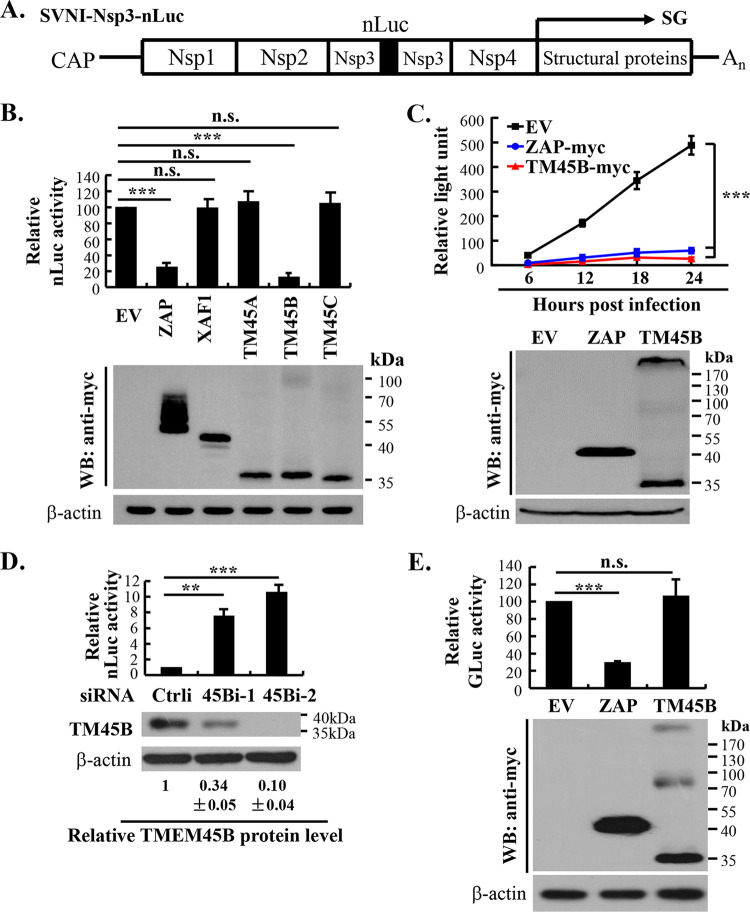
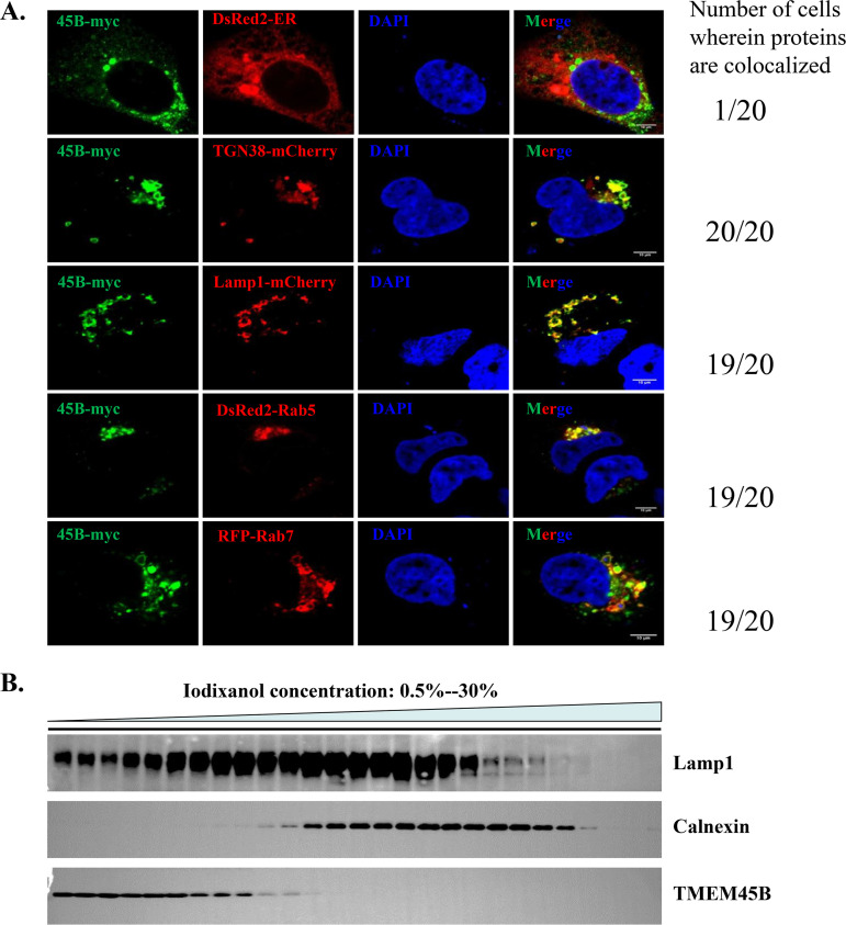
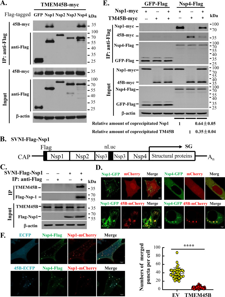
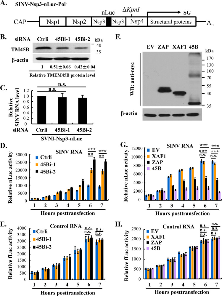
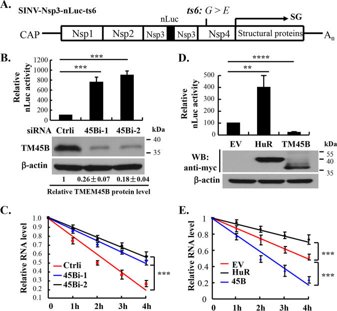
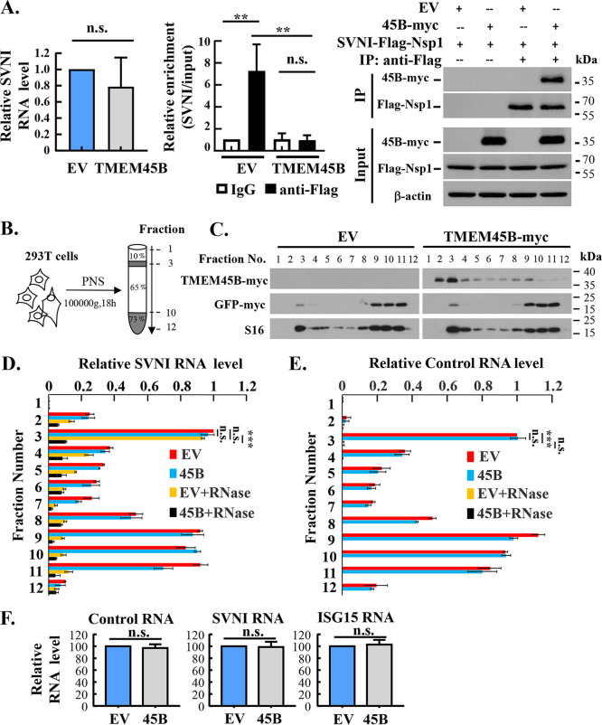
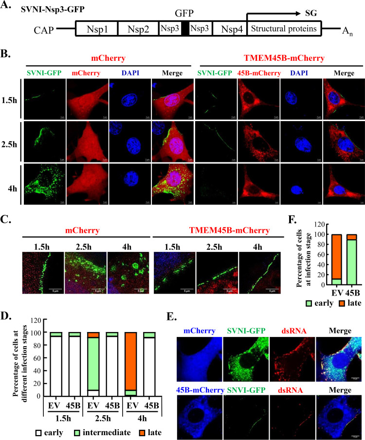
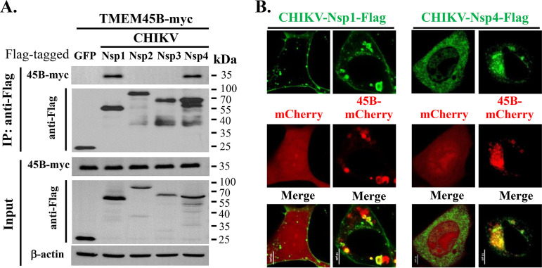
Alphavirus infection induces the expression of type I interferons, which inhibit the viral replication by upregulating the expression of interferon-stimulated genes (ISGs). Identification and mechanistic studies of the antiviral ISGs help to better understand how the host controls viral infection and help to better understand the viral replication process. Here, we report that the ISG product TMEM45B inhibits the replication of Sindbis virus (SINV). TMEM45B is a transmembrane protein that was detected mainly in the trans-Golgi network, endosomes, and lysosomes but not obviously at the plasma membrane or endoplasmic reticulum. TMEM45B interacted with the viral nonstructural proteins Nsp1 and Nsp4 and inhibited the translation and promoted the degradation of SINV RNA. TMEM45B overexpression rendered the intracellular membrane-associated viral RNA sensitive to RNase treatment. In line with these results, the formation of cytopathic vacuoles (CPVs) was dramatically diminished in TMEM45B-expressing cells. TMEM45B also interacted with Nsp1 and Nsp4 of chikungunya virus (CHIKV), suggesting that it may also inhibit the replication of other alphaviruses. These findings identified TMEM45B as an antiviral factor against alphaviruses and help to better understand the process of the viral genome replication. IMPORTANCE Alphaviruses are positive-stranded RNA viruses with more than 30 members. Infection with Old World alphaviruses, which comprise some important human pathogens such as chikungunya virus and Ross River virus, rarely results in fatal diseases but can lead to high morbidity in humans. Infection with New World alphaviruses usually causes serious encephalitis but low morbidity in humans. Alphavirus infection induces the expression of type I interferons, which subsequently upregulate hundreds of interferon-stimulated genes. Identification and characterization of host antiviral factors help to better understand how the viruses can establish effective infection. Here, we identified TMEM45B as a novel interferon-stimulated antiviral factor against Sindbis virus, a prototype alphavirus. TMEM45B interacted with viral proteins Nsp1 and Nsp4, interfered with the interaction between Nsp1 and Nsp4, and inhibited the viral replication. These findings provide insights into the detailed process of the viral replication and help to better understand the virus-host interactions.

Keywords: Sindbis virus; TMEM45B; virus host interaction.


Conflict of interest statement

The authors declare no conflict of interest.

Figures

FIG 1
FIG 1
TMEM45B inhibits SINV replication. (A) Schematic representation of SVNI-Nsp3-nLuc. The nLuc reporter was expressed in fusion with split Nsp3. SG, subgenomic RNA. (B) 293T cells were transfected with plasmids expressing the proteins indicated. At 48 h posttransfection, the cells were infected with SVNI-Nsp3-nLuc at an MOI of 0.01 PFU/cell. At 24 h postinfection, the cells were analyzed for luciferase activity (upper panel). Protein expressions were confirmed by Western blotting (WB; lower panel). EV, empty vector; TM45A, TMEM45A-myc; TM45B, TMEM45B-myc; TM45C, TMEM45C-myc; ZAP-myc, NZAP-Zeo-myc, to serve as a positive control; XAF1, XAF1-myc, an ISG product to serve as a negative control. (C) 293T cells were transfected with plasmids expressing the proteins indicated. At 48 h posttransfection, the cells were infected with SVNI-Nsp3-nLuc at an MOI of 0.01 PFU/cell. At different time points postinfection, aliquots of culture supernatants were collected to infect BHK-21 cells, which were later lysed to measure the luciferase activity as an indicator of the relative amount of the virus in the culture supernatant of the 293T cells (upper panel). Protein expressions were confirmed by Western blotting (lower panel). (D) 293A cells were transfected with the siRNAs indicated. At 48 h posttransfection, the cells were infected with SVNI-Nsp3-nLuc at an MOI of 0.01 PFU/cell. At 48 h postinfection, the cells were analyzed for protein levels and luciferase activity. The intensities of the bands of TMEM45B were determined by ImageJ and normalized to those of β-actin. The relative protein level of TMEM45B, as indicated by the normalized intensity, in the control cells was set as 1. Data presented are means ± SD of the results of three independent experiments. Ctrli, control siRNA; 45 Bi-1 and 45 Bi-2, siRNAs targeting different sites of TMEM45B. (E) 293T cells were transfected with plasmids expressing the proteins indicated. At 48 h posttransfection, the cells were infected with IAV-Gluc at an MOI of 0.01 PFU/cell. At 24 h postinfection, cells were analyzed for luciferase activity (upper panel). The relative luciferase activity in the control cells was set as 100. Protein expressions were confirmed by Western blotting (lower panel). Data presented are means ± SD of the results of three independent experiments. **, P < 0.01; ***, P < 0.001; n.s., P > 0.05.
FIG 2
FIG 2
Intracellular localization of TMEM45B. (A) 293T cells were transfected with a plasmid expressing TMEM45B-myc, together with a plasmid expressing an organelle marker fused with a red fluorescent protein. At 16 h posttransfection, the cells were fixed in paraformaldehyde. TMEM45B-myc was stained with anti-myc antibody and Alexa 488-conjugated secondary antibody (green). Fluorescence images were obtained by confocal fluorescence microscopy (left). Scale bar, 10 μm. Twenty randomly selected cells were analyzed to determine the colocalization of TMEM45B with the organelle marker (right). 45B, TMEM45B. Data presented are representative of three independent experiments. (B) The lysate of 293T cells was fractionated through iodixanol gradients as described in Materials and Methods. The cell lysate fractions were subjected to Western blotting to analyze the distribution patterns of the endogenous proteins indicated. Lamp1, marker for late endosome/lysosome; calnexin, marker for ER. Data presented are representative of three independent experiments.
FIG 3
FIG 3
TMEM45B interacts with Nsp1 and Nsp4. (A) Flag-tagged Nsp1, Nsp2, Nsp3, Nsp4, or GFP was transiently coexpressed with TMEM45B-myc in 293T cells. At 48 h posttransfection, cell lysates were immunoprecipitated (IP) with Anti-Flag affinity gel, and the precipitated proteins were analyzed by Western blotting. (B) Schematic representation of SVNI-Flag-Nsp1. SG, subgenomic RNA. (C) 293T cells were infected with SVNI-Flag-Nsp1 at an MOI of 20 PFU/cell for 6 h at 37°C. After extensive washing, the cells were lysed and immunoprecipitated with Anti-Flag affinity gel or control IgG. The precipitated proteins were analyzed by Western blotting. (D) The proteins indicated were transiently coexpressed in BHK-21 cells. At 16 h posttransfection, the cells were fixed with paraformaldehyde and analyzed by confocal fluorescence microscopy. Scale bar, 10 μm. (E) Myc-tagged Nsp1 and TMEM45B were either individually expressed or coexpressed together with Flag-tagged Nsp4 or GFP in 293T cells. At 48 h posttransfection, the cells were lysed and immunoprecipitated with Anti-Flag affinity gel. The precipitated proteins were analyzed by Western blotting. The intensities of the bands of Nsp1 or TMEM45B were determined by ImageJ and normalized to those of β-actin. The relative amount of coprecipitated Nsp1 or TMEM45B was calculated. Data presented are means ± SD of the results of three independent experiments. (F) Plasmids expressing the proteins indicated were transiently cotransfected into BHK-21 cells. At 16 h posttransfection, the cells were fixed. Nsp4-Flag was detected with anti-Flag antibody and Alexa 488-conjugated secondary antibody. The cells were analyzed by confocal fluorescence microscopy (left). Scale bar, 10 μm. The numbers of puncta with merged Nsp1 and Nsp4 signals were counted in 40 randomly selected cells (right). Data presented are means ± standard errors of the mean (SEM) of the numbers of puncta with merged signals. ****, P < 0.0001. 45B, TMEM45B.
FIG 4
FIG 4
TMEM45B inhibits SINV infection at a step after entry. (A) Schematic representation of nLuc-expressing SINV-Nsp3-nLuc-Pol. The 36-nucleotide deletion (ΔKpnI) in the coding sequence of Nsp4 of SINV strain Toto1101 resulted in a defective RNA-dependent RNA polymerase. SG, subgenomic RNA. (B) 293A cells were transfected with the siRNAs indicated, and the downregulation of TMEM45B was confirmed by Western analysis, as described in the legend to Fig. 1D. (C) The cells were incubated with SVNI-Nsp3-nLuc (10 PFU/cell) for 1 h at 4°C to allow binding without penetration of the virus. The cells were washed extensively, and the cell-associated viral RNA level was measured. (D and E) The cells as described for panel B were transfected with a mixture of in vitro-transcribed and capped SINV-Nsp3-nLuc-Pol RNA and firefly luciferase-encoding control RNA. At the time points indicated, the cells were analyzed for luciferase activities. (F to H) 293T cells were transfected with plasmids expressing the proteins indicated. At 48 h posttransfection, the cells were transfected with a mixture of the SINV-Nsp3-nLuc-Pol RNA and firefly luciferase-encoding control RNA. At the time points indicated, the cells were analyzed for protein expressions (F) and luciferase activities (G and H). EV, empty vector; ZAP and XAF1, controls as described in the legend to Fig. 1B; 45B, TMEM45B-myc. Data presented are means ± SD of the results of three independent experiments. **, P < 0.01; ***, P < 0.001; n.s., P > 0.05.
FIG 5
FIG 5
TMEM45B promotes SINV RNA degradation. (A) Schematic representation of SINV-Nsp3-nLuc-ts6. The Gly-to-Glu mutation in Nsp4 of SINV strain Toto1101 rendered the virus temperature sensitive: the virus can replicate at 28°C but not at 40°C. SG, subgenomic RNA. (B to E) Cells were transfected with the siRNAs (B and C) or plasmids (D and E) indicated. At 48 h posttransfection, the cells were incubated with SINV-Nsp3-nLuc-ts6 virus at an MOI of 20 PFU/cell for 1 h at 4°C. (B and D) The cells were shifted to 28°C and cultured for 24 h, followed by measurement of the luciferase activity and protein expression levels. The relative TMEM45B level was determined as described in the legend to Fig. 1D. (C and E) The cells were shifted to 40°C. At the time points indicated, the cells were lysed and the viral RNA levels were measured. Data presented are means ± SD of the results of three independent experiments. **, P < 0.01; ***, P < 0.001; ****, P < 0.0001; n.s., P > 0.05.
FIG 6
FIG 6
TMEM45B renders membrane-associated SINV RNA sensitive to RNase A treatment. (A) TMEM45B inhibits the interaction of the viral RNA with Nsp1. 293T cells were transfected with an empty vector or a plasmid expressing TMEM45B-myc. At 48 h posttransfection, the cells were infected with SVNI-Flag-Nsp1 at an MOI of 20 PFU/cell for 6 h at 37°C. After extensive washing, the cells were lysed. The viral RNA levels in the cell lysate were measured (left). The cell lysate was immunoprecipitated with Anti-Flag affinity gel or control IgG. Association of the viral RNA with Nsp1 or IgG was indicated by relative enrichment, which was calculated as the amount of the viral RNA in the precipitates divided by that in the input cell lysate. The relative enrichment of the viral RNA immunoprecipitated with control IgG in the cells expressing empty vector was set as 1 (middle). Precipitated proteins were analyzed by Western blotting (right). Data presented are means ± SD of the results of three independent experiments. (B) Schematic illustration of the experimental procedure. 293T cells were transfected with an empty vector or a plasmid expressing TMEM45B-myc. Plasmids expressing Renilla luciferase and GFP-myc were included to serve as controls. At 48 h posttransfection, the cells were infected with SVNI-Nsp3-nLuc at an MOI of 20 PFU/cell for 1.5 h at 37°C. After extensive washing, the cells were lysed in hypotonic buffer and the lysates were centrifuged. The postnuclear supernatant (PNS) was analyzed by the membrane flotation assay (see Materials and Methods for details). (C) Protein levels in each fraction were analyzed by Western blotting. S16, a ribosomal protein. (D and E) PNS was subjected to the membrane flotation assay, and fractions were collected. One aliquot of each fraction was treated with RNase A. The RNA in the samples with or without RNase A treatment was extracted and quantitated. The relative RNA level in fraction 3 of the cells transfected with the empty vector was set as 1. Control RNA, the Renilla luciferase-encoding RNA expressed from the transfected plasmid. (F) The levels of the control RNA, SVNI RNA, and ISG15 RNA in PNS were measured. The relative RNA level in the empty vector-transfected cells was set as 100. Data presented are means ± SD of the results of two independent experiments. **, P< 0.01; ***, P< 0.001; n.s., P > 0.05. 45B, TMEM45B.
FIG 7
FIG 7
The formation of SINV-induced cytopathic vacuoles is diminished in TMEM45B-expressing cells. (A) Schematic representation of GFP-expressing virus SVNI-Nsp3-GFP. SG, subgenomic RNA. (B to D) BHK-21 cells were transfected with a plasmid expressing mCherry or TMEM45B-mCherry. At 24 h posttransfection, the cells were incubated with SVNI-Nsp3-GFP at an MOI of 500 PFU/cell at 4°C for 1 h and then shifted to 37°C. At the time points indicated, the cells were fixed and analyzed by confocal fluorescence microscopy or structure illumination microscopy. (B) Confocal microscopy analysis of the cells. Scale bar, 5 μm. Data presented are representative of three independent experiments. (C) Structure illumination microscopy analysis of the cells. Scale bar, 5 μm. Data presented are representative of three independent experiments. (D) Fifty randomly selected cells from panel B were analyzed for the infection stage at 1.5 h, 2.5 h, and 4 h. Based on GFP localization, the cells were classified into three categories of infection stages: early (punctate fluorescence or continuous fluorescence at the plasma membrane), intermediate (diffuse cytoplasmic fluorescence near the plasma membrane), and late (cytoplasmic granular fluorescence). (E) BHK-21 cells were transfected with a plasmid expressing mCherry or TMEM45B-mCherry. At 24 h posttransfection, the cells were incubated with SVNI-Nsp3-GFP at an MOI of 500 PFU/cell at 4°C for 1 h and then shifted to 37°C. At 6 h postinfection, the cells were stained with anti-dsRNA antibody and Alexa 680-conjugated secondary antibody and analyzed by confocal fluorescence microscopy. Scale bar, 10 μm. Data presented are representative of three independent experiments. (F) Fifty randomly selected cells at 6 h postinfection in panel E were analyzed for infection stage, as described above. EV, mCherry; 45B, TMEM45B-mCherry.
FIG 8
FIG 8
TMEM45B interacts with CHIKV Nsp1 and Nsp4. (A) Flag-tagged CHIKV Nsp1, Nsp2, Nsp3, Nsp4, or GFP was transiently coexpressed with TMEM45B-myc in 293T cells. At 48 h posttransfection, cell lysates were immunoprecipitated with Anti-Flag affinity gel, and the precipitated proteins were analyzed by Western blotting. (B) The proteins indicated were transiently coexpressed with mCherry or TMEM45B-mCherry in BHK-21 cells. At 16 h posttransfection, cells were fixed in paraformaldehyde, stained with anti-Flag antibody and Alexa 488-conjugated secondary antibody, and analyzed by confocal fluorescence microscopy. Scale bar, 10 μm. Data presented are representative of three independent experiments. 45B, TMEM45B.

Similar articles

Cited by

References

    1. Forrester NL, Palacios G, Tesh RB, Savji N, Guzman H, Sherman M, Weaver SC, Lipkin WI. 2012. Genome-scale phylogeny of the alphavirus genus suggests a marine origin. J Virol 86:2729–2738. 10.1128/JVI.05591-11. - DOI - PMC - PubMed
    1. Nasar F, Palacios G, Gorchakov RV, Guzman H, Da Rosa AP, Savji N, Popov VL, Sherman MB, Lipkin WI, Tesh RB, Weaver SC. 2012. Eilat virus, a unique alphavirus with host range restricted to insects by RNA replication. Proc Natl Acad Sci USA 109:14622–14627. 10.1073/pnas.1204787109. - DOI - PMC - PubMed
    1. Jose J, Snyder JE, Kuhn RJ. 2009. A structural and functional perspective of alphavirus replication and assembly. Future Microbiol 4:837–856. 10.2217/fmb.09.59. - DOI - PMC - PubMed
    1. DeTulleo L, Kirchhausen T. 1998. The clathrin endocytic pathway in viral infection. EMBO J 17:4585–4593. 10.1093/emboj/17.16.4585. - DOI - PMC - PubMed
    1. Hammar L, Markarian S, Haag L, Lankinen H, Salmi A, Cheng RH. 2003. Prefusion rearrangements resulting in fusion peptide exposure in Semliki Forest virus. J Biol Chem 278:7189–7198. 10.1074/jbc.M206015200. - DOI - PubMed

Publication types

MeSH terms

LinkOut - more resources