

Volumetric Imaging of Neural Activity by Light Field Microscopy
- PMID: 35939199
- PMCID: PMC9723040
- DOI: 10.1007/s12264-022-00923-9
Volumetric Imaging of Neural Activity by Light Field Microscopy
Abstract
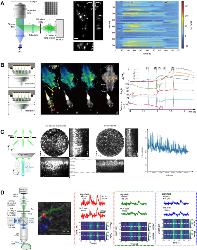
Recording the highly diverse and dynamic activities in large populations of neurons in behaving animals is crucial for a better understanding of how the brain works. To meet this challenge, extensive efforts have been devoted to developing functional fluorescent indicators and optical imaging techniques to optically monitor neural activity. Indeed, optical imaging potentially has extremely high throughput due to its non-invasive access to large brain regions and capability to sample neurons at high density, but the readout speed, such as the scanning speed in two-photon scanning microscopy, is often limited by various practical considerations. Among different imaging methods, light field microscopy features a highly parallelized 3D fluorescence imaging scheme and therefore promises a novel and faster strategy for functional imaging of neural activity. Here, we briefly review the working principles of various types of light field microscopes and their recent developments and applications in neuroscience studies. We also discuss strategies and considerations of optimizing light field microscopy for different experimental purposes, with illustrative examples in imaging zebrafish and mouse brains.
Keywords: Brain activity; Calcium imaging; Light field microscopy; Voltage imaging; Volumetric imaging.
© 2022. Center for Excellence in Brain Science and Intelligence Technology, Chinese Academy of Sciences.
Conflict of interest statement
The authors report no declarations of interest.
Figures

Similar articles
-
Light-field microscopy for fast volumetric brain imaging.J Neurosci Methods. 2021 Mar 15;352:109083. doi: 10.1016/j.jneumeth.2021.109083. Epub 2021 Jan 20. J Neurosci Methods. 2021. PMID: 33484746
-
Imaging volumetric dynamics at high speed in mouse and zebrafish brain with confocal light field microscopy.Nat Biotechnol. 2021 Jan;39(1):74-83. doi: 10.1038/s41587-020-0628-7. Epub 2020 Aug 10. Nat Biotechnol. 2021. PMID: 32778840
-
Light-Sheet Microscopy in Neuroscience.Annu Rev Neurosci. 2019 Jul 8;42:295-313. doi: 10.1146/annurev-neuro-070918-050357. Annu Rev Neurosci. 2019. PMID: 31283896 Free PMC article. Review.
-
Fast functional imaging of multiple brain regions in intact zebrafish larvae using selective plane illumination microscopy.Front Neural Circuits. 2013 Apr 9;7:65. doi: 10.3389/fncir.2013.00065. eCollection 2013. Front Neural Circuits. 2013. PMID: 23576959 Free PMC article.
-
Breaking trade-offs: Development of fast, high-resolution, wide-field two-photon microscopes to reveal the computational principles of the brain.Neurosci Res. 2022 Jun;179:3-14. doi: 10.1016/j.neures.2022.03.010. Epub 2022 Apr 4. Neurosci Res. 2022. PMID: 35390357 Review.
Cited by
-
Population imaging of internal state circuits relevant to psychiatric disease: a review.Neurophotonics. 2025 Jan;12(Suppl 1):S14607. doi: 10.1117/1.NPh.12.S1.S14607. Epub 2025 Jan 28. Neurophotonics. 2025. PMID: 39872404 Free PMC article. Review.
-
Neural Network Mechanisms Underlying General Anesthesia: Cortical and Subcortical Nuclei.Neurosci Bull. 2024 Dec;40(12):1995-2011. doi: 10.1007/s12264-024-01286-z. Epub 2024 Aug 21. Neurosci Bull. 2024. PMID: 39168960 Review.
-
Ultrafast optical imaging techniques for exploring rapid neuronal dynamics.Neurophotonics. 2025 Jan;12(Suppl 1):S14608. doi: 10.1117/1.NPh.12.S1.S14608. Epub 2025 Feb 27. Neurophotonics. 2025. PMID: 40017464 Free PMC article. Review.
-
Role of Neural Circuits in Cognitive Impairment.Neurochem Res. 2024 Dec 7;50(1):49. doi: 10.1007/s11064-024-04309-3. Neurochem Res. 2024. PMID: 39644416 Review.
References
Publication types
MeSH terms
LinkOut - more resources
Full Text Sources
