. 2022 Sep;19(5):1588-1602.
doi: 10.1007/s13311-022-01283-y. Epub 2022 Aug 8.

A Brain-Targeting Bispecific-Multivalent Antibody Clears Soluble Amyloid-Beta Aggregates in Alzheimer's Disease Mice

Affiliations

A Brain-Targeting Bispecific-Multivalent Antibody Clears Soluble Amyloid-Beta Aggregates in Alzheimer's Disease Mice

Fadi Rofo et al. Neurotherapeutics. 2022 Sep.

Abstract

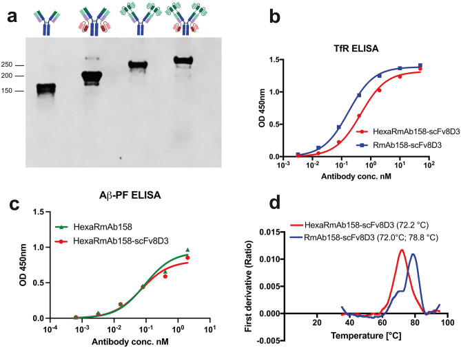
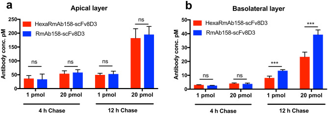
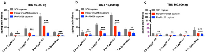
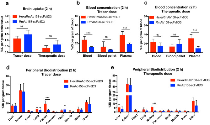
Amyloid-β (Aβ) oligomers and protofibrils are suggested to be the most neurotoxic Aβ species in Alzheimer's disease (AD). Hence, antibodies with strong and selective binding to these soluble Aβ aggregates are of therapeutic potential. We have recently introduced HexaRmAb158, a multivalent antibody with additional Aβ-binding sites in the form of single-chain fragment variables (scFv) on the N-terminal ends of Aβ protofibril selective antibody (RmAb158). Due to the additional binding sites and the short distance between them, HexaRmAb158 displayed a slow dissociation from protofibrils and strong binding to oligomers in vitro. In the current study, we aimed at investigating the therapeutic potential of this antibody format in vivo using mouse models of AD. To enhance BBB delivery, the transferrin receptor (TfR) binding moiety (scFv8D3) was added, forming the bispecific-multivalent antibody (HexaRmAb158-scFv8D3). The new antibody displayed a weaker TfR binding compared to the previously developed RmAb158-scFv8D3 and was less efficiently transcytosed in a cell-based BBB model. HexaRmAb158 detected soluble Aβ aggregates derived from brains of tg-ArcSwe and AppNL-G-F mice more efficiently compared to RmAb158. When intravenously injected, HexaRmAb158-scFv8D3 was actively transported over the BBB into the brain in vivo. Brain uptake was marginally lower than that of RmAb158-scFv8D3, but significantly higher than observed for conventional IgG antibodies. Both antibody formats displayed similar brain retention (72 h post injection) and equal capacity in clearing soluble Aβ aggregates in tg-ArcSwe mice. In conclusion, we demonstrate a bispecific-multivalent antibody format capable of passing the BBB and targeting a wide-range of sizes of soluble Aβ aggregates.

Keywords: Aβ; BBB; Bispecific; Mouse models; Multivalent antibodies; Oligomers.


Conflict of interest statement

The authors declare no competing interests.

Figures

Fig. 1
Fig. 1
Design of the four recombinant antibodies. a RmAb158. b RmAb158-scFv8D3 where scFv8D3 is added to the C-terminal end of the RmAb158 light chains. c HexaRmAb158, where scFvs consisting of the heavy and light variable domains of RmAb158 are added to the N-terminal ends of RmAb158’s heavy and light chains. d HexaRmab158-scFv8D3, where scFv8D3 is added to the C-terminal ends of the HexaRmAb158 light chains
Fig. 2
Fig. 2
In vitro characterization of HexaRmAb158-scFv8D3. a SDS-PAGE showing a band at around 150 kDa for RmAb158, 200 kDa for RmAb158-scFv8D3, 250 kDa for Hexa-RmAb158 and 300 kDa for HexaRmAb158-scFv8D3. The complete SDS-PAGE can be found in Supplementary Fig. 5. b Indirect ELISA displaying binding of HexaRmAb158-scFv8D3 to mouse TfR. Binding to mouse TfR was twofold weaker with HexaRmAb158-scFv8D3 (EC50 = 6.3 nM) compared to RmAb158-scFv8D3 (EC50 = 3.2 nM). c Indirect ELISA displaying binding of HexaRmAb158-scFv8D3 to Aβ protofibrils. Similar binding to Aβ protofibrils was observed for HexaRmAb158 and HexaRmAb158-scFv8D3, demonstrating retained functionality despite the additional binding sites of HexaRmAb158-scFv8D3. d Structural stability of HexaRmAb158-scFv8D3 and RmAb158-scFv8D3 under thermal stress measured with Tycho nt.6 instrument
Fig. 3
Fig. 3
Graphical representations comparing the concentration of RmAb158-scFv8D3 and HexaRmAb158-scFv8D3 in the apical (a) and basolateral (b) compartments of the in vitro BBB transcytosis assay. Statistically significant decreases in the transport across the cEND cells into the basolateral layer were detected for HexaRmAb158-scFv8D3 compared to RmAb158-scFv8D3, both at low (1 pmol) and high (20 pmol) doses, only after 12 h chase. Results are presented as mean ± SD and analysed with unpaired t-test. (*: p < 0.05, **: p < 0.01, ***: p < 0.001, n.s: p > 0.05) (n = 6 per treatment)
Fig. 4
Fig. 4
HexaRmAb158 and RmAb158 sandwich ELISA for the measurement of soluble Aβ aggregates in brain homogenates from AppNL−G−F mice (2.5, 5 and 9 months old) and tg-ArcSwe mice (7 months old), using 3D6 ELISA as a control. a HexaRmAb158 ELISA detected significantly higher concentrations of soluble Aβ aggregates compared to RmAb158 ELISA in TBS treated brain homogenates. b HexaRmAb158 ELISA detected significantly higher concentrations of Aβ compared to RmAb158 ELISA in TBS-Triton (TBS-T) brain homogenates from 5-month-old and 9-month-old AppNL−G−F mice. c No significant differences in Aβ concentrations were detected by HexaRmAb158 and RmAb158 ELISAs in TBS-brain homogenates ultra-centrifuged at 100 000 × g. HexaRmAb158 displayed a trend (p = 0.06) towards increased detection of Aβ aggregates in tg-ArcSwe brain TBS homogenates ultra-centrifuged at 100 000 × g. Results are presented as mean ± SD. To detect statistically significant differences between HexaRmAb158 and RmAb158 ELISAs, unpaired t test was applied. (*: p < 0.05, **: p < 0.01, ***: p < 0.001, n.s: p > 0.05) (n = 6 for 2.5 m AppNL−G−F, n = 10 for 5 m AppNL−G−F, n = 7 for 9 m App.NL−G−F, n = 6 for 7 m tg-ArcSwe)
Fig. 5
Fig. 5
Comparison of [125I]HexaRmAb158-scFv8D3 and [125I]RmAb158-scFv8D3 concentrations in the brain and blood of 6-month-old C57Bl/6 WT mice. a Brain concentrations of [125I]HexaRmAb158-scFv8D3 and [125I]RmAb158-scFv8D3, expressed as percentage of injected dose (% ID) per gram brain tissue, 2 h post injection. b Statistically significant increase in blood and plasma concentrations of [125I]HexaRmAb158-scFv8D3 compared to [125I]RmAb158-scFv8D3, 2 h post injection, when given as a tracer dose. c Statistically significant increase in plasma concentrations of [125I]HexaRmAb158-scFv8D3 compared to [125I]RmAb158-scFv8D3, 2 h post injection, when given as a therapeutic dose. d and e Significantly higher concentrations of [125I]HexaRmAb158-scFv8D3 in the kidneys compared to [125I]RmAb158-scFv8D3, 2 h post injection, given at tracer and therapeutic doses respectively. Results are presented as mean ± SD and analysed with unpaired t test. (*: p < 0.05, **: p < 0.01, ***: p < 0.001, n.s.: p > 0.05) (n = 5 per group)
Fig. 6
Fig. 6
Biodistribution of [125I]HexaRmAb158-scFv8D3 and [125I]RmAb158-scFv8D3 in 10–11 months old tg-ArcSwe mice at 72 h post injection of a therapeutic dose of 24 nmol/kg. a Brain retention of [125I]HexaRmAb158-scFv8D3 and [125I]RmAb158-scFv8D3 in tg-ArcSwe mice, expressed as percentage of injected dose (% ID) per gram brain tissue, 72 h post injection. No statistically significant differences were detected between the two groups. b Blood concentrations of the two antibodies measured 1–72 h post injection. c Blood concentrations of [125I]HexaRmAb158-scFv8D3 were lower compared those of [125I]RmAb158-scFv8D3 at 72 h post injection. d Similar retention of [125I]HexaRmAb158-scFv8D3 and [125I]RmAb158-scFv8D3 in the peripheral organs, 72 h post injection. A trend towards a lower retention in the spleen was detected for [125I]HexaRmAb158-scFv8D3. Results are presented as mean ± SD and analysed with unpaired t-test. (*: p < 0.05, **: p < 0.01, ***: p < 0.001, n.s.: p > 0.05) (n = 5 per group)
Fig. 7
Fig. 7
Concentration of soluble Aβ aggregates in the brain of tg-ArcSwe mice following treatment with therapeutic doses of 24 nmol/kg of HexaRmAb158-scFv8D3 or RmAb158-scFv8D3, using PBS as a negative control. a No significant differences were detected in concentration of soluble Aβ aggregates among the groups in TBS and TBS-T brain extracts centrifuged at 16,000 xg. b Concentration of soluble Aβ aggregates were significantly reduced in TBS soluble brain extracts ultra-centrifuged at 100,000 × g following treatment with HexaRmAb158-scFv8D3 and RmAb158-scFv8D3 compared to PBS. c Concentration of soluble Aβ aggregates were significantly reduced in TBS soluble brain extracts ultra-centrifuged at 200,000 × g following treatment with HexaRmAb158-scFv8D3 and RmAb158-scFv8D3 compared to PBS controls. d No significant differences were detected in total Aβ38, Aβ40 and Aβ42 concentration among the groups in FA brain extracts centrifuged at 16,000 xg. Results are presented as mean ± SD and analysed with one-way ANOVA followed by Bonferroni’s post-hoc analysis. (*: p < 0.05, **: p < 0.01, ***: p < 0.001, n.s.: p > 0.05), (n = 4/PBS, n = 5/HexaRmAb158-scFv8D3, n = 5/RmAb158-scFv8D3)

Similar articles

Cited by

References

    1. Chen GF, Xu TH, Yan Y, Zhou YR, Jiang Y, Melcher K, et al. Amyloid beta: structure, biology and structure-based therapeutic development. Acta Pharmacol Sin. 2017;38(9):1205–1235. - PMC - PubMed
    1. Hardy JA, Higgins GA. Alzheimer’s disease: the amyloid cascade hypothesis. Science. 1992;256(5054):184–185. - PubMed
    1. Long JM, Holtzman DM. Alzheimer disease: an update on pathobiology and treatment strategies. Cell. 2019;179(2):312–339. - PMC - PubMed
    1. Esparza TJ, Wildburger NC, Jiang H, Gangolli M, Cairns NJ, Bateman RJ, et al. Soluble amyloid-beta aggregates from human Alzheimer’s disease brains. Sci Rep. 2016;5(6):38187. - PMC - PubMed
    1. Ferreira ST, Lourenco MV, Oliveira MM, De Felice FG. Soluble amyloid-β oligomers as synaptotoxins leading to cognitive impairment in Alzheimer’s disease. Front Cell Neurosci. 2015;9:191. - PMC - PubMed

Publication types