. 2022 Aug 8;18(8):e1010763.
doi: 10.1371/journal.ppat.1010763. eCollection 2022 Aug.

TMEM41B and VMP1 modulate cellular lipid and energy metabolism for facilitating dengue virus infection

Affiliations

TMEM41B and VMP1 modulate cellular lipid and energy metabolism for facilitating dengue virus infection

Meisam Yousefi et al. PLoS Pathog. .

Abstract

Transmembrane Protein 41B (TMEM41B) and Vacuole Membrane Protein 1 (VMP1) are two ER-associated lipid scramblases that play a role in autophagosome formation and cellular lipid metabolism. TMEM41B is also a recently validated host factor required by flaviviruses and coronaviruses. However, the exact underlying mechanism of TMEM41B in promoting viral infections remains an open question. Here, we validated that both TMEM41B and VMP1 are essential host dependency factors for all four serotypes of dengue virus (DENV) and human coronavirus OC43 (HCoV-OC43), but not chikungunya virus (CHIKV). While HCoV-OC43 failed to replicate entirely in both TMEM41B- and VMP1-deficient cells, we detected diminished levels of DENV infections in these cell lines, which were accompanied by upregulation of the innate immune dsRNA sensors, RIG-I and MDA5. Nonetheless, this upregulation did not correspondingly induce the downstream effector TBK1 activation and Interferon-beta expression. Despite low levels of DENV replication, classical DENV replication organelles were undetectable in the infected TMEM41B-deficient cells, suggesting that the upregulation of the dsRNA sensors is likely a consequence of aberrant viral replication rather than a causal factor for reduced DENV infection. Intriguingly, we uncovered that the inhibitory effect of TMEM41B deficiency on DENV replication, but not HCoV-OC43, can be partially reversed using exogenous fatty acid supplements. In contrast, VMP1 deficiency cannot be rescued using the metabolite treatment. In line with the observed phenotypes, we found that both TMEM41B- and VMP1-deficient cells harbor higher levels of compromised mitochondria, especially in VMP1 deficiency which results in severe dysregulations of mitochondrial beta-oxidation. Using a metabolomic profiling approach, we revealed distinctive global dysregulations of the cellular metabolome, particularly lipidome, in TMEM41B- and VMP1-deficient cells. Our findings highlight a central role for TMEM41B and VMP1 in modulating multiple cellular pathways, including lipid mobilization, mitochondrial beta-oxidation, and global metabolic regulations, to facilitate the replication of flaviviruses and coronaviruses.


Conflict of interest statement

The authors have declared that no competing interests exist.

Figures
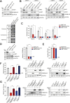
Fig 1
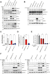
Fig 1. TMEM41B and VMP1 are host dependency factors for flaviviruses and human coronaviruses, but not alphaviruses.
(A) Multiple rounds of virus infection in 293FT WT, TMEM41B KO clone #1, and VMP1 KO clone #1 cells. (Top) Western blotting analysis to detect TMEM41B and VMP1 protein levels in the WT and KO cells. (Bottom) Crystal violet staining to visualize surviving cells after cytolytic virus infection. Non-infected controls are indicated as “mock”. (B, C) Validation of TMEM41B and VMP1 as host dependency factors across all DENV serotypes. For (B), cells were infected by either DENV1 (MOI = 1 PFU/cell), DENV2 (MOI = 0.1 PFU/cell), DENV3 (MOI = 3 PFU/cell), or DENV4 (MOI = 5 PFU/cell) for 72 hours. Accumulation of DENV NS3 proteins was detected by western blot. For (C), cells were infected at MOI of 1 for 72 hours. Released progeny virions in the supernatant were assessed by plaque assay. (D) HCoV-OC43 protein accumulation (Left) and progeny virion levels (Right) in 293FT WT and KO cells. Cells were infected with HCoV-OC43 at MOI of 0.5 for 48 hours. Released progeny virions in the supernatants were harvested for titration by TCID50 assay, and infected cell lysates were analyzed by western blotting. (E) Production of CHIKV progeny virions from WT and KO cells. Cells were infected with CHIKV at MOI of 0.1 for 24 hours, and released virions were harvested for titration by plaque assay. (F) cDNA complementation rescue of DENV infection. (Left) Production of DENV2 infectious particles from KO cells complemented with corresponding cDNAs, i.e., mCherry (control), TMEM41B, or VMP1. (Right) Western blotting analysis of DENV2 NS3 accumulation in WT, KO, and cDNA-complemented cells. Cells were infected at MOI of 0.1 for 72 hours. Released virions in the supernatants were harvested for titration by plaque assay, and cell lysates were analyzed by western blotting. (G) Validation of TMEM41B and VMP1 as DENV host factors in A549 cells. (Left) Production of DENV1 infectious particles from A549 WT, TMEM41B KO clone #1, and VMP1 KO clone #1 cells complemented with corresponding cDNAs. (Right) Western blotting analysis of DENV1 NS3 accumulation in WT, KO, and cDNA-complemented cells. Cells were infected at MOI of 0.5 for 32 hours. GAPDH was used as a loading control for all blots. All data shown represent results from at least two independent experiments. Error bars represent mean +/- SEM, n = 3. * indicates p-value < 0.05 as determined by two-tailed unpaired t-test or one-way ANOVA. LOD indicates the limit of detection.
Fig 2
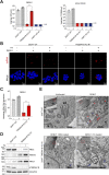
Fig 2. Upregulation of dsRNA sensors in 293FT TMEM41B KO and VMP1 KO cells upon DENV infection.
(A) Western blotting analysis to detect RIG-I, MDA5, TBK1, and p-TBK1 protein levels in 293FT WT cells, TMEM41B KO clone #1, and VMP1 KO clone #1, upon DENV infection. Cells were infected with DENV1 at MOI of 1 and lysates were harvested at 48 hours post-infection. (B) Western blotting analysis to detect RIG-I and MDA5 protein levels in 293FT WT, TMEM41B KO clone #1, and VMP1 KO clone #1 cells, upon HCoV-OC43 infection. Cells were infected with HCoV-OC43 at MOI of 0.5 and lysates were harvested at 48 hours post-infection. (C,D) IFN-β mRNA (C) and DENV RNA (D) abundance in uninfected and DENV1 infected (MOI of 1, 48 hours post-infection) WT and KO cells. Values demonstrated are relative to the expression levels in uninfected WT cells (C), or DENV infected WT cells (D). (E,F) DENV viral protein accumulation and infectious viral particles produced in 293FT WT, TMEM41B KO clone #1, and VMP1 KO clone #1 cells, upon further knocking out of RIG-I and MDA5. Infection assays were carried out at MOI of 1 and (E) the amount of infectious virus in the supernatant was determined at 48 hours post-infection by plaque assay, and (F) protein levels of the indicated proteins were determined by western blotting analysis. GAPDH was used as a loading control for all blots. 18S rRNA was used as the control for the RT-qPCR experiments. All data shown represent results from at least two independent experiments. Single-dashed lines divide blots from two gels loaded with the same set of lysates. Double-dashed lines divide blots from gels belonging to separate experiments using the same experimental conditions. Error bars represent mean +/- SEM, n = 3. * indicates p-value < 0.05 as determined by one-way ANOVA. LOD indicates the limit of detection.
Fig 3
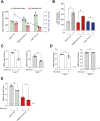
Fig 3. Exogenous fatty acid complementation can partially rescue DENV infection in 293FT TMEM41B KO cells but not VMP1.
(A) Infectious viral particles produced in 293FT WT, TMEM41B KO clone #1, and VMP1 KO clone #1 cells upon FA supplementation. DENV1 infection was carried out at MOI of 1 and progeny viruses were harvested at 72 hours post-infection. HCoV-OC43 infection was done at MOI of 0.5 and progeny viruses were harvested at 48 hours post-infection. Cells were treated with a 1:1 mixture of oleic and linoleic acid (FA), or BSA. (B) Confocal microscopy imaging of WT and TMEM41B KO clone #1 cells infected with DENV1 and supplemented with FAs or BSA; dsRNA is stained in red and cell nuclei are displayed in blue. Image scales are indicated in the merged images. (C) RT-qPCR analysis of DENV RNA levels in infected (MOI of 1, 48 hours post-infection) WT and KO cells, upon treatment with FA or BSA. Values demonstrated are relative to DENV RNA load in infected WT cells. 18S rRNA has been used as the internal control. (D) Western blotting analysis to compare DENV viral protein accumulation in WT and TMEM41B KO cells, with and without FA supplementation. Cells were infected with DENV1 at MOI of 1 and lysates were harvested 48 hours post-infection. GAPDH was used as a loading control. (E) TEM imaging of DENV1-infected (MOI of 5, 48 hours post-infection) 293FT WT and TMEM41B KO clone #1. Virion-like particles and classical DENV ROs are indicated with blue and red arrowheads, respectively. Nuclei are identified by “N”. The ER structures are indicated as “ER”. Scale bars are as indicated. All data shown represent results from at least two independent experiments. Error bars represent mean +/- SEM, n = 3. * indicates p-value < 0.05 as determined by two-tailed unpaired t-test or one-way ANOVA. LOD indicates the limit of detection.
Fig 4
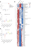
Fig 4. Impaired mitochondrial beta-oxidation contributes to reduced DENV infection in 293FT TMEM41B KO and VMP1 KO cells.
(A) Comparison of total (MitoView Green) vs functionally active (MitoView 633) mitochondria level in 293 FT WT, TMEM41B KO clone #1, and VMP1 KO clone #1 cells. Blue dashed line indicates the ratio of MitoView 633 to MitoView Green signal level. (B) Beta-oxidation associated ATP production in the WT and clonal KO cells upon FA supplementation, measured by extracellular flux analysis. (C) Impact of Etomoxir treatment on production of infectious DENV particles in 293FT WT and Huh7 WT cells. Cells were infected with DENV1 at MOI of 1 and treated with 200 μM Etomoxir for 48 hours. (D) Impact of Etomoxir treatment on production of infectious CHIKV particles in 293FT WT and Huh7 WT cells. Cells were infected with CHIKV at MOI of 0.1 and treated with 200 μM Etomoxir for 24 hours. (E) Impact of Etomoxir treatment on production of infectious DENV particles in 293FT WT cells and TMEM41B KO clone #1. Cells were infected with DENV1 at MOI of 1 and treated with 200 μM Etomoxir for 48 hours. All data shown represent results from at least two independent experiments. Error bars represent mean +/- SEM, n = 3. * indicates p-value < 0.05 as determined by two-tailed unpaired t-test or one-way ANOVA. LOD indicates the limit of detection.
Fig 5
Fig 5. TMEM41B and VMP1 deficiency impose a diverse and distinctive impact on cellular metabolism in 293FT.
(A) Principal Components Analysis (PCA) for the metabolomic datasets of 293FT WT, TMEM41B clone #1, and VMP1 KO clone #1 cells. First two principal components are demonstrated on the x- and y-axis. (B) Heatmap of altered metabolites in the 293FT WT and KO clones. Z-score normalized values of metabolite levels were used to plot the heatmap (see Materials and Methods). Color scheme depicts the relative abundance of metabolites with red and blue indicating higher and lower levels, respectively. Columns indicate different samples, each with 3 replicates. (C) Relative fold changes of metabolites level in TMEM41B KO clone #1 cells in comparison to 293FT WT. Values associated with metabolites from the same family are grouped together in each boxplot. Negative values represent metabolites with decreased abundance in the KO cells, as described in the Materials and Methods. (D) Relative fold changes of metabolites level in VMP1 KO #1 cells in comparison to 293FT WT. Values associated with metabolites from the same family are grouped together in each boxplot. Negative values represent metabolites with decreased abundance in the KO cells, as described in the Materials and Methods.
Fig 6
Fig 6. The schematic model for multiple roles of TMEM41B and VMP1 in DENV infection.
TMEM41B and VMP1 modulate ER homeostasis, lipid mobilization, mitochondrial beta-oxidation, and global metabolomic regulations to facilitate productive DENV infection. This figure was created with Biorender.com.

Similar articles

Cited by

References

    1. Guzman MG, Harris E. Dengue. Lancet. 2015;385: 453–465. doi: 10.1016/S0140-6736(14)60572-9 - DOI - PubMed
    1. Halstead SB. Dengvaxia sensitizes seronegatives to vaccine enhanced disease regardless of age. Vaccine. 2017;35: 6355–6358. doi: 10.1016/j.vaccine.2017.09.089 - DOI - PubMed
    1. Gubler DJ, Halstead SB. Is Dengvaxia a useful vaccine for dengue endemic areas? BMJ. 2019;367: l5710. doi: 10.1136/bmj.l5710 - DOI - PubMed
    1. Puschnik AS, Majzoub K, Ooi YS, Carette JE. A CRISPR toolbox to study virus-host interactions. Nat Rev Microbiol. 2017;15: 351–364. doi: 10.1038/nrmicro.2017.29 - DOI - PMC - PubMed
    1. Marceau CD, Puschnik AS, Majzoub K, Ooi YS, Brewer SM, Fuchs G, et al.. Genetic dissection of Flaviviridae host factors through genome-scale CRISPR screens. Nature. 2016;535: 159–63. doi: 10.1038/nature18631 - DOI - PMC - PubMed

Publication types