. 2022 Aug 16;119(33):e2117904119.
doi: 10.1073/pnas.2117904119. Epub 2022 Aug 8.

Lactobacillus crispatus Limits Bladder Uropathogenic E. coli Infection by Triggering a Host Type I Interferon Response

Affiliations

Lactobacillus crispatus Limits Bladder Uropathogenic E. coli Infection by Triggering a Host Type I Interferon Response

Chang Hyun Song et al. Proc Natl Acad Sci U S A. .

Abstract

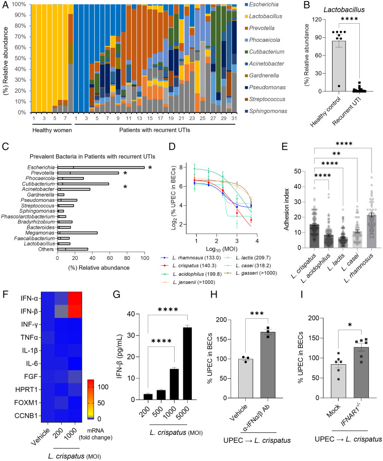
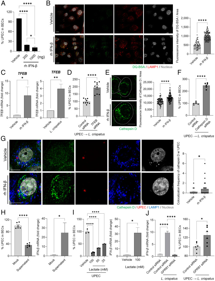
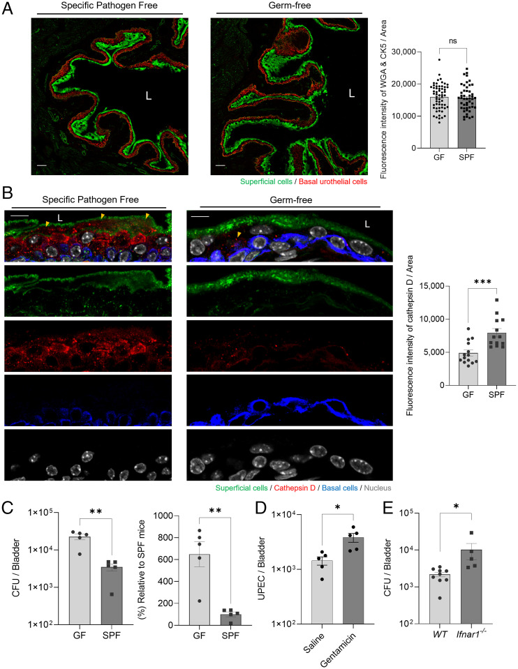
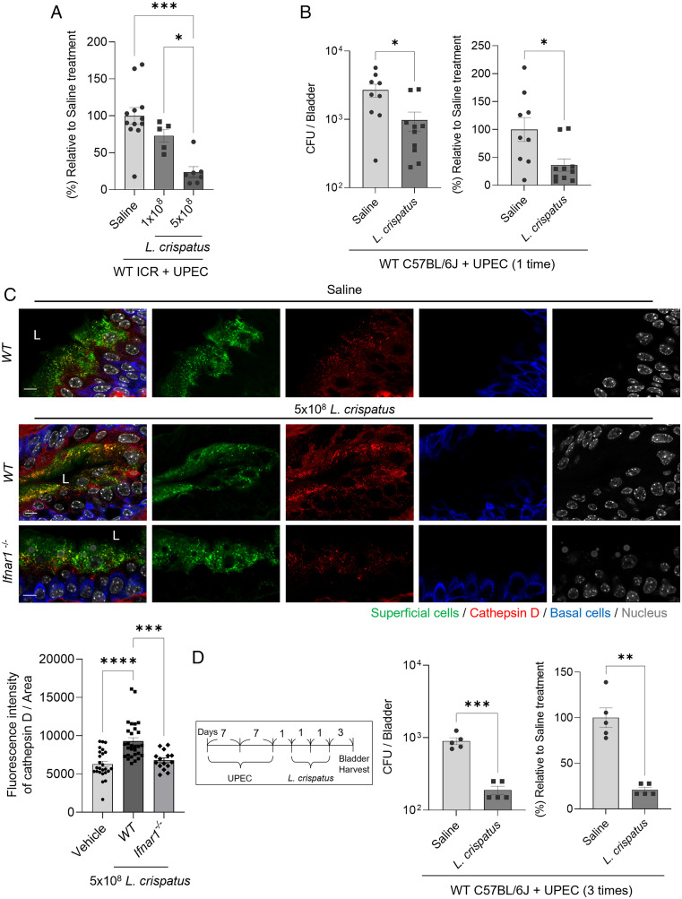
Many urinary tract infections (UTIs) are recurrent because uropathogens persist within the bladder epithelial cells (BECs) for extended periods between bouts of infection. Because persistent uropathogens are intracellular, they are often refractive to antibiotic treatment. The recent discovery of endogenous Lactobacillus spp. in the bladders of healthy humans raised the question of whether these endogenous bacteria directly or indirectly impact intracellular bacterial burden in the bladder. Here, we report that in contrast to healthy women, female patients experiencing recurrent UTIs have a bladder population of Lactobacilli that is markedly reduced. Exposing infected human BECs to L. crispatus in vitro markedly reduced the intracellular uropathogenic Escherichia coli (UPEC) load. The adherence of Lactobacilli to BECs was found to result in increased type I interferon (IFN) production, which in turn enhanced the expression of cathepsin D within lysosomes harboring UPECs. This lysosomal cathepsin D-mediated UPEC killing was diminished in germ-free mice and type I IFN receptor-deficient mice. Secreted metabolites of L. crispatus seemed to be responsible for the increased expression of type I IFN in human BECs. Intravesicular administration of Lactobacilli into UPEC-infected murine bladders markedly reduced their intracellular bacterial load suggesting that components of the endogenous microflora can have therapeutic effects against UTIs.

Keywords: Lactobacilli; commensal bacteria; type I interferon; urinary tract infection.


Conflict of interest statement

The authors declare no competing interest.

Figures

Fig. 1.
Fig. 1.
Lactobacilli, prevalent in healthy women, decreased intracellular UPEC load by triggering the expression of type I IFN in BECs. (A) Distribution of bacterial genera in the urine of healthy women or patients with recurrent UTIs. Color-coded columns indicate bacterial genera. (B) Comparison of the relative population of Lactobacilli in healthy women (n = 8) or patients with recurrent UTIs (n = 31). Each data point represents one person. (C) Floating bar graphs indicate relative bacterial abundance in the urine of patients with recurrent UTIs. The line in each bar represents the mean value. Except for Prevotella, Escherichia has a statistically higher abundance. Prevotella outnumbers all other bacteria except Phocaeicola and Cutibacterium. (D) Intracellular UPEC killing by Lactobacilli. UPEC CI5 strain infected human 5637 BECs (MOI 200). After 6 h, infected cells were treated with Lactobacilli at four different MOI (5000, 1000, 200, and 40). The intracellular number of UPECs in treated BECs was counted after overnight incubation. The numbers in parenthesis are the MOI values for 50% reduction (IC50) which were interpolated from the curves. (E) Adhesion of five Lactobacilli strains (adhered Lactobacillus/BEC) was compared by treating 5636 BECs with Lactobacilli at MOI 1000. After 2 h of incubation, cells were thoroughly washed and stained using a Gram staining solution. (F) Human 5637 BECs were treated with L. crispatus at 200 or 1000 MOI for 6 h. qRT-PCR was used to assess changes in messenger RNA (mRNA) expression. (G) Human IFN-β secreted by human 5637 BECs in response to L. crispatus exposure was assessed by ELISA. (H) Neutralization of type I IFN following treatment with specific antibodies. UPEC-infected cells were pretreated with anti-IFN-α/β antibodies before exposure to L. crispatus. The intracellular numbers of UPEC in pretreated BECs was assessed after 16 h. (I) Increased UPEC load on IFNAR1−/−. The IFNAR gene of human 5637 BECs was deleted by using the CRISPR/Cas9 system (pX459). IFNAR1−/− or mock control BECs (empty pX459) were infected with UPEC CI5 strain (MOI 200) for 6 h, and then treated with L. crispatus (MOI 500). The intracellular UPEC number was counted overnight. Error bars in each bar graph show mean ± SEM. Each experiment (DI) was independently repeated two to three times with similar results. Data were analyzed by unpaired two-tailed Student t test (B, H, I) or an ordinary one-way ANOVA (C, E, G) with Tukey’s multiple comparison posttest. *P < .05; **P < .01; ***P < .001; ****P < .0001. Ab, antibody.
Fig. 2.
Fig. 2.
Intracellular bactericidal action of type I IFN is mediated by lysosomal maturation. (A) Intracellular UPEC killing by type I IFN. Human 5637 BECs were treated with L. crispatus at various MOI (200). After 6 h of infection, the bacterial burden was measured 20 h after treatment with recombinant human (rh) IFN-β (200 or 1000 ng). (B) Lysosomal maturation by IFN-β. Human 5637 BECs were treated with rh IFN-β (500 ng). After 24 h of treatment, cells were exposed to DQ-BSA (green) for 6 h and then immunostained using an anti-LAMP1 (red) antibody for confocal microscopic imaging. (B, Right) The mean fluorescence intensity of DQ-BSA was measured in random fields. (C) After treatment with rh IFN-β (500 ng) for 6 h, the expression of TFEB mRNA in cells treated with IFN-β or L. crispatus was compared to that in vehicle-treated control using qPCR. (D) TFEB mediated L. crispatus-induced intracellular UPEC killing in BECs. After human BECs were transfected with control or TFEB siRNA, UPEC-infected BECs were exposed to L. crispatus. The intracellular number of UPEC in treated BECs was assessed after 16 h. (E) Enhanced cathepsin D expression was assessed by treating BECs with rh IFN-β (500 ng) for 24 h followed by anti-cathepsin D (green) immunostaining for confocal imaging. The dotted line indicates the periphery of BECs. (E, Right) The fluorescence intensity of cathepsin D was measured in random fields. (F) Cathepsin D regulates the size of intracellular UPEC population. After human 5637 BECs were transfected with control or cathepsin D siRNA, UPEC-infected BECs were exposed to L. crispatus. The intracellular number of UPEC in treated BECs was assessed after 16 h. (G) UPEC-infected BECs were treated for 12 h with IFN-β (500 ng) and then immunostained for cathepsin D (green), UPEC (red), and LAMP1 (blue). (G, Right) The frequency of colocalization between cathepsin D and UPEC was measured in random fields. (H) Effects of CFS of L. crispatus on human BECs. (H, Left) UPEC-infected human BECs were CFS or mock treated for 16 h, and the bacterial load was determined. (H, Right) The relative change of IFN-β mRNA expression by human BECs was analyzed using qPCR after 6 h of exposure to CFS of L. crispatus. (I) Effects of lactate on human BECs. (I, Left) UPEC-infected human BECs were treated with lactate in a dose-dependent manner or mock treated for 16 h; thereafter, the intracellular bacterial load was determined. (I, Right) The relative change in expression levels of IFN-β mRNA in human BECs was assessed using qPCR after 6 h of treatment with lactate (100 mM). (J) The lactate effect on BECs is mediated by GPR81. After human 5637 BECs were transfected with control or GPR81 siRNA, (J, Left) the relative change in IFN-β mRNA expression levels was assessed using qPCR after 6 h of treatment with L. crispatus. (J, Right) UPEC-infected BECs were exposed to L. crispatus (MOI 1000), and the intracellular number of UPECs in treated BECs was assessed after 16 h. Error bars in each bar graph show mean ± SEM. Each experiment (AJ) was independently repeated two to three times with similar results. Data were analyzed by unpaired two-tailed Student t test (BJ) or an ordinary one-way ANOVA (A and I) with Tukey’s multiple comparison posttest. *P < .05; **P < .01; ****P < .0001. Scale bars: 10 μm.
Fig. 3.
Fig. 3.
Host protective role of endogenous microbiome mediated by upregulation of cathepsin D expression. (A) Comparison of the expression of BEC markers in C57BL/6J GF SPF mice. Immunostaining of superficial epithelial cells (green; wheat-germ-agglutinin-fluorescein isothiocyanate [WGA-FITC]) and basal urothelial cells (red; cytokeratin 5 [CK5]) in bladders of C57BL/6J GF or SPF mice. Scale bars: 50 μm. “L” denotes bladder lumen. (A, Right) The fluorescence intensity of WGA and CK5 was measured in random fields. (B) Bladders sectioned from GF or SPF mice were stained using anti-cathepsin D, CK5 antibodies, or WGA-FITC. Yellow arrowheads indicate cathepsin D expression in BECs. Representative images were chosen. (B, Right) The fluorescence intensity of cathepsin D was measured in random fields. Scale bars: 10 μm. (C) Intravesical infection of GF or SPF mice with UPEC CI5 (1 × 10⁸ CFU). Bladders from each group were harvested after 24 h to count the bacterial burden. (D) Host protective role of gentamicin-sensitive endogenous microbiome in the bladder. WT C57BL/6J mice were administered gentamicin (10 mg/kg) intraperitoneally and (1 mg/kg) intravesically three times one day apart. Three days after the last treatment, the bacterial load of UPEC-infected mouse bladders was examined. (E) Intravesical infection of WT or Ifnar1−/− mice with UPEC CI5 (1 × 10⁸ CFU). Bladders from each group were harvested after 24 h to count the bacterial burden. Each data point represents one mouse. Error bars in each bar graph show mean ± SEM. Each experiment (AE) was independently repeated, and representative results were presented. Data were analyzed by unpaired two-tailed Student t test (AE). *P < .05; **P < .01; ***P < .001. ns, not significant.
Fig. 4.
Fig. 4.
Reduced bacterial burden in UPEC-infected bladders mediated by treatment with L. crispatus. (A) Intravesical infection of ICR female mice with UPEC CI5 strain (1 × 10⁸ CFU). After 24 h, infected mice were intravesically inoculated with L. crispatus (1 × 10⁸ or 5 × 10⁸ CFU). The bladders fromeach group of mice were harvested after 24 h. (B) Intravesical infection of C57BL/6J mice with UPEC CI5 strain (1 × 10⁸ CFU). After 24 h, infected mice were intravesically inoculated with L. crispatus (5 × 10⁸ CFU). The bladders from each group of mice were harvested after 24 h. (C) Intravesical inoculation of L. crispatus (5 × 10⁸ CFU) or saline into C57BL/6J mice or Ifnar1−/− mice. After 24 h, WGA (superficial epithelial cell), anti-cathepsin D, or anti-cytokeratin 5 (basal urothelial cell) antibodies were used to stain each bladder, and representative images were chosen. (D) The UPEC CI5 strain (1 × 10⁸ CFU) was used to intravesically infect C57BL/6J mice three times over a period of 7 d. L. crispatus (5 × 10⁸ CFU) was intravesically instilled three times after 24 h from the last infection. The bladders from each group of mice were harvested to assess the bacterial burden. Each data point represents one mouse. Error bars in each bar graph show mean ± SEM. Each experiment (AD) was independently repeated two to three times with similar results. Data were analyzed by an ordinary one-way ANOVA (A and C) with Tukey’s multiple comparison posttest or unpaired two-tailed Student t test (B and D). *P < .05; **P < .01; ***P < .001; ****P < .0001.

Comment in

  • Uro-Science.
    Atala A. Atala A. J Urol. 2023 Apr;209(4):801-802. doi: 10.1097/JU.0000000000003161. Epub 2023 Jan 19. J Urol. 2023. PMID: 36655472 No abstract available.

References

    1. Langille M. G., et al. , Predictive functional profiling of microbial communities using 16S rRNA marker gene sequences. Nat. Biotechnol. 31, 814–821 (2013). - PMC - PubMed
    1. Thomas-White K., et al. , Culturing of female bladder bacteria reveals an interconnected urogenital microbiota. Nat. Commun. 9, 1557 (2018). - PMC - PubMed
    1. Pearce M. M., et al. , The female urinary microbiome: A comparison of women with and without urgency urinary incontinence. MBio 5, e01283-e14 (2014). - PMC - PubMed
    1. Wolfe A. J., Brubaker L., Urobiome updates: Advances in urinary microbiome research. Nat. Rev. Urol. 16, 73–74 (2019). - PMC - PubMed
    1. Nickel J. C., et al. ; MAPP Research Network, A culture-independent analysis of the microbiota of female interstitial cystitis/bladder pain syndrome participants in the MAPP Research Network. J. Clin. Med. 8, 415 (2019). - PMC - PubMed

Publication types

MeSH terms