. 2022 Aug 16;119(33):e2204706119.
doi: 10.1073/pnas.2204706119. Epub 2022 Aug 8.

Oropouche orthobunyavirus infection is mediated by the cellular host factor Lrp1

Affiliations

Oropouche orthobunyavirus infection is mediated by the cellular host factor Lrp1

Madeline M Schwarz et al. Proc Natl Acad Sci U S A. .

Abstract

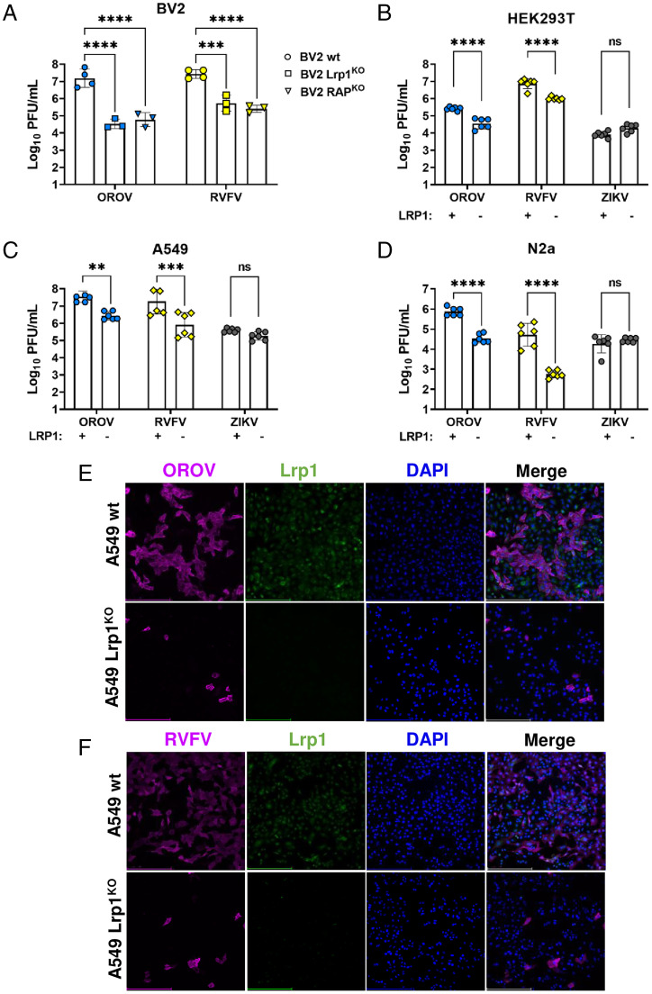
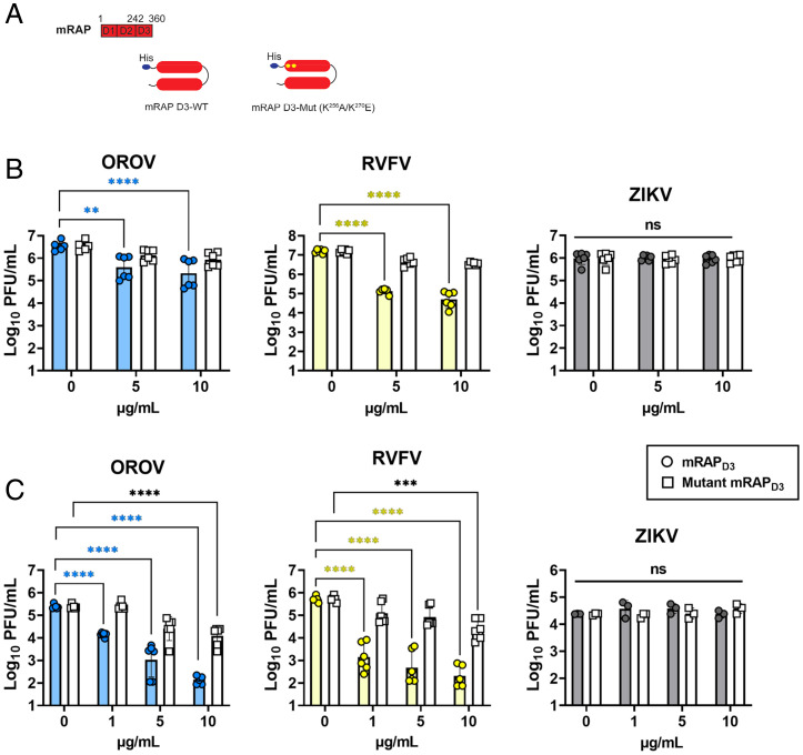
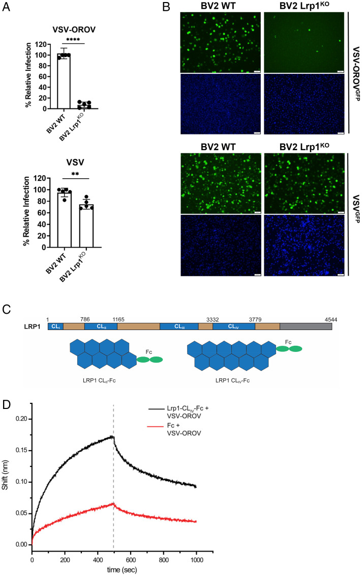
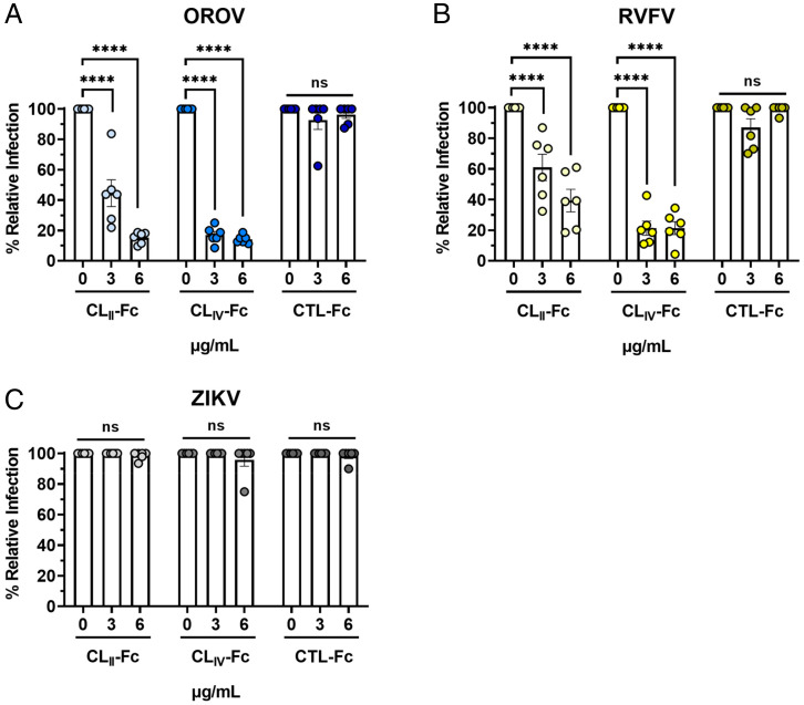
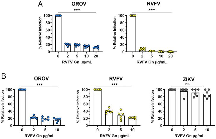
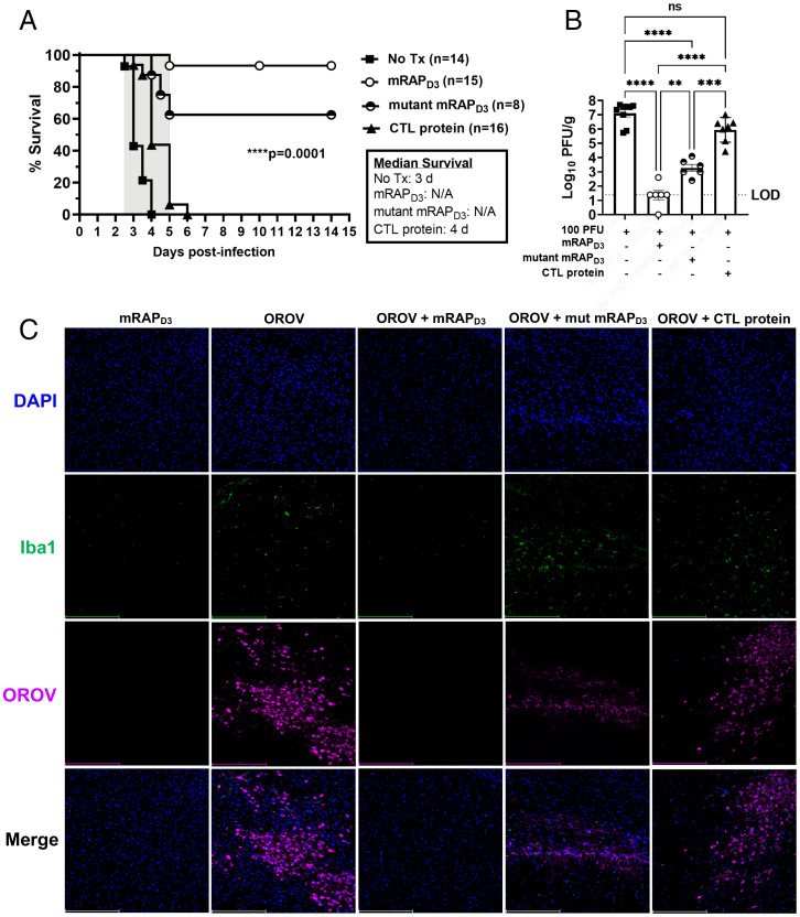
Oropouche orthobunyavirus (OROV; Peribunyaviridae) is a mosquito-transmitted virus that causes widespread human febrile illness in South America, with occasional progression to neurologic effects. Host factors mediating the cellular entry of OROV are undefined. Here, we show that OROV uses the host protein low-density lipoprotein-related protein 1 (Lrp1) for efficient cellular infection. Cells from evolutionarily distinct species lacking Lrp1 were less permissive to OROV infection than cells with Lrp1. Treatment of cells with either the high-affinity Lrp1 ligand receptor-associated protein (RAP) or recombinant ectodomain truncations of Lrp1 significantly reduced OROV infection. In addition, chimeric vesicular stomatitis virus (VSV) expressing OROV glycoproteins (VSV-OROV) bound to the Lrp1 ectodomain in vitro. Furthermore, we demonstrate the biological relevance of the OROV-Lrp1 interaction in a proof-of-concept mouse study in which treatment of mice with RAP at the time of infection reduced tissue viral load and promoted survival from an otherwise lethal infection. These results with OROV, along with the recent finding of Lrp1 as an entry factor for Rift Valley fever virus, highlight the broader significance of Lrp1 in cellular infection by diverse bunyaviruses. Shared strategies for entry, such as the critical function of Lrp1 defined here, provide a foundation for the development of pan-bunyaviral therapeutics.

Keywords: Lrp1; Oropouche virus; bunyavirus; entry factor; lipoprotein.


Conflict of interest statement

The authors declare no competing interest.

Figures

Fig. 1.
Fig. 1.
OROV and RVFV show reduced infection in multiple cell lines that are KO for Lrp1. (A) Infection of Lrp1 KO and RAP KO cell lines described in Ganaie et al. (11), with RVFV and OROV at MOI 0.1. Infection of WT and Lrp1 KO versions of (B) HEK293T, (C) A549, and (D) N2a cells with OROV, RVFV, and ZIKV at MOI 0.1. OROV and RVFV samples were harvested at 24 hpi, and infectious virus was measured by viral plaque assay. ZIKV samples were harvested at 48 hpi and viral RNA (vRNA) was evaluated by qRT-PCR. Fluorescent microscopy (20×) of (E) OROV and (F) RVFV infection of A549 WT and Lrp1 KO cells at MOI 0.1 at 24 hpi. Scale bars, 250 μm. Statistical significance was determined using an unpaired t test on log-transformed data. Experiments were repeated three times. **P < 0.01; ***P < 0.001; ****P < 0.0001.
Fig. 2.
Fig. 2.
The Lrp1-binding chaperone RAP can inhibit OROV infection of Vero E6 cells and undifferentiated SH-SY5Y cells. (A) RAP is a 39-kDa ER-resident protein consisting of 3 domains (D1-3) that chaperones LDLR family proteins, including LRP1. Recombinant mRAPD3 and mutant mRAPD3 (K256A and K270E) were expressed and purified from BL21 (DE3) cells using an N-terminal His-tag. mRAPD3 or mutant mRAPD3 was added to (B) Vero E6 nonhuman primate cells or (C) SH-SY5Y human neuroblastoma cells 1 h before infection with MOI 0.1 of RVFV, OROV, or ZIKV. Samples were harvested at 24 hpi (for RVFV and OROV) or 48 hpi (for ZIKV), and infectious virus was measured by plaque assay or qRT-PCR. Statistical significance was determined using two-way ANOVA on log-transformed data. Experiments were repeated three times. **P < 0.01; ****P < 0.0001.
Fig. 3.
Fig. 3.
Lrp1 KO reduces VSV-OROV infection in BV2 cells and VSV-OROV binds to Lrp1 CLIV. BV2 WT and BV2 Lrp1 KO cells were infected with MOI 1 of VSV or MOI of 5 of VSV-OROV. Samples were collected at 6 and 8 hpi to be processed by (A) flow cytometry or (B) imaging by fluorescent microscopy (20×). Scale bars, 50 μm. (C) LRP1 consists of a 515-kDa extracellular alpha chain (blue/tan) and an 85-kDa intracellular beta chain (not shown) connected by a transmembrane domain (gray). The alpha chain is further divided into four complement-type repeat clusters (CLI-IV; blue), and epidermal growth factor (EGF)-like and YWTD domains (tan). Recombinant Fc-fused LRP1 CLII and CLIV were expressed and purified from Expi293 cells for the experiments presented here. (D) AHC sensors coated with either Fc or Lrp1-CLIV-Fc and incubated with VSV-OROV particles. Sensograms show the over-time association and dissociation of virus particles to coated sensors. Significance was determined using an unpaired t test. Experiments were repeated two times. **P < 0.01; ****P < 0.0001.
Fig. 4.
Fig. 4.
Soluble Fc-bound Lrp1 CLII and CLIV inhibit cellular infection by OROV. (A) Soluble Fc-bound CLII, CLIV, or Fc control proteins were added to Vero E6 cells 1 h before infection with (A) OROV, (B) RVFV, or (C) ZIKV at MOI 0.1. Samples were harvested at 24 hpi (OROV and RVFV) or 48 hpi (ZIKV), and virus was measured by plaque assay and qRT-PCR. Data are expressed as a percentage of untreated control titers. Statistical significance was determined using two-way ANOVA. Experiments were repeated two times. ****P < 0.0001.
Fig. 5.
Fig. 5.
RVFV Gn inhibits cellular infection by OROV. (A) RVFV Gn was added to BV2 mouse microglia cells 1 h before infection with MOI 0.1 of OROV or RVFV ZH501. Samples were collected at 24 hpi and processed by viral plaque assay. (B) RVFV Gn was added to Vero E6 nonhuman primate cells 1 h before infection with MOI 0.1 of OROV, RVFV, or ZIKV. Samples were harvested at 24 hpi (for RVFV and OROV) or 48 hpi (for ZIKV) and infectious virus was measured by plaque assay and qRT-PCR. Data are expressed as a percentage of untreated control titers. Statistical significance was determined using one-way ANOVA. Experiments were repeated two times. ***P < 0.001.
Fig. 6.
Fig. 6.
mRAPD3 protects mice from lethal OROV IC infection and significantly reduces infectious virus in the brain at 3 dpi. (A) Mice were infected with 100 PFU of OROV IC alone or in combination with either mRAPD3, mutant mRAPD3, or the control protein VP30. They were monitored for 15 d to determine percentage of survival in each group. (B) A subset of mice from each group was euthanized at 3 dpi to collect brain tissue, which was processed by viral plaque assay. (C) Immunofluorescent microscopy of brain tissues (cerebral cortex) from mice euthanized at 3 dpi (20×). Scale bars, 250 μm. Statistical significance was determined using a Mantel-Cox test for survival and two-way ANOVA for log-transformed data. Experiments were repeated four times. **P < 0.01; ***P < 0.001; ****P < 0.0001. No tx, No treatment; mut, mutant.

Similar articles

Cited by

References

    1. Abudurexiti A., et al. , Taxonomy of the order Bunyavirales: Update 2019. Arch. Virol. 164, 1949–1965 (2019). - PMC - PubMed
    1. Barr J. N., Weber F., Schmaljohn C. S., “Bunyavirales: The viruses and their replication” in Fields Virology Volume 1: Emerging Viruses, Knipe D. M., Howley P., Whelan S. P., Eds. (Wolters Kluwer, Philadelphia, 2020), chap. 16. pp 707–749.
    1. McMullan L. K., et al. , A new phlebovirus associated with severe febrile illness in Missouri. N. Engl. J. Med. 367, 834–841 (2012). - PubMed
    1. Sakkas H., Bozidis P., Franks A., Papadopoulou C., Oropouche fever: A review. Viruses 10, 175 (2018). - PMC - PubMed
    1. Mourãão M. P., et al. , Oropouche fever outbreak, Manaus, Brazil, 2007-2008. Emerg. Infect. Dis. 15, 2063–2064 (2009). - PMC - PubMed

Publication types

MeSH terms

Substances