. 2022 Aug 16;119(33):e2123097119.
doi: 10.1073/pnas.2123097119. Epub 2022 Aug 8.

Reciprocal interplay between OTULIN-LUBAC determines genotoxic and inflammatory NF-κB signal responses

Affiliations

Reciprocal interplay between OTULIN-LUBAC determines genotoxic and inflammatory NF-κB signal responses

Mingqi Li et al. Proc Natl Acad Sci U S A. .

Abstract

Targeting nuclear factor-kappa B (NF-κB) represents a highly viable strategy against chemoresistance in cancers as well as cell death. Ubiquitination, including linear ubiquitination mediated by the linear ubiquitin chain assembly complex (LUBAC), is emerging as a crucial mechanism of overactivated NF-κB signaling. Ovarian tumor family deubiquitinase OTULIN is the only linear linkage-specific deubiquitinase; however, the molecular mechanisms of how it counteracts LUBAC-mediated NF-κB activation have been largely unknown. Here, we identify Lys64/66 of OTULIN for linear ubiquitination facilitated in a LUBAC-dependent manner as a necessary event required for OTULIN-LUBAC interaction under unstressed conditions, which becomes deubiquitinated by OTULIN itself in response to genotoxic stress. Furthermore, this self-deubiquitination of OTULIN occurs intermolecularly, mediated by OTULIN dimerization, resulting in the subsequent dissociation of OTULIN from the LUBAC complex and NF-κB overactivation. Oxidative stress induces OTULIN dimerization via cysteine-mediated covalent disulfide bonds. Our study reveals that the status of the physical interaction between OTULIN and LUBAC is a crucial determining factor for the genotoxic NF-κB signaling, as measured by cell survival and proliferation, while OTULIN loss of function resulting from its dimerization and deubiquitination leads to a dissociation of OTULIN from the LUBAC complex. Of note, similar molecular mechanisms apply to the inflammatory NF-κB signaling in response to tumor necrosis factor α. Hence, a fuller understanding of the detailed molecular mechanisms underlying the disruption of the OTULIN-LUBAC interaction will be instrumental for developing future therapeutic strategies against cancer chemoresistance and necroptotic processes pertinent to numerous human diseases.

Keywords: LUBAC; NF-κB; OTULIN; deubiquitinases; inflammation.


Conflict of interest statement

The authors declare no competing interest.

Figures

Fig. 1.
Fig. 1.
LUBAC (HOIP) and OTULIN play opposing roles in genotoxic NF-κB activation. (A) HOIP-KO HEK293T cells were reconstituted with an HOIP WT or C885S mutant. After transfection for 48 h, cells were treated with TNFα (T, 10 ng/mL, 15 min) or Etop (E, 10 μM, 2 h). Whole-cell lysates were collected later and subjected to immunoblotting with antibodies as indicated. (B) Gel shift analysis of NF-κB activation by using Igκ probe in MDA-MB-231 cells pretreated with or without LUBAC inhibitor gliotoxin (1 μM) for 24 h and subsequently treated with CPT (10 μM, 2 h) or Dox (2 μg/mL, 2 h). Total cell lysates were analyzed by immunoblotting with antibody against α-tubulin. (C) Parental and OTULIN-KO MDA-MB-231 cells were exposed to IR (10 Gy, +) or were not (-), then were left in the incubator for 2 h. NF-κB activation and protein expression levels of cells were analyzed by gel shift assay and immunoblotting with antibodies as indicated. (D) OTULIN-KO HEK293T cells were reconstituted with OTULIN WT or C129S mutant. After transfection for 48 h, cells were treated with TNFα (T, 10 ng/mL, 15 min) or Etop (E, 10 μM, 2 h). Whole-cell lysates were collected later and subjected to immunoblotting with antibodies as indicated. (E) Immunoblotting analysis of protein expression levels in parental (P) and OTULIN-KO MDA-MB-231 cells treated with Dox (2 μg/mL) for indicated times. (F) Parental, OTULIN-KO, and OTULIN-reconstituted HEK293T cells were cotransfected with κB-Fluc/hRluc-TK reporter constructs. After transfection for 24 h, cells were treated with or without Etop (10 μM, 6 h), and cell extracts were prepared to determine luciferase activity. The activities of firefly and Renilla luciferases were evaluated sequentially from a single sample in the dual-luciferase reporter assay. Results were plotted as firefly normalized to Renilla luciferase activity. The relative luciferase activities compared with the control (lane 1) are shown as mean ± SEM from the experiments performed in triplicate. Two-way ANOVA followed by Tukey’s posthoc test was used to determine statistical significance for multiple comparisons. ***P < 0.001; ****P < 0.0001; ns, not significant.
Fig. 2.
Fig. 2.
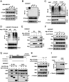
OTULIN is linearly ubiquitinated by LUBAC (HOIP), which is required for the OTULIN–LUBAC/HOIP interaction under an unstressed condition. (A) Workflow of the identification of ubiquitination (GG)-modified peptides of enriched OTULIN. (B) MS/MS spectrum showing the identification of an OTULIN peptide carrying ubiquitination (GG) on lysine (K) 64. Top, MS/MS fragmentation of the peptide (AADEIEK(-GG)EKELLIHER). Bottom, a series of black (b) and blue (y) product ions are detected in the 3-charge MS/MS spectrum, providing high confidence in the identification and localization of the ubiquitin modification. The x axis represents the mass-to-charge (m/z) ratio, and the y axis shows the relative intensity of different peaks. The monoisotopic precursor mass value [M+3H]3+ of the ubiquitinated peptide is shown. (C) Linear ubiquitination of immunoprecipitated HA-OTULIN WT, K64/66R, K34R, and K34/64/66R mutants in HEK293T cells. HEK293T cells were transfected with HA-OTULIN WT or indicated mutants. After 48 h, cell lysates were immunoprecipitated with anti-HA antibody and immunoblotted with anti-linear ubiquitin and anti-HA antibodies. (D) Different types of ubiquitination of immunoprecipitated HA-OTULIN WT and K64/66R mutant in HEK293T cells. HEK293T cells were transfected with HA-OTULIN WT or its K64/66R mutant. After 48 h, cell lysates were immunoprecipitated with anti-HA antibody and immunoblotted with antibodies for M1-Ub chains, K48-Ub chains, K63-Ub chains, and HA-tag (hemagglutinin tag). (E) Linear ubiquitination of immunoprecipitated endogenous OTULIN in parental and HOIP-KO HEK293T cells. Cell lysates were immunoprecipitated with anti-OTULIN antibody and immunoblotted with indicated antibodies. (F) Linear ubiquitination of immunoprecipitated HA-OTULIN in HEK293T cells. HOIP-KO HEK293T cells were cotransfected with HA-OTULIN and Myc-HOIP WT or its C885S mutant. After 48 h, cell lysates were immunoprecipitated with anti-HA antibody and immunoblotted with indicated antibodies. (G) Co-IP analysis of the interaction between Myc-HOIP and HA-OTULIN WT, C129S, Y56F, or K64/66R mutant in HEK293T cells. HEK293T cells were cotransfected with Myc-HOIP and indicated HA-OTULIN constructs in HEK293T cells. After 48 h, cell lysates were immunoprecipitated with anti-Myc antibody and immunoblotted with antibodies for HA-tag and Myc-tag. (H) Linear ubiquitination of immunoprecipitated Myc-OTULIN in OTULIN-KO MDA-MB-231 cells. Cells were transfected with or without Myc-OTULIN WT, C129S, Y56F, or K64/66R mutants. After 48 h, cell lysates were immunoprecipitated with anti-Myc antibody and immunoblotted with the indicated antibodies. (+) indicates the samples receiving the specific transfection listed on the left side, and (-) indicates mock controls.
Fig. 3.
Fig. 3.
Genotoxic stress induces OTULIN dimerization, OTULIN self-deubiquitination, and the OTULIN–HOIP complex dissociation. (A) Co-IP analysis of the interaction between endogenous OTULIN and HOIP in MDA-MB-231 cells treated with dimethyl sulfoxide (DMSO) (-), Dox (2 μg/mL, 2h), CBP (10 μg/mL, 2h) or exposed to IR (10 Gy) and left in the incubator for 2 h. Cell lysates were immunoprecipitated with anti-OTULIN antibody and immunoblotted with antibodies for HOIP and OTULIN. (B) Linear ubiquitination of immunoprecipitated endogenous OTULIN in HEK293T cells treated with DMSO (-), Etop (10 μM, 2h), CPT (10 μM, 2h) or exposed to IR (10 Gy) and left in the incubator for 2 h. Cell lysates were immunoprecipitated with anti-OTULIN antibody and immunoblotted with anti-linear ubiquitin and anti-OTULIN antibodies. (C) Linear ubiquitination of immunoprecipitated endogenous OTULIN in CYLD+/+ and CYLD−/− MEFs treated with Etop (10 μM) or Dox (2 μg/mL) for 2 h or left untreated (-). Cell lysates were immunoprecipitated with anti-OTULIN antibody and immunoblotted with indicated antibodies. (D) Linear ubiquitination of immunoprecipitated Myc-OTULIN in HEK293T cells. OTULIN-KO HEK293T cells were reconstituted with Myc-OTULIN WT or its C129S mutant. After 48 h, cells were treated with or without Etop (10 μM, 2 h). Cell lysates were immunoprecipitated with anti-Myc antibody and immunoblotted with anti-linear ubiquitin and anti-Myc antibodies. (E) Co-IP analysis of the interaction between Myc-OTULIN and HA-OTULIN in HEK293T cells. Cells were cotransfected with Myc-OTULIN and HA-OTULIN as indicated. After 48 h, cell lysates were immunoprecipitated with anti-HA or anti-Myc antibody and immunoblotted with indicated antibodies. (F) Co-IP analysis of the interaction between Myc-OTULIN and HA-OTULIN in HEK293T cells. Cells were cotransfected with Myc-OTULIN and HA-OTULIN WT, C129S, Y56F, or the K64/66R mutant. After 48 h, cell lysates were immunoprecipitated with anti-Myc antibody and immunoblotted with antibodies as indicated. (G) Top, mapping of OTULIN domain required for the OTULIN dimerization. Bottom, interactions between GFP-OTULIN and indicated HA-OTULIN constructs were analyzed by Co-IP assay in HEK293T cells. Cells were cotransfected with GFP-OTULIN and full-length (FL) HA-OTULIN, C terminus (C) of OTULIN, or N terminus (N) of OTULIN. After 48 h, cell lysates were immunoprecipitated with anti-HA antibody and immunoblotted with indicated antibodies. (H) Co-IP analysis of the interaction between Myc-OTULIN and GFP-OTULIN in HEK293T cells. Cells were cotransfected with Myc-OTULIN and GFP-OTULIN as indicated for 48 h and subsequently treated with Etop (10 μM, 2 h) or CPT (10 μM) or left untreated (-). Cell lysates were immunoprecipitated with anti-Myc antibody and immunoblotted with indicated antibodies. (I) DSS cross-linking assay reveals the formation of OTULIN dimers by Western blot analysis. HEK293T cells were treated with or without Etop (10 μM, 2 h) followed by the cross-linking assay. Cells were collected and incubated in 500 μL cold PBS containing DSS (1 mM) for 30 min at RT, which was quenched by 10 mM Tris⋅HCl (pH 7.5) for 15 min at RT. Cells were obtained by 200 g centrifugation for cell lysis. Cell lysates were immunoblotted with indicated antibodies. (+) indicates the samples receiving the specific treatment/transfection listed on the left side, and (-) indicates mock controls.
Fig. 4.
Fig. 4.
Oxidative stress mediates OTULIN dimerization by forming disulfide bonds via C17 and C47. (A) Co-IP analysis of the interaction between Myc-OTULIN and HA-OTULIN in MDA-MB-231. After the cotransfection with Myc-OTULIN and HA-OTULIN for 48 h, cells were treated with H2O2 (0, 10, or 50 μM) for 15 min. Cell lysates were immunoprecipitated with anti-Myc antibody and immunoblotted with anti-HA and anti-Myc antibodies. (B) Western blot analysis of OTULIN dimerization in MDA-MB-231 cells. Cells were treated with (+) or without (-) H2O2 (50 μM, 15 min), subjected to cell lysis with or without DTT (10 mM), and boiled with SDS loading buffer (no 2-ME). Cell lysates were immunoblotted with indicated antibodies. (C) Top, the locations of C17 and C47 in OTULIN. Bottom, the amino acid sequence alignments of the OTULIN N terminus (i.e., 11–50aa) shows the conservation of C17 and C47 in various species. (D) Immunoprecipitation analysis of the biotin-labeled HA-OTULIN (WT or C17/47A) in HEK293T cells treated with Etop (2 μM) for indicated time points and lysed with DCP-Bio1 (+) or wthout (-) as described in Materials and Methods. Cell lysates were immunoprecipitated with anti-HA antibody and immunoblotted with indicated antibodies. (E) Liquid chromatography-tandem MS analysis of Cys alkylation. HEK293T was treated with Etop (2 μM) for 2 h. The percentage of IAM-labeled Cys was calculated by the abundances of Cys with carbamidomethyl divided by the total abundances of corresponding Cys (× 100%). (F) Western blot analysis of OTULIN dimerization. OTULIN-KO MDA-MB-231 cells were reconstituted with indicated OTULIN constructs (48 h) and then treated with Dox (2 μg/mL, 2 h). Cell lysates were immunoblotted by indicated antibodies without DTT or 2-ME. (G) Linear ubiquitination of immunoprecipitated HA-OTULIN in MDA-MB-231 cells treated as in F. Cell lysates were immunoprecipitated with anti-HA antibody and immunoblotted with indicated antibodies. (H) Co-IP analysis of the interaction between HOIP and HA-OTULIN in MDA-MB-231 cells treated as in F. Cell lysates were immunoprecipitated with anti-HA antibody and immunoblotted with anti-HA and anti-HOIP antibodies. (I) Western blot analysis of OTULIN Tyr56 phosphorylation. OTULIN-KO MDA-MB-231 cells were reconstituted with HA-OTULIN WT or its C17/47A mutant (48 h) and then treated with (+) or without (-) Dox (2 μg/mL, 2 h). Cell lysates were immunoblotted with indicated antibodies.
Fig. 5.
Fig. 5.
Analysis of OTULIN dimers, linear ubiquitination, and OTULIN-HOIP interaction in TNFα-treated cells. (A) Linear ubiquitination of immunoprecipitated HA-OTULIN. OTULIN-KO HEK293T cells were reconstituted with HA-OTULIN WT or C129S mutant (48 h) and then treated with (+) or without (-) TNFα (10 ng/mL, 15 min). Cell lysates were immunoprecipitated with anti-HA antibody and immunoblotted with indicated antibodies. (B) Linear ubiquitination of immunoprecipitated OTULIN in CYLD+/+ and CYLD−/− cells treated as in A. Cell lysates were immunoprecipitated with anti-OTULIN antibody and immunoblotted with indicated antibodies. (C) Immunoprecipitation analysis of the biotin-labeled HA-OTULIN (WT or C17/47A) in HEK293T cells treated with TNFα (10 ng/mL) for indicated time points and lysed with DCP-Bio1 (+) or without (-) as described in Materials and Methods. Cell lysates were immunoprecipitated with anti-HA antibody and immunoblotted with indicated antibodies. (D) Western blot analysis of OTULIN dimerization. MDA-MB-231 cells were treated with TNFα (10 ng/mL) for indicated time points. Cell lysates were immunoblotted by indicated antibodies without DTT or 2-ME. (E) Linear ubiquitination of immunoprecipitated OTULIN in MDA-MB-231 cells treated as in D. Cell lysates were immunoprecipitated with anti-OTULIN antibody and immunoblotted with indicated antibodies. (F) Co-IP analysis of the interaction between OTULIN and HOIP in MDA-MB-231 cells treated as in D. Cell lysates were immunoprecipitated with anti-OTULIN antibody and immunoblotted with antibodies for OTULIN and HOIP.
Fig. 6.
Fig. 6.
Analysis of OTULIN dimers, linear ubiquitination, and OTULIN-HOIP interaction in the breast cancer clinical samples. (A) Western blot analysis of OTULIN dimerization in breast cancer (BC) and adjacent nontumorous (N) tissue samples from six patients. Cell lysates were immunoblotted by indicated antibodies without DTT or 2-ME. (B) Linear ubiquitination and phosphorylation of immunoprecipitated OTULIN in breast cancer (BC) and adjacent nontumorous (N) tissue samples from six patients. Protein expression levels of total OTULIN and p-OTULIN are shown in the input. Cell lysates were immunoprecipitated with a low amount of Dynabeads protein A (10 μL) and anti-OTULIN antibody (2 μL) to make them saturated and immunoblotted with indicated antibodies. (C) Co-IP analysis of the interaction between HOIP and OTULIN in breast cancer (BC) and adjacent nontumorous (N) tissue samples from six patients. Protein expression levels of OTULIN, HOIP, and p-P65 are shown in the input. Cell lysates were immunoprecipitated with a low amount of Dynabeads protein A (10 μL) and anti-HOIP antibody (2 μL) to make them saturated and immunoblotted with indicated antibodies. (D) Quantification graph based on densitometry of the Western blot data from (A, B, and C). The data are presented as the mean ± SEM from three clinical samples. Statistical analysis was performed by a two-way ANOVA with Tukey’s correction for multiple comparisons. *P < 0.05; **P < 0.01; ***P < 0.001; ****P < 0.0001; ns, not significant. (E) Graphical abstract demonstrating the crucial roles OTULIN plays in NF-κB–mediated chemotherapy resistance and inflammation. Under unstressed conditions, OTULIN binds to HOIP, which limits the linear ubiquitination of the substrate molecules such as NEMO, governing a normal NF-κB activation. Under genotoxic or inflammatory conditions, OTULIN molecules dimerize, promoting Tyr56 phosphorylation and its self-deubiquitination, resulting in its dissociation from HOIP. Ultimately, OTULIN loss of function leads to an aberrant NF-κB activation and subsequent chemotherapy resistance or inflammatory symptoms. Pt, patient. Red arrows indicate the same individual samples that show loss of function of OTULIN (i.e., correlated loss of OTULIN–HOIP interaction and OTULIN deubiquitination).

Similar articles

Cited by

References

    1. Karin M., Nuclear factor-kappaB in cancer development and progression. Nature 441, 431–436 (2006). - PubMed
    1. Liu T., Zhang L., Joo D., Sun S. C., NF-κB signaling in inflammation. Signal Transduct. Target. Ther. 2, 17023 (2017). - PMC - PubMed
    1. Zhang Q., Lenardo M. J., Baltimore D., 30 years of NF-κB: A blossoming of relevance to human pathobiology. Cell 168, 37–57 (2017). - PMC - PubMed
    1. Hojyo S., et al. , How COVID-19 induces cytokine storm with high mortality. Inflamm. Regen. 40, 37 (2020). - PMC - PubMed
    1. Hunter T., The age of crosstalk: Phosphorylation, ubiquitination, and beyond. Mol. Cell 28, 730–738 (2007). - PubMed

Publication types

LinkOut - more resources