. 2022 Aug 16;119(33):e2117903119.
doi: 10.1073/pnas.2117903119. Epub 2022 Aug 8.

Noradrenaline activation of hippocampal dopamine D1 receptors promotes antidepressant effects

Affiliations

Noradrenaline activation of hippocampal dopamine D1 receptors promotes antidepressant effects

Katsunori Kobayashi et al. Proc Natl Acad Sci U S A. .

Abstract

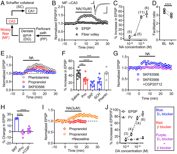
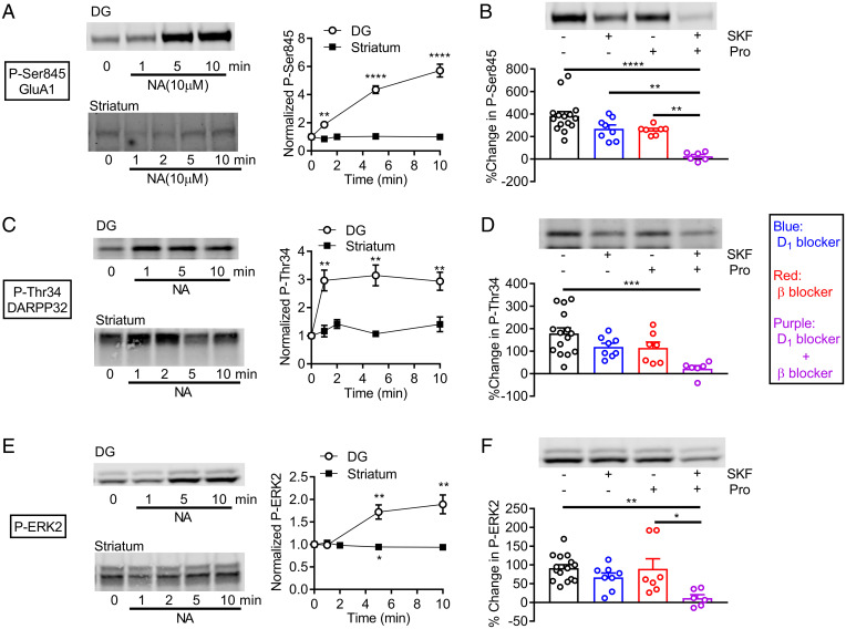
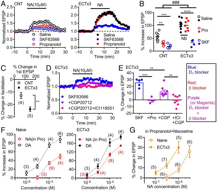
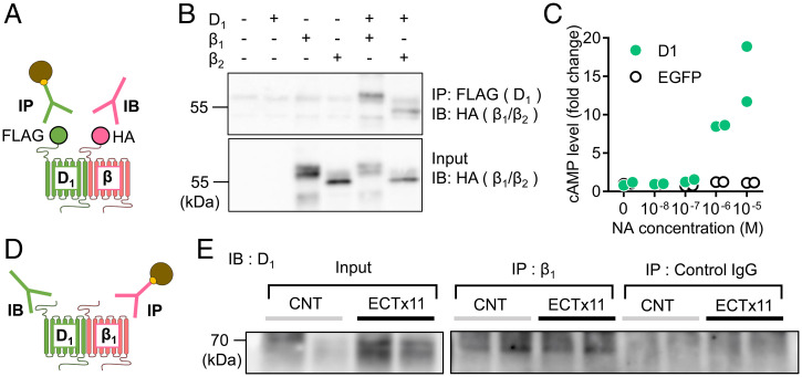
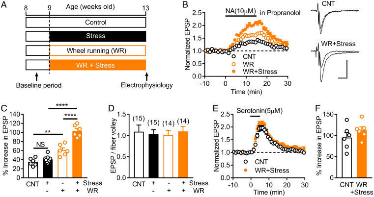
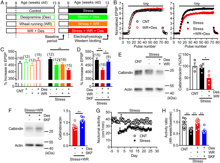
Dopamine D1 receptors (D1Rs) in the hippocampal dentate gyrus (DG) are essential for antidepressant effects. However, the midbrain dopaminergic neurons, the major source of dopamine in the brain, only sparsely project to DG, suggesting possible activation of DG D1Rs by endogenous substances other than dopamine. We have examined this possibility using electrophysiological and biochemical techniques and found robust activation of D1Rs in mouse DG neurons by noradrenaline. Noradrenaline at the micromolar range potentiated synaptic transmission at the DG output and increased the phosphorylation of protein kinase A substrates in DG via activation of D1Rs and β adrenergic receptors. Neuronal excitation preferentially enhanced noradrenaline-induced synaptic potentiation mediated by D1Rs with minor effects on β-receptor-dependent potentiation. Increased voluntary exercise by wheel running also enhanced noradrenaline-induced, D1R-mediated synaptic potentiation, suggesting a distinct functional role of the noradrenaline-D1R signaling. We then examined the role of this signaling in antidepressant effects using mice exposed to chronic restraint stress. In the stressed mice, an antidepressant acting on the noradrenergic system induced a mature-to-immature change in the DG neuron phenotype, a previously proposed cellular substrate for antidepressant action. This effect was evident only in mice subjected to wheel running and blocked by a D1R antagonist. These results suggest a critical role of noradrenaline-induced activation of D1Rs in antidepressant effects in DG. Experience-dependent regulation of noradrenaline-D1R signaling may determine responsiveness to antidepressant drugs in depressive disorders.

Keywords: dentate gyrus; exercise; monoamine; mossy fiber; stress.


Conflict of interest statement

The authors declare no competing interest.

Figures

Fig. 1.
Fig. 1.
Activation of D1 receptors by noradrenaline potentiates mossy fiber synaptic transmission in the hippocampus. (A) Schematic diagram of hippocampal excitatory circuit highlighting mossy fiber (MF)–CA3 connection. (B) Effects of noradrenaline (NA) applied at the bar on presynaptic fiber volley (FV) and EPSPs at MF–CA3 synapse. Sample traces show averaged field potentials before and during NA application (scale bars: 10 ms, 0.2 mV). (C) Dependence of synaptic potentiation on NA concentrations. (D) Decrease in triple-pulse facilitation at 200-ms intervals by NA. BL: baseline. Paired t test (t5 = 11.77, ****P < 0.0001). (E) NA-induced synaptic potentiation in D1 antagonist SKF83566 (SKF) (200 nM), β antagonist propranolol (Pro) (10 μM) or α antagonist phentolamine (Phento) (20 μM). (F) Summary of antagonist effects on NA-induced synaptic potentiation. SCH: SCH23390 (D1 antagonist, 50 nM). One-way ANOVA (F4,33 = 10.2, P < 0.0001) followed by Dunnett's test (**P = 0.0085, ***P = 0.0009, ****P = 0.0001). (G) Block of SKF-resistant potentiation by β1 antagonist CGP20712 (CGP) (100 nM). (H) Summary of β antagonist effects on SKF-resistant potentiation. One-way ANOVA (F2,11 = 35.66, P < 0.0001) followed by Dunnett's test (****P = 0.0001). (I) D1-receptor–dependent synaptic potentiation by low micromolar NA in presence (n = 6) and absence (n = 3) of nisoxetine (1 μM). (J) Dependence of FV and EPSP potentiation on dopamine (DA) concentrations. The number of data is shown in the graph in C and J. Data are presented as means ± SEM in all figures with or without individual values.
Fig. 2.
Fig. 2.
D1-receptor–dependent protein phosphorylation by noradrenaline in the dentate gyrus. (A) Effects of NA on GluA1 phosphorylation in dentate gyrus (DG) (n = 7 for each point) and striatum (n = 4). Typical immunoblots for detection of phospho-Ser845 (left) and quantified data (right). One-sample t test (**P = 0.0019, ****P < 0.0001, compared with 1). (B) Effects of SKF83566 (500 nM) and propranolol on NA-induced GluA1 phosphorylation. Typical immunoblots (top) and quantified data (bottom). Two-way ANOVA (SKF effect, F1,32 = 19.28, P = 0.0001; Pro effect, F1,32 = 22.07, P < 0.0001) followed by Tukey's test (**P < 0.005, ****P < 0.0001). (C) Effects of noradrenaline on DARPP32 phosphorylation (phospho-Thr34) in slices of DG and striatum. One-sample t test (**P < 0.005). DG: n = 6, striatum: n = 4. (D) Antagonist effects on NA-induced phosphorylation of DARPP32 in DG. Two-way ANOVA (SKF effect, F1,32 = 8.549, P = 0.0063; Pro effect, F1,32 = 9.497, P = 0.0042) followed by Tukey's test (***P = 0.0006). (E) Effects of noradrenaline on ERK2 phosphorylation in slices of DG and striatum. One-sample t test (*P < 0.05, **P < 0.01). DG: n = 7, striatum: n = 4. (F) Antagonist effects on NA-induced phosphorylation of ERK2 in DG. Two-way ANOVA (SKF effect, F1,32 = 11.65, P = 0.0018) followed by Tukey's test (*P = 0.0123, **P = 0.0025). Phospho-proteins were normalized to total proteins and then data were normalized to values obtained with time 0 (A, C, and E) or control without noradrenaline (B, D, and F).
Fig. 3.
Fig. 3.
Activity-dependent enhancement of noradrenaline–D1 receptor signaling. (A) Effects of NA in control mice (CNT) and mice treated with three times of ECT (ECTx3) in normal saline or in antagonists. (B) Summary of data shown in A. Two-way ANOVA (ECT effect, F1,35 = 83.22, P < 0.0001; drug effect, F2,35 = 42.98, P < 0.0001; interaction, F2,35 = 10.13, ###P = 0.0003) followed by Tukey's test (NS: not significant, **P = 0.0083, ***P = 0.0002, ****P < 0.0001). (C) Effects of ECTx3 on reduction of triple-pulse facilitation by NA (t7 = 3.644, **P = 0.082). (D) Effects of β receptor antagonists on SKF-resistant component of NA-induced potentiation in ECT-treated mice. (E) Summary of β antagonist effects on SKF-resistant component of NA-induced potentiation. CGP (100–200 nM), ICI: ICI118551 (β2 antagonist, 100–200 nM). One-way ANOVA (F3,21 = 17.37, P < 0.0001) followed by Bonferroni's test (**P < 0.01, ***P = 0.0001). (F) Dependence of D1-mediated synaptic potentiation on NA and DA concentrations in naive and ECT-treated mice. The DA data in naive mice are the same as those in Fig. 1J. (G) Dependence of D1-mediated potentiation on NA concentrations in nisoxetine. The number of data is shown in the graph in C, F, and G.
Fig. 4.
Fig. 4.
Interaction between D1 and β receptors. (A) Schematic diagram showing experimental design of coimmunoprecipitation assay using tagged receptors. IP: immunoprecipitation, IB: immunoblot. (B) Coimmunoprecipitation of FLAG-tagged D1 and HA-tagged β1 or β2 receptors expressed in HEK293T cells. (C) NA-induced cAMP production in HEK293T cells expressing EGFP or D1 receptors (n = 2 each). (D) Experimental design of coimmunoprecipitation assay of native receptors. (E) Coimmunoprecipitation of hippocampal D1 and β1 receptors in control and mice treated with 11 times of ECT (ECTx11). IgG: immunoglobulin G. Experiments were repeated three times with similar results (SI Appendix, Fig. S5B).
Fig. 5.
Fig. 5.
Synergistic augmentation of noradrenaline–D1 receptor signaling by chronic stress and exercise. (A) Timeline of chronic restraint stress and increased voluntary exercise by wheel running (WR). (B) Effects of chronic restraint stress and WR on NA-induced MF synaptic potentiation mediated by D1 receptors. Recordings were made in the presence of propranolol. Sample traces show averaged field potentials before and during NA application (scale bars: 10 ms, 0.2 mV). (C) Summary of effects of stress and WR on D1-mediated potentiation. Two-way ANOVA (stress effect, F1,23 = 35.29, P < 0.0001; WR effect, F1,23 = 122.9, P < 0.0001; interaction, F1,23 = 21.36, P = 0.0001) followed by Tukey's test (**P = 0.0012, ****P < 0.0001). (D) Lack of effects of stress and WR on the basal MF synaptic efficacy assessed by EPSP to fiber volley ratios. The number of data is shown in the graph. (E) Effects of stress and WR on serotonin-induced MF synaptic potentiation. (F) Summary of serotonin-induced potentiation.
Fig. 6.
Fig. 6.
Experience-dependent augmentation of noradrenaline–D1 receptor signaling facilitates effects of noradrenergic antidepressant. (A) Timeline of restraint stress, WR, and desipramine (Des) treatment. (B) Effects of chronic Des (30 mg/kg/day for 4 wk) and WR on 1-Hz frequency facilitation at MF synapse in nonstressed (left) and stressed (right) mice. (C) Summary of effects of Des and WR on frequency facilitation in nonstressed and stressed mice. Two-way ANOVA (stress effect, F1,106 = 1.757, P = 0.1879; treatment effect, F3,106 = 1.657, P = 0.1809; interaction, F3,106 = 4.424, P = 0.0057) followed by Dunnett's test (**P = 0.0011). (D) Effects of SKF (1 mg/kg/day) on reduction of frequency facilitation caused by Des and WR in stressed mice. One-way ANOVA (F2,39 = 7.508, P = 0.0017) followed by Dunnett's test (*P = 0.0247, **P = 0.0015). (E) Effects of Des and WR on calbindin expression in DG. Typical immunoblots (left) and quantified data (right). One-way ANOVA (F2,8 = 5.809, P = 0.0276) followed by Dunnett's test (*P = 0.0399). (F) Effects of SKF on calbindin expression in DG of Des-treated mice (t13 = 3.164, **P = 0.0075). (G) Effects of chronic stress on nocturnal home cage activity. (H) D1-receptor–dependent reversal of stress-induced decrease in home cage activity by Des and WR. One-way ANOVA (F3,28 = 9.789, P = 0.0001) followed by Bonferroni's test (**P < 0.01). The number of data is shown in the graph in C and D.

Similar articles

Cited by

References

    1. da Silva W. C., Köhler C. C., Radiske A., Cammarota M., D1/D5 dopamine receptors modulate spatial memory formation. Neurobiol. Learn. Mem. 97, 271–275 (2012). - PubMed
    1. Hansen N., Manahan-Vaughan D., Dopamine D1/D5 receptors mediate informational saliency that promotes persistent hippocampal long-term plasticity. Cereb. Cortex 24, 845–858 (2014). - PMC - PubMed
    1. Kobayashi K., Haneda E., Higuchi M., Suhara T., Suzuki H., Chronic fluoxetine selectively upregulates dopamine D1-like receptors in the hippocampus. Neuropsychopharmacology 37, 1500–1508 (2012). - PMC - PubMed
    1. Kobayashi K., et al. , Rapid and lasting enhancement of dopaminergic modulation at the hippocampal mossy fiber synapse by electroconvulsive treatment. J. Neurophysiol. 117, 284–289 (2017). - PMC - PubMed
    1. Shuto T., et al. , Obligatory roles of dopamine D1 receptors in the dentate gyrus in antidepressant actions of a selective serotonin reuptake inhibitor, fluoxetine. Mol. Psychiatry 25, 1229–1244 (2020). - PMC - PubMed

Publication types

MeSH terms

LinkOut - more resources