. 2022 Aug 16;119(33):e2208004119.
doi: 10.1073/pnas.2208004119. Epub 2022 Aug 8.

Cohesin ATPase activities regulate DNA binding and coiled-coil configuration

Affiliations

Cohesin ATPase activities regulate DNA binding and coiled-coil configuration

Xingya Xu et al. Proc Natl Acad Sci U S A. .

Abstract

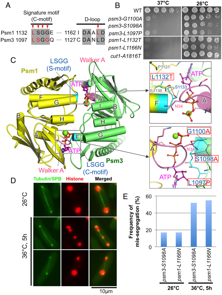
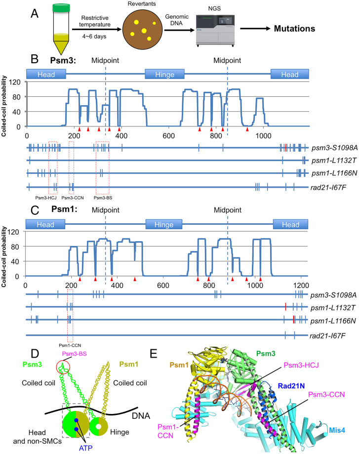
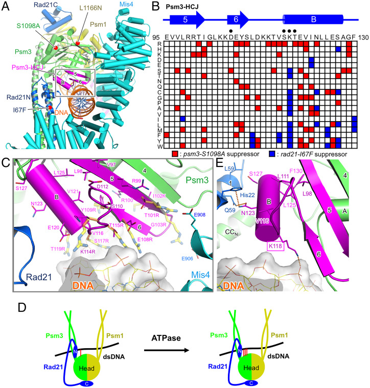
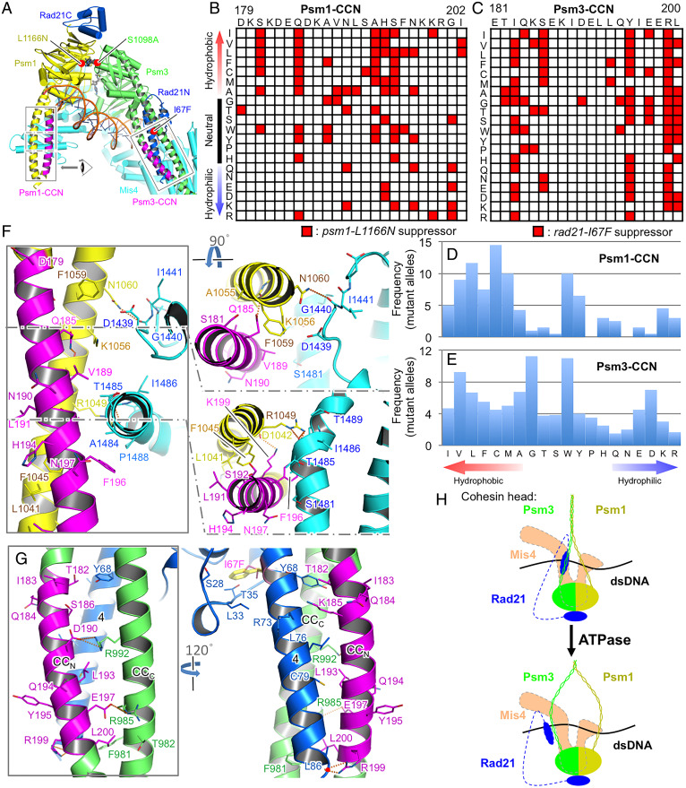
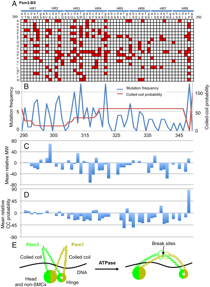
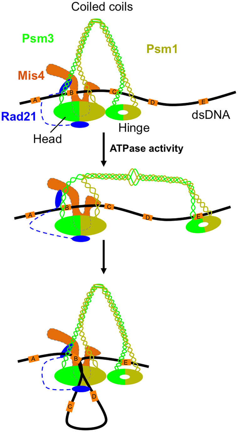
The cohesin complex is required for sister chromatid cohesion and genome compaction. Cohesin coiled coils (CCs) can fold at break sites near midpoints to bring head and hinge domains, located at opposite ends of coiled coils, into proximity. Whether ATPase activities in the head play a role in this conformational change is yet to be known. Here, we dissected functions of cohesin ATPase activities in cohesin dynamics in Schizosaccharomyces pombe. Isolation and characterization of cohesin ATPase temperature-sensitive (ts) mutants indicate that both ATPase domains are required for proper chromosome segregation. Unbiased screening of spontaneous suppressor mutations rescuing the temperature lethality of cohesin ATPase mutants identified several suppressor hotspots in cohesin that located outside of ATPase domains. Then, we performed comprehensive saturation mutagenesis targeted to these suppressor hotspots. Large numbers of the identified suppressor mutations indicated several different ways to compensate for the ATPase mutants: 1) Substitutions to amino acids with smaller side chains in coiled coils at break sites around midpoints may enable folding and extension of coiled coils more easily; 2) substitutions to arginine in the DNA binding region of the head may enhance DNA binding; or 3) substitutions to hydrophobic amino acids in coiled coils, connecting the head and interacting with other subunits, may alter conformation of coiled coils close to the head. These results reflect serial structural changes in cohesin driven by its ATPase activities potentially for packaging DNAs.

Keywords: ATPase; DNA binding; cohesin; coiled coil; suppressor screen.


Conflict of interest statement

The authors declare no competing interest.

Figures

Fig. 1.
Fig. 1.
Cohesin ATPase ts mutants. (A) Alignment around signature motifs and D loops of S. pombe cohesin ATPase domains. Amino acids selected for mutagenesis are indicated by red arrowheads. Amino acids involved in the ts mutants are colored red. (B) Spot test results of the five ATPase ts mutants. (C) Structual details of the signature motifs (PDB code: 6YUF). ATP molecules were built based on the cryogenic electron microscopy structure of the ATP-bound cohesin complex (PDB code: 6ZZ6). The signature and Walker A motifs are colored in cyan and violet, respectively. Mg2+ ions are shown as lime spheres. Hydrogen bonds are represented with dashed lines. Helices in the head are shown as cylinders. The backbone of the signature motif runs along the triphosphate group of adenosime triphosphate (ATP) to form its binding surface. The side chains of leucine and serine residues in the signature motif stabilize the ATP binding surface with hydrogen bonds or hydrophobic interactions. (D) Chromosome missegregation phenotypes observed in psm3-S1098A. (E) Frequency of chromosome missegregation events.
Fig. 2.
Fig. 2.
Suppressor mutations in cohesin SMC subunits. (A) A strategy to identify spontaneous suppressor mutations for the ATPase ts mutants. (B and C) Location of suppressor mutations of cohesin ATPase ts mutants or rad21-I67F identified in Psm3/SMC3 (B) and Psm1/SMC1 (C), are indicated by vertical bars. Locations of suppressor hotspots (Psm3-HCJ, Psm3-CCN, Psm1-CCN, and Psm3-BS), selected for targeted saturation mutagenesis, are indicated by red dashed rectangles. Potential break sites are indicated by red arrowheads. (D) A cartoon exhibiting a folded form of the cohesin complex. Position of Psm3-BS is indicated. (E) Structural view of the targeted regions (magenta) that were selected for saturation mutagenesis.
Fig. 3.
Fig. 3.
ATPase activities affect DNA binding by head. (A) Structural view of Psm3-HCJ (magenta). (B) Single amino acid substitutions that were identified as suppressors of psm3-S1098A (red) or rad21-I67F (blue) in Psm3-HCJ. Columns depict positions along the primary sequence, and rows indicate a mutation to one of the 20 amino acids. Amino acids that may bind DNA are indicated by “•.” (C) Structural detail of the suppressor sites of psm3-S1098A in Psm3-HCJ (magenta). Sticks in Psm3-HCJ represent the suppressor sites. Arg/Lys mutations identified as the suppressors are manually modeled and shown in yellow transparent sticks. (D) A cartoon exhibiting that cohesin ATPase activities may enhance the head’s interaction with DNA. Interactions between the head and DNA are indicated by red bars. (E) Structural view of Psm3-K118’s interaction with DNA.
Fig. 4.
Fig. 4.
ATPase activities affect coiled-coil interactions with associated subunits at the head. (A) Structural view of Psm1-CCN and Psm3-CCN (PDB code: 6YUF). (B and C) Data matrices presenting single amino acid substitutions that were identified as suppressors of psm1-L1166N in Psm1-CCN (B) and suppressors of rad21-I67F in Psm3-CCN (C). (D and E) Frequency of the 20 amino acids in mutant alleles obtained in Psm1-CCN (D) and Psm3-CCN (E). (F) Structural view of psm1-L1166N suppressors in Psm1-CCN. The suppressing residues and the ones interacting with these suppressing residues are represented by sticks. Hydrogen bonds and salt bridges are shown in orange broken lines. (G) Structural view of rad21-I67F suppressors in Psm3-CCN. I67 of Rad21/SCC1 faces inside the N-terminal domain of Rad21/SCC1 to form a hydrophobic core with A20, S28, L33, and T35 near the interface with Psm3-CCN. (H) A cartoon exhibiting that cohesin ATPase activities may drive a change in coiled-coil orientation at the head.
Fig. 5.
Fig. 5.
Suppressors around a coiled-coil break site. (A) Data matrix showing single amino acid substitutions in Psm3-BS that rescue the temperature sensitivity of psm3-S1098A. The eight heptad repeats predicted by MARCOIL are shown above the primary sequence. (B) Numbers of single amino acid substitutions versus coiled-coil probability. (C) Mean relative molecular weight. (D) Mean relative coiled-coil probabilities. (E) A cartoon exhibiting that cohesin ATPase activities may regulate coiled-coil folding and extension at break sites.
Fig. 6.
Fig. 6.
Potential ATP-driven conformational changes. Cohesin loader Mis4/SCC2/NIPBL loads cohesin onto chromatin and arched coiled coils hold chromatin. ATPhydrolysis in the cohesin ATPase domains would trigger several structural changes: (1) to enhance DNA binding by the head domain, (2) to open coiled coils at the headthrough destabilizing the interactions of coiled coils with Rad21N and Mis4C, (3) to temporarily extend the folded coiled coils and separate the hinge from the head. Then, coiled coils fold back again to bring the head and hinge together, which also brings DNA elements that are far away proximal. AE indicate five DNA elements in the genome.

Similar articles

Cited by

References

    1. Uhlmann F., SMC complexes: From DNA to chromosomes. Nat. Rev. Mol. Cell Biol. 17, 399–412 (2016). - PubMed
    1. Yatskevich S., Rhodes J., Nasmyth K., Organization of chromosomal DNA by SMC complexes. Annu. Rev. Genet. 53, 445–482 (2019). - PubMed
    1. Arumugam P., et al. , ATP hydrolysis is required for cohesin’s association with chromosomes. Curr. Biol. 13, 1941–1953 (2003). - PubMed
    1. Haering C. H., Löwe J., Hochwagen A., Nasmyth K., Molecular architecture of SMC proteins and the yeast cohesin complex. Mol. Cell 9, 773–788 (2002). - PubMed
    1. Lammens A., Schele A., Hopfner K. P., Structural biochemistry of ATP-driven dimerization and DNA-stimulated activation of SMC ATPases. Curr. Biol. 14, 1778–1782 (2004). - PubMed

Publication types

MeSH terms

LinkOut - more resources