

Central nervous system (CNS) inflammatory demyelinating diseases (IDDs) associated with COVID-19: A case series and review
- PMID: 35939945
- PMCID: PMC9343076
- DOI: 10.1016/j.jneuroim.2022.577939
Central nervous system (CNS) inflammatory demyelinating diseases (IDDs) associated with COVID-19: A case series and review
Abstract
Background: Over the past two years, SARS-CoV-2 has frequently been documented with various post and para-infectious complications, including cerebrovascular, neuromuscular, and some demyelinating conditions such as acute disseminated encephalomyelitis (ADEM). We report two rare neurological manifestations post-COVID-19 infection; multiple sclerosis (MS) and myelin oligodendrocyte glycoprotein antibody-associated disease (MOGAD). Further, we reviewed other CNS inflammatory demyelinating diseases (IDDs) associated with SARS-CoV-2, including optic neuritis (ON) and neuromyelitis optica spectrum disorders (NMOSD).
Methods: A descriptive analysis and literature search of Google Scholar and PubMed was conducted by two independent reviewers from December 1st, 2019, to March 30th, 2022, and included all the case studies of MS, MOGAD, NMOSD, and ON associated with COVID-19 infection.
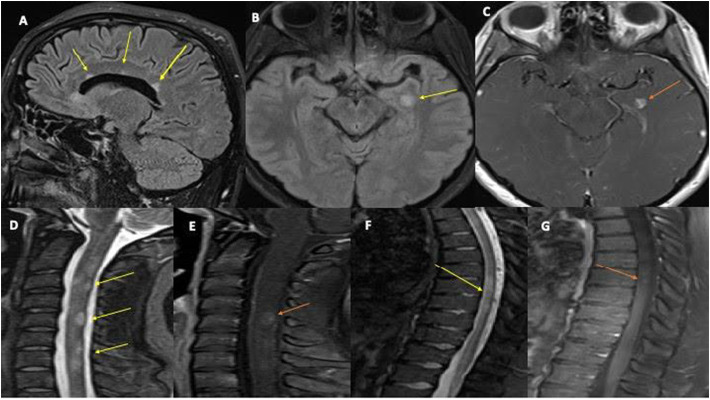
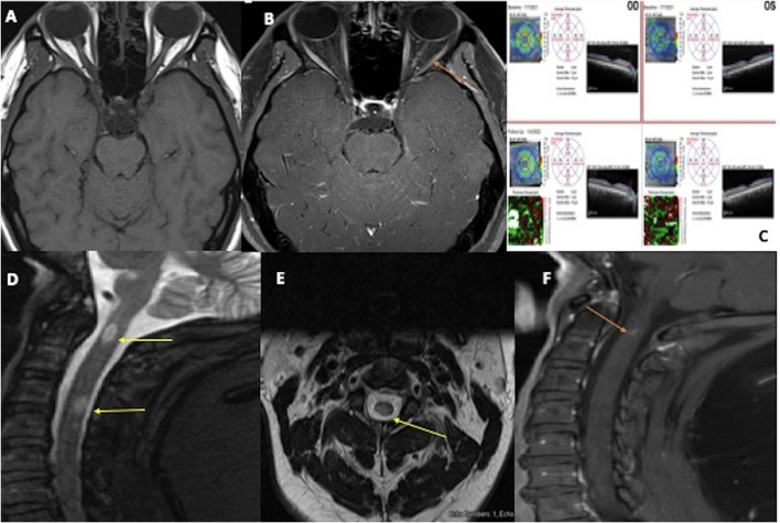
Case presentations: Case 1 (MS) was a 24-year-old female with paresthesia and bilateral weakness one week after COVID-19 symptom onset who showed demyelinating plaques and 12 isolated oligoclonal bands (OCBs). Case 2 (MOGAD) was a 41-year-old male with encephalomyelitis 16 days after COVID-19, who later developed MOG-antibody-associated optic neuritis.
Results: Out of 18 cases, NMOSD was the most common post-COVID manifestation (7, 39%), followed by MOGAD (5, 28%), MS (4, 22%), and isolated ON (2, 11%). The median duration between the onset of COVID-19 symptom onset and neurological symptoms was 14 days. 61% of these were male, with a mean age of 35 years. IVMP was the treatment of choice, and nearly all patients made a full recovery, with zero fatalities.
Conclusions: Although these neurological sequelae are few, physicians must be cognizant of their underlying pathophysiology and associated clinical and neuro-diagnostic findings when treating COVID-19 patients with atypical presentations.
Keywords: CNS IDD; COVID-19; MOGAD; MS; NMOSD; Optic neuritis;; SARS-CoV-2; Virus-induced demyelination.
Copyright © 2022 Elsevier B.V. All rights reserved.
Conflict of interest statement
Declaration of Competing Interest The authors declare that the research was conducted in the absence of any commercial or financial relationships that could be construed as a potential conflict of interest.
Figures
References
-
- Barone S., Rapisarda L., Manzo L., Mechelli A., Pascarella A., Bruno P., Pasquale M., Trimboli M., Valentino P., Gambardella A. A case of neuromyelitis optica spectrum disorder (NMOSD) and acute myositis following SARS-CoV-2 infection. J. Neurol. Sci. 2021 Oct;429:119862. doi: 10.1016/j.jns.2021.119862. Epub 2021 Oct 8. PMCID: PMC8498504. - DOI
-
- Bar-Or A., Pender M.P., Khanna R., Steinman L., Hartung H.P., Maniar T., Croze E., Aftab B.T., Giovannoni G., Joshi M.A. Epstein-Barr virus in multiple sclerosis: theory and emerging immunotherapies. Trends Mol. Med. 2020 Mar;26(3):296–310. doi: 10.1016/j.molmed.2019.11.003. Epub 2019 Dec 17. Erratum in: Trends Mol Med. 2021 Apr;27(4):410-411. PMID: 31862243; PMCID: PMC7106557. - DOI - PMC - PubMed
Publication types
MeSH terms
Substances
Grants and funding
LinkOut - more resources
Full Text Sources
Medical
Miscellaneous
