. 2022 Sep;609(7928):754-760.
doi: 10.1038/s41586-022-05163-5. Epub 2022 Aug 8.

DOCK2 is involved in the host genetics and biology of severe COVID-19

Ho Namkoong #  1 Ryuya Edahiro #  2   3 Tomomi Takano  4 Hiroshi Nishihara  5 Yuya Shirai  2   3 Kyuto Sonehara  2   6 Hiromu Tanaka  7 Shuhei Azekawa  7 Yohei Mikami  8 Ho Lee  7 Takanori Hasegawa  9 Koji Okudela  10 Daisuke Okuzaki  11 Daisuke Motooka  12 Masahiro Kanai  13 Tatsuhiko Naito  2 Kenichi Yamamoto  2 Qingbo S Wang  2 Ryunosuke Saiki  14 Rino Ishihara  8 Yuta Matsubara  8 Junko Hamamoto  7 Hiroyuki Hayashi  15 Yukihiro Yoshimura  16 Natsuo Tachikawa  16 Emmy Yanagita  5 Takayoshi Hyugaji  17 Eigo Shimizu  17 Kotoe Katayama  17 Yasuhiro Kato  3   18 Takayoshi Morita  3   18 Kazuhisa Takahashi  19 Norihiro Harada  19 Toshio Naito  20 Makoto Hiki  21   22 Yasushi Matsushita  23 Haruhi Takagi  19 Ryousuke Aoki  24 Ai Nakamura  19 Sonoko Harada  19   25 Hitoshi Sasano  19 Hiroki Kabata  7 Katsunori Masaki  7 Hirofumi Kamata  7 Shinnosuke Ikemura  7 Shotaro Chubachi  7 Satoshi Okamori  7 Hideki Terai  7 Atsuho Morita  7 Takanori Asakura  7 Junichi Sasaki  26 Hiroshi Morisaki  27 Yoshifumi Uwamino  28 Kosaku Nanki  8 Sho Uchida  1 Shunsuke Uno  1 Tomoyasu Nishimura  1   29 Takashi Ishiguro  30 Taisuke Isono  30 Shun Shibata  30 Yuma Matsui  30 Chiaki Hosoda  30 Kenji Takano  30 Takashi Nishida  30 Yoichi Kobayashi  30 Yotaro Takaku  30 Noboru Takayanagi  30 Soichiro Ueda  31 Ai Tada  31 Masayoshi Miyawaki  31 Masaomi Yamamoto  31 Eriko Yoshida  31 Reina Hayashi  31 Tomoki Nagasaka  31 Sawako Arai  31 Yutaro Kaneko  31 Kana Sasaki  31 Etsuko Tagaya  32 Masatoshi Kawana  33 Ken Arimura  32 Kunihiko Takahashi  9 Tatsuhiko Anzai  9 Satoshi Ito  9 Akifumi Endo  34 Yuji Uchimura  35 Yasunari Miyazaki  36 Takayuki Honda  36 Tomoya Tateishi  36 Shuji Tohda  37 Naoya Ichimura  37 Kazunari Sonobe  37 Chihiro Tani Sassa  37 Jun Nakajima  37 Yasushi Nakano  38 Yukiko Nakajima  38 Ryusuke Anan  38 Ryosuke Arai  38 Yuko Kurihara  38 Yuko Harada  38 Kazumi Nishio  38 Tetsuya Ueda  39 Masanori Azuma  39 Ryuichi Saito  39 Toshikatsu Sado  39 Yoshimune Miyazaki  39 Ryuichi Sato  39 Yuki Haruta  39 Tadao Nagasaki  39 Yoshinori Yasui  40 Yoshinori Hasegawa  39 Yoshikazu Mutoh  41 Tomoki Kimura  42 Tomonori Sato  42 Reoto Takei  42 Satoshi Hagimoto  42 Yoichiro Noguchi  42 Yasuhiko Yamano  42 Hajime Sasano  42 Sho Ota  42 Yasushi Nakamori  43 Kazuhisa Yoshiya  43 Fukuki Saito  43 Tomoyuki Yoshihara  43 Daiki Wada  43 Hiromu Iwamura  43 Syuji Kanayama  43 Shuhei Maruyama  43 Takashi Yoshiyama  44 Ken Ohta  44 Hiroyuki Kokuto  44 Hideo Ogata  44 Yoshiaki Tanaka  44 Kenichi Arakawa  44 Masafumi Shimoda  44 Takeshi Osawa  44 Hiroki Tateno  45 Isano Hase  45 Shuichi Yoshida  45 Shoji Suzuki  45 Miki Kawada  46 Hirohisa Horinouchi  47 Fumitake Saito  48 Keiko Mitamura  49 Masao Hagihara  50 Junichi Ochi  48 Tomoyuki Uchida  50 Rie Baba  51 Daisuke Arai  51 Takayuki Ogura  51 Hidenori Takahashi  51 Shigehiro Hagiwara  51 Genta Nagao  51 Shunichiro Konishi  51 Ichiro Nakachi  51 Koji Murakami  52 Mitsuhiro Yamada  52 Hisatoshi Sugiura  52 Hirohito Sano  52 Shuichiro Matsumoto  52 Nozomu Kimura  52 Yoshinao Ono  52 Hiroaki Baba  53 Yusuke Suzuki  54 Sohei Nakayama  54 Keita Masuzawa  54 Shinichi Namba  2 Ken Suzuki  2 Yoko Naito  12 Yu-Chen Liu  11 Ayako Takuwa  11 Fuminori Sugihara  55 James B Wing  56   57 Shuhei Sakakibara  58 Nobuyuki Hizawa  59 Takayuki Shiroyama  3 Satoru Miyawaki  60 Yusuke Kawamura  61 Akiyoshi Nakayama  61 Hirotaka Matsuo  61 Yuichi Maeda  3 Takuro Nii  3 Yoshimi Noda  3 Takayuki Niitsu  3 Yuichi Adachi  3 Takatoshi Enomoto  3 Saori Amiya  3 Reina Hara  3 Yuta Yamaguchi  3   18 Teruaki Murakami  3   18 Tomoki Kuge  3 Kinnosuke Matsumoto  3 Yuji Yamamoto  3 Makoto Yamamoto  3 Midori Yoneda  3 Toshihiro Kishikawa  2   62   63 Shuhei Yamada  64 Shuhei Kawabata  64 Noriyuki Kijima  64 Masatoshi Takagaki  64 Noah Sasa  2   62 Yuya Ueno  62 Motoyuki Suzuki  62 Norihiko Takemoto  62 Hirotaka Eguchi  62 Takahito Fukusumi  62 Takao Imai  62 Munehisa Fukushima  62   65 Haruhiko Kishima  64 Hidenori Inohara  62 Kazunori Tomono  66 Kazuto Kato  67 Meiko Takahashi  68 Fumihiko Matsuda  68 Haruhiko Hirata  3 Yoshito Takeda  3 Hidefumi Koh  69 Tadashi Manabe  69 Yohei Funatsu  69 Fumimaro Ito  69 Takahiro Fukui  69 Keisuke Shinozuka  69 Sumiko Kohashi  69 Masatoshi Miyazaki  69 Tomohisa Shoko  70 Mitsuaki Kojima  70 Tomohiro Adachi  70 Motonao Ishikawa  71 Kenichiro Takahashi  72 Takashi Inoue  73 Toshiyuki Hirano  73 Keigo Kobayashi  73 Hatsuyo Takaoka  73 Kazuyoshi Watanabe  74 Naoki Miyazawa  75 Yasuhiro Kimura  75 Reiko Sado  75 Hideyasu Sugimoto  75 Akane Kamiya  76 Naota Kuwahara  77 Akiko Fujiwara  77 Tomohiro Matsunaga  77 Yoko Sato  77 Takenori Okada  77 Yoshihiro Hirai  78 Hidetoshi Kawashima  78 Atsuya Narita  78 Kazuki Niwa  79 Yoshiyuki Sekikawa  79 Koichi Nishi  80 Masaru Nishitsuji  80 Mayuko Tani  80 Junya Suzuki  80 Hiroki Nakatsumi  80 Takashi Ogura  81 Hideya Kitamura  81 Eri Hagiwara  81 Kota Murohashi  81 Hiroko Okabayashi  81 Takao Mochimaru  82   83 Shigenari Nukaga  82 Ryosuke Satomi  82 Yoshitaka Oyamada  82   83 Nobuaki Mori  84 Tomoya Baba  85 Yasutaka Fukui  85 Mitsuru Odate  85 Shuko Mashimo  85 Yasushi Makino  85 Kazuma Yagi  86 Mizuha Hashiguchi  86 Junko Kagyo  86 Tetsuya Shiomi  86 Satoshi Fuke  87 Hiroshi Saito  87 Tomoya Tsuchida  88 Shigeki Fujitani  89 Mumon Takita  89 Daiki Morikawa  89 Toru Yoshida  89 Takehiro Izumo  90 Minoru Inomata  90 Naoyuki Kuse  90 Nobuyasu Awano  90 Mari Tone  90 Akihiro Ito  91 Yoshihiko Nakamura  92 Kota Hoshino  92 Junichi Maruyama  92 Hiroyasu Ishikura  92 Tohru Takata  93 Toshio Odani  94 Masaru Amishima  95 Takeshi Hattori  95 Yasuo Shichinohe  96 Takashi Kagaya  97 Toshiyuki Kita  97 Kazuhide Ohta  97 Satoru Sakagami  97 Kiyoshi Koshida  97 Kentaro Hayashi  98 Tetsuo Shimizu  98 Yutaka Kozu  98 Hisato Hiranuma  98 Yasuhiro Gon  98 Namiki Izumi  99 Kaoru Nagata  99 Ken Ueda  99 Reiko Taki  99 Satoko Hanada  99 Kodai Kawamura  100 Kazuya Ichikado  100 Kenta Nishiyama  100 Hiroyuki Muranaka  100 Kazunori Nakamura  100 Naozumi Hashimoto  101 Keiko Wakahara  101 Koji Sakamoto  101 Norihito Omote  101 Akira Ando  101 Nobuhiro Kodama  102 Yasunari Kaneyama  102 Shunsuke Maeda  102 Takashige Kuraki  103 Takemasa Matsumoto  103 Koutaro Yokote  104 Taka-Aki Nakada  105 Ryuzo Abe  105 Taku Oshima  105 Tadanaga Shimada  105 Masahiro Harada  106 Takeshi Takahashi  106 Hiroshi Ono  106 Toshihiro Sakurai  106 Takayuki Shibusawa  106 Yoshifumi Kimizuka  107 Akihiko Kawana  107 Tomoya Sano  107 Chie Watanabe  107 Ryohei Suematsu  107 Hisako Sageshima  108 Ayumi Yoshifuji  109 Kazuto Ito  109 Saeko Takahashi  110 Kota Ishioka  110 Morio Nakamura  110 Makoto Masuda  111 Aya Wakabayashi  111 Hiroki Watanabe  111 Suguru Ueda  111 Masanori Nishikawa  111 Yusuke Chihara  112 Mayumi Takeuchi  112 Keisuke Onoi  112 Jun Shinozuka  112 Atsushi Sueyoshi  112 Yoji Nagasaki  113 Masaki Okamoto  114   115 Sayoko Ishihara  116 Masatoshi Shimo  116 Yoshihisa Tokunaga  114   115 Yu Kusaka  117 Takehiko Ohba  117 Susumu Isogai  117 Aki Ogawa  117 Takuya Inoue  117 Satoru Fukuyama  118 Yoshihiro Eriguchi  119 Akiko Yonekawa  119 Keiko Kan-O  118 Koichiro Matsumoto  118 Kensuke Kanaoka  120 Shoichi Ihara  120 Kiyoshi Komuta  120 Yoshiaki Inoue  121 Shigeru Chiba  122 Kunihiro Yamagata  123 Yuji Hiramatsu  124 Hirayasu Kai  123 Koichiro Asano  125 Tsuyoshi Oguma  125 Yoko Ito  125 Satoru Hashimoto  126 Masaki Yamasaki  126 Yu Kasamatsu  127 Yuko Komase  128 Naoya Hida  128 Takahiro Tsuburai  128 Baku Oyama  128 Minoru Takada  129 Hidenori Kanda  129 Yuichiro Kitagawa  130 Tetsuya Fukuta  130 Takahito Miyake  130 Shozo Yoshida  130 Shinji Ogura  130 Shinji Abe  131 Yuta Kono  131 Yuki Togashi  131 Hiroyuki Takoi  131 Ryota Kikuchi  131 Shinichi Ogawa  132 Tomouki Ogata  132 Shoichiro Ishihara  132 Arihiko Kanehiro  133   134 Shinji Ozaki  133 Yasuko Fuchimoto  133 Sae Wada  133 Nobukazu Fujimoto  133 Kei Nishiyama  135 Mariko Terashima  136 Satoru Beppu  136 Kosuke Yoshida  136 Osamu Narumoto  137 Hideaki Nagai  137 Nobuharu Ooshima  137 Mitsuru Motegi  138 Akira Umeda  139 Kazuya Miyagawa  140 Hisato Shimada  141 Mayu Endo  142 Yoshiyuki Ohira  139 Masafumi Watanabe  143 Sumito Inoue  143 Akira Igarashi  143 Masamichi Sato  143 Hironori Sagara  144 Akihiko Tanaka  144 Shin Ohta  144 Tomoyuki Kimura  144 Yoko Shibata  145 Yoshinori Tanino  145 Takefumi Nikaido  145 Hiroyuki Minemura  145 Yuki Sato  145 Yuichiro Yamada  146 Takuya Hashino  146 Masato Shinoki  146 Hajime Iwagoe  147 Hiroshi Takahashi  148 Kazuhiko Fujii  148 Hiroto Kishi  148 Masayuki Kanai  149 Tomonori Imamura  149 Tatsuya Yamashita  149 Masakiyo Yatomi  150 Toshitaka Maeno  150 Shinichi Hayashi  151 Mai Takahashi  151 Mizuki Kuramochi  151 Isamu Kamimaki  151 Yoshiteru Tominaga  151 Tomoo Ishii  152 Mitsuyoshi Utsugi  153 Akihiro Ono  153 Toru Tanaka  154 Takeru Kashiwada  154 Kazue Fujita  154 Yoshinobu Saito  154 Masahiro Seike  154 Hiroko Watanabe  155 Hiroto Matsuse  156 Norio Kodaka  156 Chihiro Nakano  156 Takeshi Oshio  156 Takatomo Hirouchi  156 Shohei Makino  157 Moritoki Egi  157 Biobank Japan ProjectYosuke Omae  158 Yasuhito Nannya  14 Takafumi Ueno  159 Kazuhiko Katayama  160 Masumi Ai  161 Yoshinori Fukui  162 Atsushi Kumanogoh  3   6   18   57 Toshiro Sato  163 Naoki Hasegawa  1 Katsushi Tokunaga  158 Makoto Ishii  7 Ryuji Koike  164 Yuko Kitagawa  165 Akinori Kimura  166 Seiya Imoto  17 Satoru Miyano  9 Seishi Ogawa  14   167   168 Takanori Kanai  8   169 Koichi Fukunaga  170 Yukinori Okada  171   172   173   174   175   176
Collaborators, Affiliations

DOCK2 is involved in the host genetics and biology of severe COVID-19

Ho Namkoong et al. Nature. 2022 Sep.

Abstract

Identifying the host genetic factors underlying severe COVID-19 is an emerging challenge1-5. Here we conducted a genome-wide association study (GWAS) involving 2,393 cases of COVID-19 in a cohort of Japanese individuals collected during the initial waves of the pandemic, with 3,289 unaffected controls. We identified a variant on chromosome 5 at 5q35 (rs60200309-A), close to the dedicator of cytokinesis 2 gene (DOCK2), which was associated with severe COVID-19 in patients less than 65 years of age. This risk allele was prevalent in East Asian individuals but rare in Europeans, highlighting the value of genome-wide association studies in non-European populations. RNA-sequencing analysis of 473 bulk peripheral blood samples identified decreased expression of DOCK2 associated with the risk allele in these younger patients. DOCK2 expression was suppressed in patients with severe cases of COVID-19. Single-cell RNA-sequencing analysis (n = 61 individuals) identified cell-type-specific downregulation of DOCK2 and a COVID-19-specific decreasing effect of the risk allele on DOCK2 expression in non-classical monocytes. Immunohistochemistry of lung specimens from patients with severe COVID-19 pneumonia showed suppressed DOCK2 expression. Moreover, inhibition of DOCK2 function with CPYPP increased the severity of pneumonia in a Syrian hamster model of SARS-CoV-2 infection, characterized by weight loss, lung oedema, enhanced viral loads, impaired macrophage recruitment and dysregulated type I interferon responses. We conclude that DOCK2 has an important role in the host immune response to SARS-CoV-2 infection and the development of severe COVID-19, and could be further explored as a potential biomarker and/or therapeutic target.


Conflict of interest statement

The authors declare no competing interests.

Figures

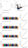
Fig. 1
Fig. 1. GWAS in a Japanese population stratified by COVID-19 severity and age.
a, Forest plots of the risk of COVID-19-associated variants in a Japanese population. Error bars indicate the 95% confidence interval. b, Manhattan plot of the GWAS on severe COVID-19 in young patients (those less than 65 years of age) (440 cases and 2,377 controls). Uncorrected P values from the GWAS analysis are shown. The dotted line represents the genome-wide significance threshold of P < 5.0 × 10−8. Manhattan and quantile–quantile plots of all GWAS results are presented in Extended Data Fig. 2. MT, mitochondrial. c, Regional association plot at the DOCK2 locus. Dots represent SNPs coloured according to linkage disequilibrium (r2) with the lead SNP of rs60200309. FAM196B is also known as INSYN2B. d, Allele frequency spectra of the rs60200309-A allele in the 1000 Genomes Project Phase3v5 database.
Fig. 2
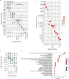
Fig. 2. Cell-type- and tissue-specific expression of DOCK2 and its downregulation in severe COVID-19.
a, eQTL effect of the COVID-19 risk variant (rs60200309) on DOCK2 expression levels using bulk RNA-seq of peripheral blood. The risk allele (rs60200309-A) decreases DOCK2 levels in patients with COVID-19 aged below 65 years. TPM, transcripts per kilobase million. b,c, Differential expression analysis of DOCK2 with varying COVID-19 severity. DOCK2 expression levels were quantified by qPCR and normalized to GAPDH expression. b, Comparison between severe and non-severe COVID-19 cases. c, Comparison between most severe, severe, mild and asymptomatic cases of COVID-19. dk, scRNA-seq in PBMCs from individuals with severe COVID-19 (n = 30) and healthy controls (n = 31). d, Uniform manifold approximation and projection (UMAP) visualization of all 394,526 cells. e, Projection of DOCK2 gene expression. Innate immune cell clusters are outlined with a red dashed line. f, Percentage of DOCK2-expressing cells and DOCK2 expression levels. g, Expression change with severe COVID-19 in six major cell types. h, Visualization and annotation of the innate immune cell clusters. ik, DOCK2 expression and expression changes with severe COVID-19 in the innate immune cell clusters. i, Projection of DOCK2 gene expression. j, Percentage of DOCK2-expressing cells and DOCK2 expression levels. k, Expression change with severe COVID-19 in five cell types. l, COVID-19 context-specific decreasing eQTL effect of the DOCK2 risk variant in non-classical monocytes. m,n, Immunohistochemical analysis of DOCK2. Lung and hilar lymph nodes were obtained from patients with COVID-19 pneumonia (m) or controls without COVID-19 or pneumonia (n), and stained with anti-DOCK2 polyclonal antibody. Results for all samples are shown in Extended Data Fig. 9. Scale bars, 0.25 mm. In ac,l, boxes denote the interquartile range (IQR) and the median is shown as horizontal bars; whiskers extend to 1.5 times the IQR; outliers are shown as individual points in ac and all samples are shown as individual points in l. Uncorrected P values are shown in (ac,g,k,l). cDC, conventional dendritic cells; cMono, classical monocytes; intMono, intermediate monocytes; Mono, monocytes; ncMono, non-classical monocytes; NK, natural killer cells; Pro T, proliferative T cells; Treg, T regulatory cells.
Fig. 3
Fig. 3. In vivo suppression of DOCK2 in a Syrian hamster model of SARS-CoV-2 infection.
a, Changes in body weight of hamsters infected with SARS-CoV-2. b, Representative images of lungs collected after euthanizing the hamsters at 11 dpi. c, Lung weight changes after infection. The number of samples (n) is indicated. d, Representative lung histopathology and immunohistochemistry of the infected hamsters at 6 dpi. Outlined areas are expanded to the right of each image. Right, lung tissue was stained with the anti-CD68 mouse monoclonal antibody to highlight alveolar macrophages. e, SARS-CoV-2 viral loads in the organs of the infected hamsters at 3 and 6 dpi. f, Lung cytokine expression assays of the infected animals. Ip-10 is also known as CXCL10. In (a) and (c), the error bars represent standard error of the mean, and P values were determined with two-sided Welch’s t-test; *P < 0.05; **P < 0.01; ***P < 0.001. In (e) and (f), boxes denote the IQR, and the median is shown as horizontal bars. Whiskers extend to 1.5 times the IQR, and all animals are shown as individual points. P values were determined with two-sided Wilcoxon rank sum test.
Extended Data Fig. 1
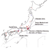
Extended Data Fig. 1. Japan COVID-19 Task Force.
Japan COVID-19 Task Force is a nation-wide consortium to overcome COVID-19 pandemic in Japan, which was established in early 2020. Japan COVID-19 Task Force consists of > 100 hospitals (red dots) led by core academic institutes (blue labels), and collected DNA, RNA, and plasma from the COVID-19 cases along with detailed clinical information. The figure was originally created using sf and ggplot2 R packages based on Global Map Japan version 2.1 Vector data (Geospatial Information Authority of Japan).
Extended Data Fig. 2
Extended Data Fig. 2. A principal component analysis plot of the GWAS participants and Manhattan and quantile-quantile plots of the GWAS.
(a, b) A principal component analysis (PCA) plot of the GWAS participants (COVID-19 cases and controls) along with and without International HapMap populations (a and b, respectively). (c) Manhattan plots and quantile-quantile plots of the Japanese GWAS of COVID-19. Uncorrected P values from GWAS analysis are shown. Dotted lines represent the genome-wide significance threshold of P < 5.0 × 10⁻⁸.
Extended Data Fig. 3
Extended Data Fig. 3. Regional association plots of the HLA imputation analysis.
Regional association plots of the HLA imputation analysis results. Dots represent SNPs and HLA variants with colors according to the legend. Uncorrected P values from HLA imputation analysis are shown. Dotted lines represent the genome-wide significance threshold of P < 5.0 × 10⁻⁸. HLA genes with the most significant associations in each of the case-control phenotypes are indicated.
Extended Data Fig. 4
Extended Data Fig. 4. ABO blood type associations with COVID-19 in Japanese and cross-population Mendelian randomization analysis of the COVID-19 GWAS.
(a) Odds ratios of the ABO blood types in the Japanese population are indicated. Dots represent the odds ratios and bars represent the 95 % confidence intervals. P values are uncorrected. Detailed results are presented in Supplementary Table 5. (b) Forest plots of the Mendelian randomization (MR) analysis results of causal inference on the COVID-19 GWAS in Japanese (left panel) and Europeans (right panel). Since effect sizes (= beta) of MR are not scalable among phenotypes and populations, normalized beta is indicated. For each phenotype and population, the standard error for the COVID-19 GWAS with the largest sample size (i.e., “COVID-19 vs control” for Japanese and “Self-reported COVID-19 vs control (C2)” for Europeans) was set to be 0.1. Dots represent the effect size normalized beta estimates and bars represent the 95 % confidence intervals. P values are uncorrected. The abbreviations of the exposure phenotypes and the detailed MR results are given in Supplementary Table 6 and Supplementary Table 7. BMI; body mass index, T2D; type 2 diabetes, CPD; cigarettes per day, CAD; cardiovascular disease, SBP; systolic blood pressure, DBP; diastolic blood pressure, eGFR; estimated glomerular filtration rate, UA; serum uric acids, RA; rheumatoid arthritis, SLE; systemic lupus erythematosus.
Extended Data Fig. 5
Extended Data Fig. 5. Effect size comparisons of the COVID-19 risk loci between the discovery GWAS and the replication study.
Co-plots of the odds ratios and 95% confidence intervals between the discovery GWAS cohort and replication cohort. To focus on the differences in the cases collected in different pandemic waves (initial waves for GWAS and latter waves for the replication), same controls as GWAS were currently used for the cases in the replication. A regression coefficient was estimated based on logarithm of odds ratios. Dots represent the odds ratios and bars represent the 95 % confidence intervals.
Extended Data Fig. 6
Extended Data Fig. 6. Colocalization analysis of the GWAS and eQTL signals at the DOCK2 locus.
Regional colocalization plots of the GWAS signals (severe and younger COVID-19 cases vs controls) and the eQTL signals on DOCK2 expression in the COVID-19 patients at the DOCK2 locus. CLPP; colocalization posterior probability. The eQTL effects of the variants around DOCK2 region are given in Supplementary Table 10.
Extended Data Fig. 7
Extended Data Fig. 7. Cell type definition and gene ontology enrichment analysis of DOCK2 co-expression gene module in the PBMC single cell analysis.
(a) Violin plots showing the expression distribution of selected canonical cell markers in the 12 clusters of PBMC. The rows represent selected marker genes and the columns represent clusters with the same color as in Fig. 2d. (b) Violin plots showing the expression distribution of selected canonical cell markers in the 5 clusters of innate immune cell clusters, shown in the same color as in Fig. 2h. (c) Tile plot showing percentage concordance between the manually annotated 12 clusters and Azimuth annotation. (d) The top 25 enriched biological processes by gene ontology (GO) analysis of DOCK2 co-expression gene module identified by weighted gene co-expression network analysis (WGCNA) in the non-classical monocytes of COVID-19 patients, where DOCK2 showed the highest cell type-specific expression profile. The color of the dots represents the adjusted P values.
Extended Data Fig. 8
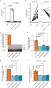
Extended Data Fig. 8. Biological impacts of DOCK2 downregulation in primary cells and DOCK2 knockdown and Interferon-α production assay in THP-1 Blue ISG cells.
(a) The impact of DOCK2 downregulation on interferon-α (IFN-α) production ability in pDC. Sorted pDC were stimulated with CpG and/or CPYPP. Data shows means ± s.e.m. (n = 3 per group). Differences of IFN-α production ability between the groups were evaluated using two-sided paired t-test. (b) The impact of DOCK2 downregulation on chemotaxis in CD3+ T cells. CD3+ T cells were stimulated with CXCL12 or CXCL12 + CPYPP (n = 19 per group). Differences of chemotaxis between the groups were evaluated using two-sided paired t-test. (c, d) Knockdown of DOCK2 by CRISPR system was confirmed by western blotting (c) and qRT-PCR. (d) Semi-quantitative staining density measure was determined using ImageJ (NIH). Data shows means ± s.e.m. (n = 3 per group). Data are compared to control group. P values were determined with One-way ANOVA followed by Dunnett’s post hoc test. (e, f) Activity ratio of SEAP reporter to no treatment group. Reporter was activated by 50 ng/ml LPS (e) or 50 μg/ml polyIC (f). Data shows means ± s.e.m. (n = 3 per group). Data are compared to control group. P values were determined with One-way ANOVA followed by Dunnett’s post hoc test.
Extended Data Fig. 9
Extended Data Fig. 9. Immunohistochemical analysis for DOCK2.
Lung and hilar lymph nodes were obtained from autopsied cadaver (Sample 1–3, 6, 7) or surgical specimen (Sample 4, 5), and stained by anti-DOCK2 polyclonal antibody. Sample 1–3; COVID-19 pneumonia. Sample 4-5; control. Sample 6; non-COVID-19 severe pneumonia. Sample 7; non-COVID-19 mild pneumonia.
Extended Data Fig. 10
Extended Data Fig. 10. In vivo suppression of DOCK2 in a Syrian hamster model with SARS-CoV-2 infection.
(a) Schematic timeline of the experimental procedure. (b) Changes in weight of uninfected animals. The error bars represent standard error of the mean. (c) Changes in weight of each of the infected animals, corresponding to Fig. 3a. Three CPYPP-administrated animals reaching humane endpoint were euthanized at dpi 7 and 9, lowering survival rate to 77% (=10/13), while survival of vehicle-administrated animals was 100% (=12/12). The animals were administered with CPYPP (red), or vehicle (blue). (d) Histopathological examination of the lungs of infected hamsters. Syrian hamsters were inoculated with SARS-CoV-2 with CPYPP or Vehicle. Syrian hamsters infected with CPYPP or Vehicle were euthanized on dpi 3, 6, and 11 for pathological examinations (n = 3). Shown are pathological findings in the lungs of hamsters infected with the virus on dpi 3, 6, and 11 (hematoxylin and eosin staining). Middle and Right show enlarged views of the area circled in black in Left. (Scale bars, 2.5 mm [Left], 1.0 mm [Middle], and 0.25 mm [Right].) (e) Immunohistochemistry for alveolar macrophages. Shown are immunohistochemical findings in the lungs of hamsters infected with the virus on dpi 6 (n = 3 per group). Lung tissue was stained with the anti-CD68 mouse monoclonal antibody. (Scale bars, 0.25 mm.) (f) Pathological severity scores in infected hamsters. To evaluate comprehensive histological changes, lung tissue sections were scored based on (d) pathological changes. Scores were determined based on the percentage of inflammation area of the maximum cut surface collected from each animal in each group by using the following scoring system: 0, no pathological change; 1, affected area (≤10%); 2, affected area (<50%, > 10%); 3, affected area (<90%, ≥50%); 4, (≥90%) an additional point was added when pulmonary edema and/or alveolar hemorrhage was observed. The total score is shown for individual animals. Blue dot and red dot indicate +Vehicle and +CPYPP, respectively.

Similar articles

Cited by

  • Dysuricemia.
    Nakayama A, Kurajoh M, Toyoda Y, Takada T, Ichida K, Matsuo H. Nakayama A, et al. Biomedicines. 2023 Nov 28;11(12):3169. doi: 10.3390/biomedicines11123169. Biomedicines. 2023. PMID: 38137389 Free PMC article. Review.
  • The whole blood transcriptional regulation landscape in 465 COVID-19 infected samples from Japan COVID-19 Task Force.
    Wang QS, Edahiro R, Namkoong H, Hasegawa T, Shirai Y, Sonehara K, Tanaka H, Lee H, Saiki R, Hyugaji T, Shimizu E, Katayama K, Kanai M, Naito T, Sasa N, Yamamoto K, Kato Y, Morita T, Takahashi K, Harada N, Naito T, Hiki M, Matsushita Y, Takagi H, Ichikawa M, Nakamura A, Harada S, Sandhu Y, Kabata H, Masaki K, Kamata H, Ikemura S, Chubachi S, Okamori S, Terai H, Morita A, Asakura T, Sasaki J, Morisaki H, Uwamino Y, Nanki K, Uchida S, Uno S, Nishimura T, Ishiguro T, Isono T, Shibata S, Matsui Y, Hosoda C, Takano K, Nishida T, Kobayashi Y, Takaku Y, Takayanagi N, Ueda S, Tada A, Miyawaki M, Yamamoto M, Yoshida E, Hayashi R, Nagasaka T, Arai S, Kaneko Y, Sasaki K, Tagaya E, Kawana M, Arimura K, Takahashi K, Anzai T, Ito S, Endo A, Uchimura Y, Miyazaki Y, Honda T, Tateishi T, Tohda S, Ichimura N, Sonobe K, Sassa CT, Nakajima J, Nakano Y, Nakajima Y, Anan R, Arai R, Kurihara Y, Harada Y, Nishio K, Ueda T, Azuma M, Saito R, Sado T, Miyazaki Y, Sato R, Haruta Y, Nagasaki T, Yasui Y, Hasegawa Y, Mutoh Y, Kimura T, Sato T, Takei R, Hagimoto S, Noguchi Y, Yamano Y, Sasano H, Ota S, Nakamori Y, Yoshiya K, Saito F, Yoshihara T, Wada D, Iwamura H, Kanayama S, Maruyama S, Yoshiyama T, Ohta K, Kok… See abstract for full author list ➔ Wang QS, et al. Nat Commun. 2022 Aug 22;13(1):4830. doi: 10.1038/s41467-022-32276-2. Nat Commun. 2022. PMID: 35995775 Free PMC article.
  • Quantitative multiorgan proteomics of fatal COVID-19 uncovers tissue-specific effects beyond inflammation.
    Schweizer L, Schaller T, Zwiebel M, Karayel Ö, Müller-Reif JB, Zeng WF, Dintner S, Nordmann TM, Hirschbühl K, Märkl B, Claus R, Mann M. Schweizer L, et al. EMBO Mol Med. 2023 Sep 11;15(9):e17459. doi: 10.15252/emmm.202317459. Epub 2023 Jul 31. EMBO Mol Med. 2023. PMID: 37519267 Free PMC article.
  • Rare predicted loss-of-function variants of type I IFN immunity genes are associated with life-threatening COVID-19.
    Matuozzo D, Talouarn E, Marchal A, Zhang P, Manry J, Seeleuthner Y, Zhang Y, Bolze A, Chaldebas M, Milisavljevic B, Gervais A, Bastard P, Asano T, Bizien L, Barzaghi F, Abolhassani H, Abou Tayoun A, Aiuti A, Alavi Darazam I, Allende LM, Alonso-Arias R, Arias AA, Aytekin G, Bergman P, Bondesan S, Bryceson YT, Bustos IG, Cabrera-Marante O, Carcel S, Carrera P, Casari G, Chaïbi K, Colobran R, Condino-Neto A, Covill LE, Delmonte OM, El Zein L, Flores C, Gregersen PK, Gut M, Haerynck F, Halwani R, Hancerli S, Hammarström L, Hatipoğlu N, Karbuz A, Keles S, Kyheng C, Leon-Lopez R, Franco JL, Mansouri D, Martinez-Picado J, Metin Akcan O, Migeotte I, Morange PE, Morelle G, Martin-Nalda A, Novelli G, Novelli A, Ozcelik T, Palabiyik F, Pan-Hammarström Q, de Diego RP, Planas-Serra L, Pleguezuelo DE, Prando C, Pujol A, Reyes LF, Rivière JG, Rodriguez-Gallego C, Rojas J, Rovere-Querini P, Schlüter A, Shahrooei M, Sobh A, Soler-Palacin P, Tandjaoui-Lambiotte Y, Tipu I, Tresoldi C, Troya J, van de Beek D, Zatz M, Zawadzki P, Al-Muhsen SZ, Alosaimi MF, Alsohime FM, Baris-Feldman H, Butte MJ, Constantinescu SN, Cooper MA, Dalgard CL, Fellay J, Heath JR, Lau YL, Lifton RP, Maniatis T, Mogensen TH, v… See abstract for full author list ➔ Matuozzo D, et al. Genome Med. 2023 Apr 5;15(1):22. doi: 10.1186/s13073-023-01173-8. Genome Med. 2023. PMID: 37020259 Free PMC article.
  • Quantification of escape from X chromosome inactivation with single-cell omics data reveals heterogeneity across cell types and tissues.
    Tomofuji Y, Edahiro R, Sonehara K, Shirai Y, Kock KH, Wang QS, Namba S, Moody J, Ando Y, Suzuki A, Yata T, Ogawa K, Naito T, Namkoong H, Xuan Lin QX, Buyamin EV, Tan LM, Sonthalia R, Han KY, Tanaka H, Lee H; Asian Immune Diversity Atlas (AIDA) Network; Japan COVID-19 Task Force; Biobank Japan Project; Okuno T, Liu B, Matsuda K, Fukunaga K, Mochizuki H, Park WY, Yamamoto K, Hon CC, Shin JW, Prabhakar S, Kumanogoh A, Okada Y. Tomofuji Y, et al. Cell Genom. 2024 Aug 14;4(8):100625. doi: 10.1016/j.xgen.2024.100625. Epub 2024 Jul 30. Cell Genom. 2024. PMID: 39084228 Free PMC article.

References

    1. The Severe Covid-19 GWAS Group. Genomewide association study of severe Covid-19 with respiratory failure. N. Engl. J. Med. 2020;383:1522–1534. doi: 10.1056/NEJMoa2020283. - DOI - PMC - PubMed
    1. Zeberg H, Pääbo S. The major genetic risk factor for severe COVID-19 is inherited from Neanderthals. Nature. 2020;587:610–612. doi: 10.1038/s41586-020-2818-3. - DOI - PubMed
    1. Niemi MEK, et al. Mapping the human genetic architecture of COVID-19. Nature. 2021;600:472–477. doi: 10.1038/s41586-021-03767-x. - DOI - PMC - PubMed
    1. Kosmicki JA, et al. Pan-ancestry exome-wide association analyses of COVID-19 outcomes in 586,157 individuals. Am. J. Hum. Genet. 2021;108:1350–1355. doi: 10.1016/j.ajhg.2021.05.017. - DOI - PMC - PubMed
    1. Pairo-Castineira E, et al. Genetic mechanisms of critical illness in COVID-19. Nature. 2021;591:92–98. doi: 10.1038/s41586-020-03065-y. - DOI - PubMed

MeSH terms

Substances