Review
. 2022 Sep 9;8(9):1758-1814.
doi: 10.1021/acsinfecdis.2c00204. Epub 2022 Aug 8.

Two Years into the COVID-19 Pandemic: Lessons Learned

Affiliations
Review

Two Years into the COVID-19 Pandemic: Lessons Learned

Severino Jefferson Ribeiro da Silva et al. ACS Infect Dis. .

Abstract

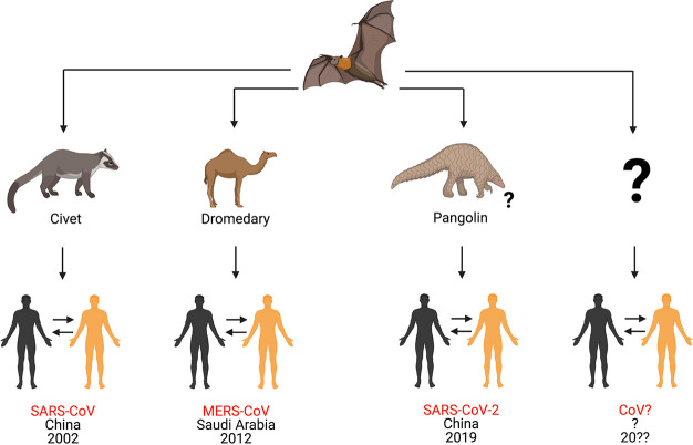
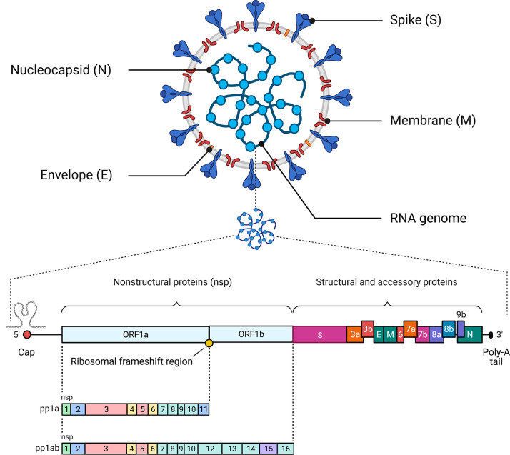
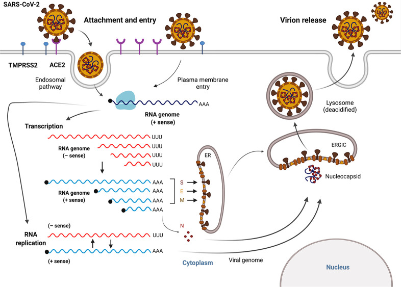
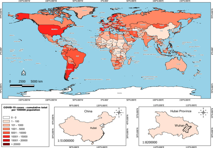
Severe acute respiratory syndrome coronavirus 2 (SARS-CoV-2) is a highly transmissible and virulent human-infecting coronavirus that emerged in late December 2019 in Wuhan, China, causing a respiratory disease called coronavirus disease 2019 (COVID-19), which has massively impacted global public health and caused widespread disruption to daily life. The crisis caused by COVID-19 has mobilized scientists and public health authorities across the world to rapidly improve our knowledge about this devastating disease, shedding light on its management and control, and spawned the development of new countermeasures. Here we provide an overview of the state of the art of knowledge gained in the last 2 years about the virus and COVID-19, including its origin and natural reservoir hosts, viral etiology, epidemiology, modes of transmission, clinical manifestations, pathophysiology, diagnosis, treatment, prevention, emerging variants, and vaccines, highlighting important differences from previously known highly pathogenic coronaviruses. We also discuss selected key discoveries from each topic and underline the gaps of knowledge for future investigations.

Keywords: SARS-CoV-2; clinical features; diagnosis; pathophysiology; prevention; reservoir hosts; transmission; treatment; vaccines; variants.


Conflict of interest statement

The authors declare no competing financial interest.

Figures

Figure 1
Figure 1
Origins of different coronaviruses. In the 21st century, three highly pathogenic betacoronaviruses have emerged from bats to cause respiratory disease in humans. In 2002, a betacoronavirus called severe acute respiratory syndrome coronavirus (SARS-CoV) emerged in Guangdong Province, China, and caused respiratory disease in humans. One decade later, another betacoronavirus called Middle East respiratory syndrome coronavirus (MERS-CoV) was reported in Saudi Arabia. Both SARS-CoV and MERS-CoV emerged from bats and were transmitted to humans via civets and dromedary camels, respectively. Later, in December 2019, a novel betacoronavirus called severe acute respiratory syndrome coronavirus 2 (SARS-CoV-2) emerged from bats and caused a pandemic disease called coronavirus disease 2019 (COVID-19). It was likely transmitted to humans by pangolins, although its origin is still being investigated. In summary, coronaviruses represent an example of emerging zoonotic viruses that have crossed the species barrier to cause disease in the human population. The possibility of a new, highly pathogenic coronavirus emerging from wild animals in the next few years cannot be ruled out. This figure was created with Biorender.com.
Figure 2
Figure 2
Schematic of the SARS-CoV-2 virus particle and genome architecture. The top panel illustrates the general structure of the SARS-CoV-2 viral particle, indicating its structural proteins and genome. The bottom panel illustrates the genome organization of SARS-CoV-2, including the 5′ cap, the region that encodes the nonstructural proteins required for viral replication (nsp1–nsp16), the region that encodes accessory and structural proteins (spike [S] protein, membrane [M] protein, envelope [E] protein, and nucleocapsid [N] proteins), and the poly-A tail. This figure was created with Biorender.com.
Figure 3
Figure 3
The SARS-CoV-2 replication cycle. SARS-CoV-2 enters the host cell via an endosomal pathway or through fusion of the viral envelope with the host cell membrane. Briefly, viral entry is initiated by binding of the RBD of the spike (S) protein to the human host cell receptor (ACE2). After the RBD–receptor interaction, the S protein undergoes proteolytic cleavage, which can be catalyzed by several host proteases, such as TMPRSS2, furin, and cathepsin B/L. Following viral entry, SARS-CoV-2 releases its genomic RNA into the cytoplasm and utilizes both the host’s and its own enzymatic machinery to replicate its genetic material and assemble new viral particles. The viral RNA genome is first translated into viral replicase polyproteins (pp1a and pp1ab), which are then cleaved into 16 nsps. In the process of genome replication and transcription mediated by the replication–transcription complex (RTC), the negative-sense (− sense) genomic RNA is synthesized and used as a template to generate a positive-sense (+ sense) genomic RNA and subgenomic RNAs. Viral assembly is aided by the interaction between viral genomic RNA and structural proteins located in the endoplasmic reticulum (ER) and ER–Golgi intermediate compartment (ERGIC). Finally, these virions are released to the plasma membrane via deacidified lysosomes and secreted from the infected cell via exocytosis. This figure was created with Biorender.com.
Figure 4
Figure 4
Epidemiology map of COVID-19. Cumulative cases of COVID-19 in all countries throughout the world. The bottom panels indicate the geographic location of Wuhan and Hubei Province in China, where the first COVID-19 cases were identified. The data were obtained from the World Health Organization (WHO).
Figure 5
Figure 5
Major modes of SARS-CoV-2 human-to-human transmission. Transmission can be through direct contact of airborne infectious particles deposited in respiratory droplets and aerosols. Indirect contact by infectious particles deposited on fomites represents another potential route for viral transmission. This figure was created with Biorender.com.
Figure 6
Figure 6
Clinical manifestations of COVID-19. Patients infected with SARS-CoV-2 can be asymptomatic, develop mild disease with diverse symptoms, or progress to severe illness. COVID-19 cases with severe complications are more frequently presented by patients from the high-risk group. This figure was created with Biorender.com.
Figure 7
Figure 7
Extrapulmonary complications from COVID-19. The extrapulmonary complications include a wide spectrum of disorders in several systems, including the neurological, cardiac, hepatic, renal, gastrointestinal, endocrine, vascular, and integumentary systems, which may occur in severe and critically ill COVID-19 patients and are linked to prolonged hospitalization and increased mortality risk., This figure was created with Biorender.com.
Figure 8
Figure 8
Gene expression of the ACE2 receptor in human tissues. The level of expression in each organ is categorized from high to low using different colors. Sources: https://www.proteinatlas.org/ENSG00000130234-ACE2/tissue and Li et al. (2020). This figure was created with Biorender.com.
Figure 9
Figure 9
Overview of different methods for COVID-19 diagnosis. SARS-CoV-2 can be directly detected in humans using molecular approaches, such as RT-qPCR, DNA sequencing, RT-LAMP, CRISPR/Cas systems, and sensors. Imaging tests, including chest computed tomography (CT), have been widely used as a complementary approach to diagnose COVID-19 patients. Additionally, human antibodies produced against SARS-CoV-2 antigens can be detected in blood samples via serological methods, including enzyme-linked immunosorbent assay (ELISA), chemiluminescence immunoassay (CLIA), immunofluorescence assay (IFA), and lateral flow assay (LFA). This figure was created with Biorender.com.
Figure 10
Figure 10
Kinetics of viral load and immune response during SARS-CoV-2 infection. During the first week after SARS-CoV-2 exposure, a period when patients are typically presymptomatic, the viral load increases and reaches its peak during the initial days after symptom onset. Seroconversion in infected patients begins in the second week after symptom onset. Three to four weeks after symptom onset, the IgM and IgG levels both reach their peaks and then begin to drop—more rapidly for IgM than for IgG. To avoid false-negative results when COVID-19 diagnostic tests are performed, the kinetics of viral load and immune response should be taken into consideration. The figure was adapted from the template in Biorender.com.
Figure 11
Figure 11
Vaccine platforms currently being developed against SARS-CoV-2 infection. Since the emergence of SARS-CoV-2, great efforts have been made by research groups and companies around the world toward the development of effective vaccines. Briefly, the graph in (A) shows current vaccine candidates in the clinical phase. Vaccine platforms currently being developed against SARS-CoV-2 infection include those based on (B) RNA, (C) DNA, (D) replicating viral vectors, (E) inactivated viruses, (F) attenuated viruses, (G) viruslike particles (VLPs), (H) nonreplicating viral vectors, (I) protein subunits, and (J) modified antigen-presenting cells (APCs). Abbreviations: S, spike protein; LNP, lipid nanoparticle; LV, lentiviral vector; DC, dendritric cell. This figure was created with Biorender.com.
Figure 12
Figure 12
SARS-CoV-2 variants of concern (VOCs). (A) Schematic of the SARS-CoV-2 genome architecture. (B) Definition of nonsynonymous mutations and deletions of each VOC. Nucleotide alterations without predicted or confirmed impact on protein structure and/or function are shown in black. Nucleotide alterations with predicted or confirmed impact on protein structure and/or function are shown in red. (C) Phenotypic characteristics of VOCs. Global distributions are according to the PANGO lineages website (https://cov-lineages.org/index.html). This figure was created with Biorender.com.

Similar articles

Cited by

References

    1. Weiss S. R.; Leibowitz J. L. Coronavirus Pathogenesis. Adv. Virus Res. 2011, 81, 85–164. 10.1016/B978-0-12-385885-6.00009-2. - DOI - PMC - PubMed
    1. Su S.; Wong G.; Shi W.; Liu J.; Lai A. C. K.; Zhou J.; Liu W.; Bi Y.; Gao G. F. Epidemiology, Genetic Recombination, and Pathogenesis of Coronaviruses. Trends Microbiol. 2016, 24 (6), 490–502. 10.1016/j.tim.2016.03.003. - DOI - PMC - PubMed
    1. Peiris J. S. M.; Guan Y.; Yuen K. Y. Severe Acute Respiratory Syndrome. Nat. Med. 2004, 10 (S12), S88–S97. 10.1038/nm1143. - DOI - PMC - PubMed
    1. Chan-Yeung M.; Xu R.-H. SARS: Epidemiology. Respirology 2003, 8 (S1), S9–S14. 10.1046/j.1440-1843.2003.00518.x. - DOI - PMC - PubMed
    1. Zaki A. M.; Van Boheemen S.; Bestebroer T. M.; Osterhaus A. D. M. E.; Fouchier R. A. M. Isolation of a Novel Coronavirus from a Man with Pneumonia in Saudi Arabia. N. Engl. J. Med. 2012, 367 (19), 1814–1820. 10.1056/NEJMoa1211721. - DOI - PubMed

Publication types