

doi: 10.1212/WNL.0000000000200836.
Epub 2022 Jun 13.
Pearls & Oy-sters: Challenges and Controversies in Wilson Disease
Affiliations
- PMID: 35940888
- PMCID: PMC9442620
- DOI: 10.1212/WNL.0000000000200836
Item in Clipboard
Pearls & Oy-sters: Challenges and Controversies in Wilson Disease
Neurology.
.
Abstract
Wilson disease (WD) is a genetic disorder of copper metabolism caused by variants in the ATP7B gene, which are inherited in an autosomal recessive pattern. Despite all the advances made on pathogenesis, cellular biology, and genetics, to date, WD remains a diagnostic and therapeutic challenge. With this series of cases, we aim to illustrate the main challenges that clinicians may encounter when dealing with patients with WD: the difficulties with clinical diagnosis, the therapeutic management of WD and the indication for advanced therapies, management during pregnancy, and genotype-phenotype correlations.
© 2022 American Academy of Neurology.
Figures

(A) Brain MRI images from case 1. Pontine hypointensities are observed in T1 sequences and hyperintensities in midbrain, pontine nucleus, tegmentum, and periaqueductal gray matter (arrows in the upper left of the figure) in fluid-attenuated inversion recovery-T2 sequences, suggesting the diagnosis of central pontine myelinolysis. It is also shown that the hyperintensity of the midbrain contrasts with the hypointensity of the red nucleus, the pars reticularis of the substantia nigra, and the aquaductus (arrows), which are relatively spared, resembling the face of a panda. Bilateral lenticular hypointensities can also be seen (arrows in the upper right of the figure). (B) Pathologic findings are shown (hematoxylin and eosin stain), both (B.a) macroscopically and (B.b–B.d) microscopically, showing central pontine vacuolization and loss of glial cells. Both findings are consistent with central pontine myelinolisis. (B.b–B.d) Vacuolization is marked with arrowheads and a star in microscopic samples. (C) Kayser-Fleischer ring is marked with an arrow.
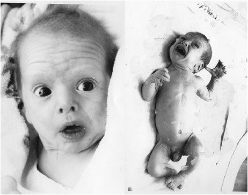
Penicillamine-induced cutis laxa syndrome in a baby boy born to a mother on d -penicillamine during pregnancy (case 3). The wrinkled appearance of baby's skin can be seen, which is particularly evident in his forehead, lips, neck, and fold areas, such as underarms and groins.
Similar articles
-
[Management of hepatolenticular degeneration during pregnancy].Zhonghua Gan Zang Bing Za Zhi. 2022 Jan 20;30(1):107-109. doi: 10.3760/cma.j.cn501113-20200807-00441. Zhonghua Gan Zang Bing Za Zhi. 2022. PMID: 35152680 Chinese.
-
The genetics of Wilson disease.Handb Clin Neurol. 2017;142:19-34. doi: 10.1016/B978-0-444-63625-6.00003-3. Handb Clin Neurol. 2017. PMID: 28433102 Free PMC article. Review.
-
Analysis of Wilson disease mutations revealed that interactions between different ATP7B mutants modify their properties.Sci Rep. 2020 Aug 10;10(1):13487. doi: 10.1038/s41598-020-70366-7. Sci Rep. 2020. PMID: 32778786 Free PMC article.
-
Wilson disease and related copper disorders.Handb Clin Neurol. 2018;147:279-292. doi: 10.1016/B978-0-444-63233-3.00018-X. Handb Clin Neurol. 2018. PMID: 29325617 Review.
-
[Progress in molecular mechanism of hepatolenticular degeneration induced by ATP7B gene mutation].Zhonghua Gan Zang Bing Za Zhi. 2020 Feb 20;28(2):188-192. doi: 10.3760/cma.j.issn.1007-3418.2020.02.019. Zhonghua Gan Zang Bing Za Zhi. 2020. PMID: 32164076 Review. Chinese.
Cited by
-
Functional Screen of Wilson Disease ATP7B Variants Reveals Residual Transport Activities.Hum Mutat. 2025 Jul 7;2025:7485658. doi: 10.1155/humu/7485658. eCollection 2025. Hum Mutat. 2025. PMID: 40661833 Free PMC article.
-
Myocardial involvement characteristics by cardiac MR imaging in neurological and non-neurological Wilson disease patients.Insights Imaging. 2024 Jan 25;15(1):24. doi: 10.1186/s13244-023-01583-7. Insights Imaging. 2024. PMID: 38270718 Free PMC article.
References
MeSH terms
Substances
LinkOut - more resources
Full Text Sources
Medical