Review
. 2023 Jan 3;15(1):a041256.
doi: 10.1101/cshperspect.a041256.

ER-Phagy: Quality and Quantity Control of the Endoplasmic Reticulum by Autophagy

Affiliations
Review

ER-Phagy: Quality and Quantity Control of the Endoplasmic Reticulum by Autophagy

Haruka Chino et al. Cold Spring Harb Perspect Biol. .

Abstract

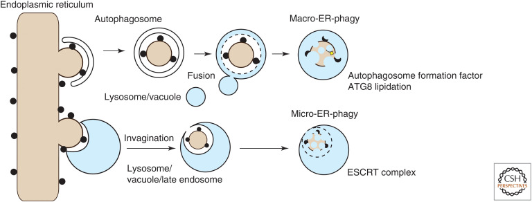
The endoplasmic reticulum (ER) is the largest organelle and has multiple roles in various cellular processes such as protein secretion, lipid synthesis, calcium storage, and organelle biogenesis. The quantity and quality of this organelle are controlled by the ubiquitin-proteasome system and autophagy (termed "ER-phagy"). ER-phagy is defined as the degradation of part of the ER by the vacuole or lysosomes, and there are at least two types of ER-phagy: macro-ER-phagy and micro-ER-phagy. In macro-ER-phagy, ER fragments are enclosed by autophagosomes, which is mediated by ER-phagy receptors. In micro-ER-phagy, a portion of the ER is engulfed directly by the vacuole or lysosomes. In these two pathways, some proteins in the ER lumen can be recognized selectively and subjected to ER-phagy. This review summarizes our current knowledge of ER-phagy, focusing on its membrane dynamics, molecular mechanisms, substrate specificity, and physiological significance.


Figures

Figure 1.
Figure 1.
Different types of endoplasmic reticulum (ER)-phagy. Macro-ER-phagy: Fragments of the ER are enclosed by autophagosomes. After fusion with lysosomes, the inner autophagosomal membrane and sequestered materials, including ER fragments, are degraded. This pathway requires the core ATG factors involved in autophagy initiation and ATG8 lipidation. Micro-ER-phagy: A small portion of the ER is engulfed directly by inward invagination of lysosomal/endosomal or vacuolar membranes and then degraded. This pathway depends on some or no ATG factors and requires the endosomal sorting complex required for transport (ESCRT) machinery for the closure step.
Figure 2.
Figure 2.
Structure and characteristics of endoplasmic reticulum (ER)-phagy receptors in mammals and yeast. Membrane topology and other characteristic features (e.g., the presence of the intrinsically disordered region [IDR] between the LC3-interacting region [LIR] and transmembrane domain) of mammalian and yeast ER-phagy receptors are shown. (a.a.) Amino acid, (AIM) Atg8-interacting motif, (ATZ) α1-antitrypsin Z variant, (NPC1) Niemann-Pick disease type C1, (PC) procollagen, (RHD) reticulon homology domain, (S.c.) Saccharomyces cerevisiae, (S.p.) Schizosaccharomyces pombe, (TMD) transmembrane domain, (UPR) unfolded protein response.
Figure 3.
Figure 3.
Various models of endoplasmic reticulum (ER)-phagy. Models of macro-ER-phagy and micro-ER-phagy in yeast and mammalian cells are depicted. Structures, ER-phagy receptors, and related factors and proteins to be degraded are shown. Please see the text for details of each process. (ERPHS) ER-phagy site, (ERES) ER exit sites.
Figure 4.
Figure 4.
Two distinct endoplasmic reticulum (ER)-phagy pathways for ATZ degradation. p62-dependent macro-ER-phagy and FAM134B-calnexin-mediated vesicular transport pathways for ATZ degradation are depicted.

References

    1. An H, Ordureau A, Paulo JA, Shoemaker CJ, Denic V, Harper JW. 2019. TEX264 is an endoplasmic reticulum-resident ATG8-interacting protein critical for ER remodeling during nutrient stress. Mol Cell 74: 891–908.e810. 10.1016/j.molcel.2019.03.034 - DOI - PMC - PubMed
    1. Antonucci L, Fagman JB, Kim JY, Todoric J, Gukovsky I, Mackey M, Ellisman MH, Karin M. 2015. Basal autophagy maintains pancreatic acinar cell homeostasis and protein synthesis and prevents ER stress. Proc Natl Acad Sci 112: E6166–E6174. 10.1073/pnas.1519384112 - DOI - PMC - PubMed
    1. Ast T, Michaelis S, Schuldiner M. 2016. The protease Ste24 clears clogged translocons. Cell 164: 103–114. 10.1016/j.cell.2015.11.053 - DOI - PMC - PubMed
    1. Axe EL, Walker SA, Manifava M, Chandra P, Roderick HL, Habermann A, Griffiths G, Ktistakis NT. 2008. Autophagosome formation from membrane compartments enriched in phosphatidylinositol 3-phosphate and dynamically connected to the endoplasmic reticulum. J Cell Biol 182: 685–701. 10.1083/jcb.200803137 - DOI - PMC - PubMed
    1. Bernales S, McDonald KL, Walter P. 2006. Autophagy counterbalances endoplasmic reticulum expansion during the unfolded protein response. PLoS Biol 4: e423. 10.1371/journal.pbio.0040423 - DOI - PMC - PubMed

Publication types

MeSH terms

Substances

LinkOut - more resources