Review
. 2022 Dec 1;14(12):a041247.
doi: 10.1101/cshperspect.a041247.

Endoplasmic Reticulum-Associated Protein Degradation

Affiliations
Review

Endoplasmic Reticulum-Associated Protein Degradation

Logesvaran Krshnan et al. Cold Spring Harb Perspect Biol. .

Abstract

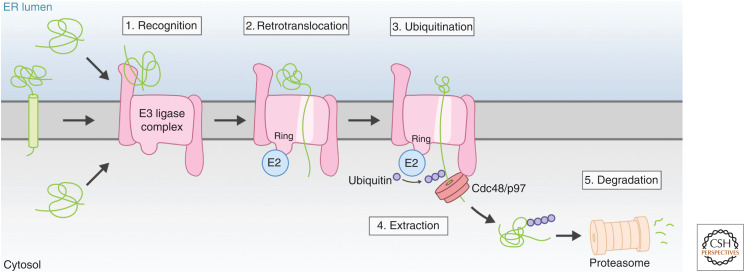
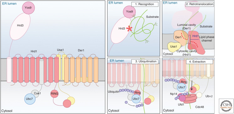
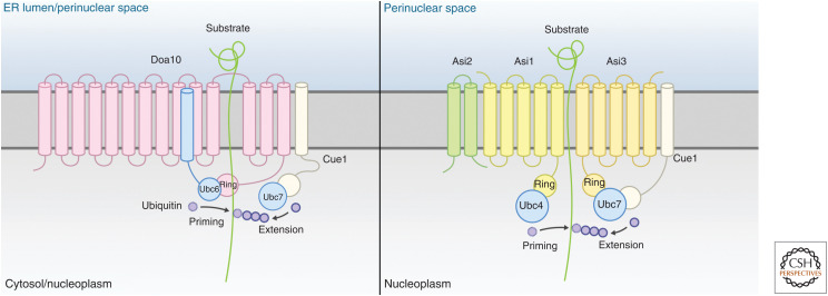
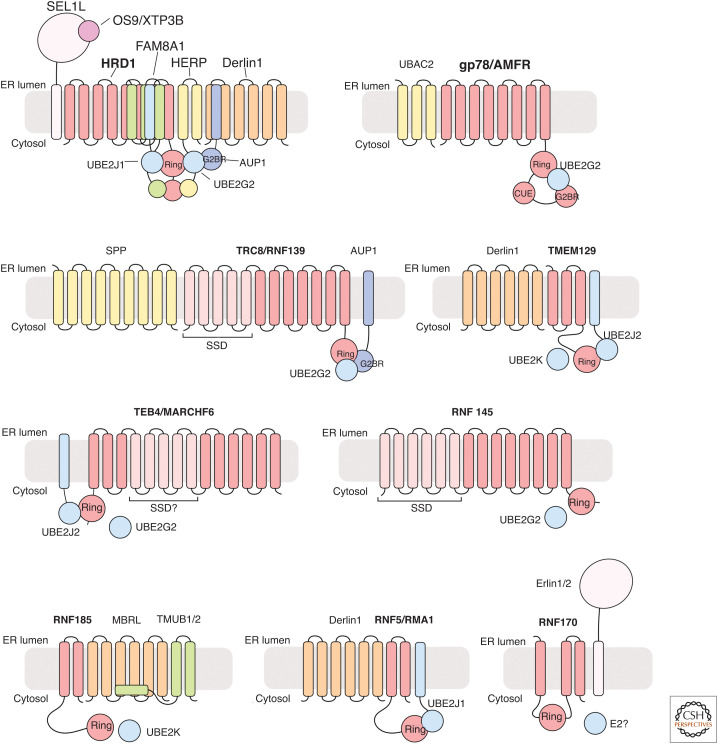
Misfolded, potentially toxic proteins in the lumen and membrane of the endoplasmic reticulum (ER) are eliminated by proteasomes in the cytosol through ER-associated degradation (ERAD). The ERAD process involves the recognition of substrates in the lumen and membrane of the ER, their translocation into the cytosol, ubiquitination, and delivery to the proteasome for degradation. These ERAD steps are performed by membrane-embedded ubiquitin-ligase complexes of different specificity that together cover a wide range of substrates. Besides misfolded proteins, ERAD further contributes to quality control by targeting unassembled and mislocalized proteins. ERAD also targets a restricted set of folded proteins to influence critical ER functions such as sterol biosynthesis, calcium homeostasis, or ER contacts with other organelles. This review describes the ubiquitin-ligase complexes and the principles guiding protein degradation by ERAD.


Figures

Figure 1.
Figure 1.
Generic organization of endoplasmic reticulum (ER)-associated degradation (ERAD) and its steps. A ubiquitin-ligase complex in the ER membrane coordinates the multiple ERAD steps starting with the recognition of substrates (shown in green). Depending on the substrate and ERAD branch involved, substrate recognition can occur on either side or within the ER membrane (1). On recognition, substrates in the lumen and membrane are retrotranslocated into the cytosolic side of the ER membrane. This step is facilitated by the membrane domains of ubiquitin-ligase complex components (2). Once exposed to the cytosolic face of the ER membrane, substrates are ubiquitinated (3). The cytosolic Cdc48/p97 complex binds to ubiquitinated substrates and facilitates late stages of retrotranslocation by extracting substrates out of the ER membrane (4). Substrates are subsequently delivered to the proteasome for degradation (5).
Figure 2.
Figure 2.
The mechanism of degradation of a luminal glycoprotein by the yeast Hrd1 complex. The yeast Hrd1 complex consists of a membrane core composed of the ubiquitin ligase Hrd1, Hrd3, Usa1, and Der1, which interact with Yos9 on the endoplasmic reticulum (ER) lumen and with the ATPase Cdc48 and its cofactors Npl4 and Ufd1 on the cytosolic side. The ubiquitin-conjugating enzyme is recruited to the membrane by Cue1 (left). The insets on the right indicate the individual steps involved in the ER-associated degradation (ERAD) of a luminal glycoprotein (see text for details).
Figure 3.
Figure 3.
The yeast Doa10 and Asi complexes and the sequential steps involved in substrate ubiquitination. The Doa10 complex (left) is composed of the ubiquitin ligase Doa10, the ubiquitin-conjugating enzymes Ubc6 and Ubc7, and the Ubc7 recruitment factor and activator Cue1. The Asi complex (right) is composed of the ubiquitin ligases Asi1 and Asi3 and the additional membrane protein Asi2. Ubiquitination of Asi substrates requires the conjugating enzymes Ubc4 and Ubc7. The scheme shows how these complexes use sequential steps for substrate (shown in green) ubiquitination. Initially the substrate is primed with a ubiquitin molecule reaction that involves the conjugating enzymes Ubc6 (Doa10 complex) or Ubc4 (Asi complex). These ubiquitin-conjugating enzymes are promiscuous and transfer ubiquitin to amine groups in lysine residues or to hydroxyl groups in serines or threonines. The initial ubiquitin serves as an acceptor for subsequent ubiquitination in a reaction that, for both complexes, depends on Ubc7.
Figure 4.
Figure 4.
Mammalian endoplasmic reticulum (ER)-associated degradation (ERAD) ubiquitin-ligase complexes. Overview of the mammalian ERAD ubiquitin-ligase complexes and their cognate E2 ubiquitin-conjugating enzymes. Ubiquitin ligases that name the complex are shown in bold. (RING) Really interesting new gene, (SSD) sterol-sensing domain, (G2BR) UBE2G2-binding region.

Similar articles

Cited by

References

    1. Adle DJ, Lee J. 2008. Expressional control of a cadmium-transporting P1B-type ATPase by a metal sensing degradation signal. J Biol Chem 283: 31460–31468. 10.1074/jbc.M806054200 - DOI - PMC - PubMed
    1. Avci D, Fuchs S, Schrul B, Fukumori A, Breker M, Frumkin I, Chen C-Y, Biniossek ML, Kremmer E, Schilling O, et al. 2014. The yeast ER-intramembrane protease Ypf1 refines nutrient sensing by regulating transporter abundance. Mol Cell 56: 630–640. 10.1016/j.molcel.2014.10.012 - DOI - PubMed
    1. Baldridge RD, Rapoport TA. 2016. Autoubiquitination of the Hrd1 ligase triggers protein retrotranslocation in ERAD. Cell 166: 394–407. 10.1016/j.cell.2016.05.048 - DOI - PMC - PubMed
    1. Ballar P, Pabuccuoglu A, Kose FA. 2011. Different p97/VCP complexes function in retrotranslocation step of mammalian ER-associated degradation (ERAD). Int J Biochem Cell Biol 43: 613–621. 10.1016/j.biocel.2010.12.021 - DOI - PubMed
    1. Bays NW, Gardner RG, Seelig LP, Joazeiro CA, Hampton RY. 2001. Hrd1p/Der3p is a membrane-anchored ubiquitin ligase required for ER-associated degradation. Nat Cell Biol 3: 24–29. 10.1038/35050524 - DOI - PubMed

Publication types

MeSH terms

LinkOut - more resources