. 2022 Aug 8;13(1):4633.
doi: 10.1038/s41467-022-32135-0.

Blocking ActRIIB and restoring appetite reverses cachexia and improves survival in mice with lung cancer

Affiliations

Blocking ActRIIB and restoring appetite reverses cachexia and improves survival in mice with lung cancer

Andre Lima Queiroz et al. Nat Commun. .

Abstract

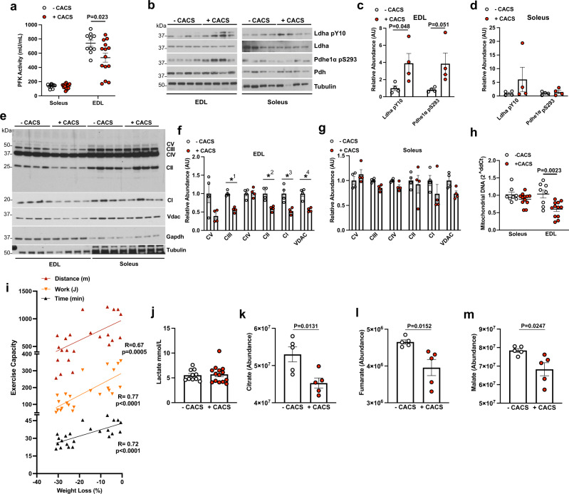
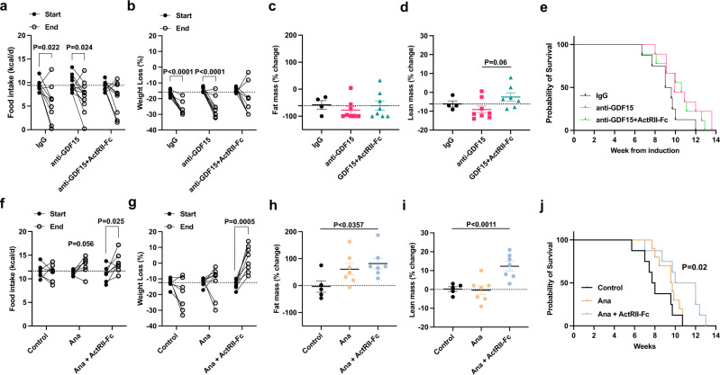
Cancer cachexia is a common, debilitating condition with limited therapeutic options. Using an established mouse model of lung cancer, we find that cachexia is characterized by reduced food intake, spontaneous activity, and energy expenditure accompanied by muscle metabolic dysfunction and atrophy. We identify Activin A as a purported driver of cachexia and treat with ActRIIB-Fc, a decoy ligand for TGF-β/activin family members, together with anamorelin (Ana), a ghrelin receptor agonist, to reverse muscle dysfunction and anorexia, respectively. Ana effectively increases food intake but only the combination of drugs increases lean mass, restores spontaneous activity, and improves overall survival. These beneficial effects are limited to female mice and are dependent on ovarian function. In agreement, high expression of Activin A in human lung adenocarcinoma correlates with unfavorable prognosis only in female patients, despite similar expression levels in both sexes. This study suggests that multimodal, sex-specific, therapies are needed to reverse cachexia.


Conflict of interest statement

L.C.C. is a founder, shareholder, and member of the scientific advisory board of Agios Pharmaceuticals and a founder and former member of the scientific advisory board of Ravenna Pharmaceuticals (previously Petra Pharmaceuticals). These companies are developing novel therapies for cancer. L.C.C. has received research funding from Ravenna Pharmaceuticals. M.D.G. reports personal fees from Novartis, Petra Pharmaceuticals, and Bayer. He has received research support from Pfizer Inc. L.C.C. and M.D.G. are inventors on patents unrelated to the scope of the current work. L.C.C. and M.D.G. are co-founders and shareholders in Faeth Therapeutics. Z.W. is a full-time employee of Pfizer Inc. All other authors report no competing interests.

Figures

Fig. 1
Fig. 1. Appetite and energy expenditure are reduced during CACS.
a Progression of weights normalized to their peak since tumor induction (n = 70). b The normalized weight of the mice in A at endpoint. c Lung mass of mice with and without CACS at endpoint (-CACS n = 23, +CACS n = 26). Lean (d) and Fat mass (e) measured 4 weeks after induction, defined as “Early”,(-CACS n = 13, +CACS n = 16), 4 to 8 weeks from induction defined as “Mid”, (-CACS n = 35, +CACS n = 48), and before euthanasia, defined as “Late” stages of disease, (-CACS n = 17, +CACS n = 16). f Total daily energy expenditure versus total body lean mass for WT (n = 29), -CACS, tumor-bearing mice without weight loss that later developed CACS (Pre-CACS), (n = 35), and +CACS (n = 19) either at 22 °C or 30 °C. The black line corresponds to WT mice at 22 °C (n = 29), Pre-CACS and - CACS mice are represented by dotted red squares (n = 35) and +CACS mice (22 °C and 30 °C, n = 19) by solid red squares. g Total daily energy expenditure adjusted by lean mass using ANCOVA using data from f. Cumulative food intake (h) in kcal and Cumulative activity (i) (distance traveled in meters), over a representative 24 h period at 22 °C of -CACS (n = 15) and + CACS (n = 5) or at 30 °C of -CACS (n = 8) and + CACS mice (n = 8). Both male and female mice were used in all panels. Graphs be, gi show mean ± SEM. Comparisons in be were made with two-tailed Student’s t-test between with +CACS and -CACS. Comparisons in g were done using one-way ANOVA followed by Tukey’s multiple comparisons test. Comparisons in h, i were made by 2-way ANOVA followed by Tukey’s multiple comparisons test. Individual data points are independent biological replicates unless otherwise stated. Source data are provided as a Source Data file.
Fig. 2
Fig. 2. Mice with CACS have impaired skeletal muscle metabolism.
a Phosphofructokinase (PFK) activity measured using Soleus lysates from mice without (-CACS, n = 10) and with cachexia (+CACS, n = 15) and extensor digitorum longus (EDL) lysates from mice without (-CACS, n = 11) and with cachexia (+CACS, n = 15). b Western blot of phosphorylated (Tyr10) and total LDHa, phosphorylated (Ser293) and total PDHe1α, and Tubulin from Soleus and EDL lysates from -CACS and +CACS mice. c Relative quantification of WB shown in B for phosphorylated (Tyr10) LDHa and phosphorylated (Ser293) PDHe1α in EDL lysates from -CACS (n = 4), and +CACS (n = 4)mice. d Relative quantification of WB shown in B for phosphorylated (Tyr10) LDHa and phosphorylated (Ser293) PDHe1α in Soleus lysates from -CACS (n = 4), and +CACS (n = 4) mice. e Western blot analysis of mitochondrial oxidative phosphorylation complexes (CI-subunit NDUFB8, CII-SDHB, CIII-UQCRC2, CIV-MTCO1, and CV-ATP5A) as well as VDAC, GAPDH, and Tubulin in lysates of Soleus and EDL muscles from -CACS and +CACS mice. f Relative quantification of WB shown in E for mitochondrial oxidative phosphorylation complexes in EDL lysates from -CACS (n = 4), and +CACS (n = 4) mice(P-value = ✱1 = 0.0001, ✱2 < 0.004, ✱3 < 0.007, ✱4 < 0.001). g Relative quantification of WB shown in E for mitochondrial oxidative phosphorylation complexes in Soleus lysates from -CACS (n = 4), and +CACS (n = 4) mice. h Relative mitochondrial DNA content in Soleus and EDL muscles of -CACS(n = 8) and +CACS(n = 8) mice. i Distance traveled (m), work performed (J), and duration (min) of maximal endurance performance test (running on a treadmill until exhaustion) of KL mice versus total body weight loss. Linear regression of each metric is shown. j Blood lactate levels of -CACS (n = 10) and +CACS(n = 14) mice at the completion of the maximal endurance performance test in I. k Citrate, (l) Fumarate, (m) Malate levels in gastrocnemius extracts from -CACS (n = 5) and +CACS(n = 5) mice by mass spectrometry. Both male and female mice were used in all panels. Graphs show mean ± SEM. a, c, d, fh, jm comparisons were made using two-tailed Student’s t-test compared with -CACS mice. Comparison in i was made using correlation analysis (R, Pearson r, and p, P-value). Individual data points are independent biological replicates unless otherwise stated. Source data are provided as a Source Data file.
Fig. 3
Fig. 3. Caloric Restriction does not recapitulate the metabolic changes observed in CACS.
Wild-type mice were calorie restricted (CR) by feeding a ~8 kcal/day diet (amount consumed by cachectic mice) until weight stabilization. a Percent of body weight change over a period of 18 days of CR (n = 8), or control diet (Fed, n = 2); The dashed line on the y-axis reflects the mean calorie consumption of mice with CACS taken from Fig. 1b. b Total body weight, Fat, and Lean mass of Fed (n = 8) and CR(n = 8) mice of a at day 18. c Total daily energy expenditure (kcal) in 24 h of Fed (n = 8) and CR(n = 8). d Cumulative activity (distance traveled in meters) over a representative 24 h period of Fed(n = 8) and CR mice(n = 8). e Distance traveled (m) and (F) Work (J) performed by Fed(n = 5) and CR (n = 4) mice performing a maximal endurance performance test (running on a treadmill until exhaustion). g Phosphofructokinase (PFK) activity in the soleus of Fed (n = 4) and CR mice (n = 8) and in the EDL(n = 4) of Fed and CR mice(n = 9). h Citrate Synthase (CS) activity in the soleus of Fed (n = 8) and CR mice (n = 22) and in the EDL(n = 8) of Fed and CR mice(n = 22). All mice used in the figure were wild-type males. Graphs show mean ± SEM. ac, fh comparisons made using two-tailed Student’s t-test compared with -CACS mice, (e) with a one-tailed Student’s t-test and d by 2-way ANOVA with Tukey’s multiple comparisons. Individual data points are independent biological replicates unless o therwise stated. Source data are provided as a Source Data file.
Fig. 4
Fig. 4. Tumors from mice with CACS have higher Activin A expression.
a Principal Component Analysis (PCA) of RNA-Seq from tumors of -CACS (n = 9) and +CACS (n = 13) mice. b Volcano plot depicting INHBA as one of the most differentially expressed genes (DEG) in the RNA-Seq of tumors from +CACS mice compared to -CACS (P < 0.01 & Log2Fold Change >1.2). c Venn diagram between the DEG identified in b and the human genes associated with poor prognosis in lung cancer from TCGA. d IL-1α levels in the serum of Fed (n = 8), -CACS (n = 10) and +CACS (n = 14) mice at the time of euthanasia. e Activin A levels in the serum of Fed (n = 8), -CACS (n = 8), +CACS (n = 17) and CR (n = 10) mice at the time of euthanasia. f GDF15 levels in the serum of Fed (n = 7), -CACS (n = 11), +CACS (n = 10), and CR (n = 10) mice at the time of euthanasia. a, b were done with samples from male mice, serum measurements in df were performed in both male and female mice. Graphs show mean ± SEM. df comparisons were made using One-way ANOVA with Tukey’s multiple comparison test. Individual data points are independent biological replicates unless otherwise stated. Source data are provided as a Source Data file.
Fig. 5
Fig. 5. Anamorelin but not GDF15 in combination with ActRIIB-Fc improves body weight, activity, and overall survival in female KL mice with CACS.
a Food intake (kcal/d) at week 0 (Start) and after 2 weeks of treatment (End) with control immunoglobulin (IgG, n = 9), anti-GDF15 mAB (n = 11) or ActRIIB-Fc decoy mAb in combination with GDF15 mAb (n = 9). b Total body weight loss at Start and End in mice treated with either IgG (n = 9), anti-GDF15 monoclonal antibody (n = 11) or anti-GDF15 mAb together with an ActRIIB-Fc decoy mAb (n = 9). Percentage of fat mass (c) and lean mass (d) change after 2 weeks of treatment with either IgG (n = 4), anti-GDF15 monoclonal antibody (n = 8) or anti-GDF15 mAb together with a decoy ActRIIB-Fc mAb (n = 8). e Kaplan-Meier (KM) plot with the probability of survival of the mice from (b). f Food intake (kcal/d) at Start and End with control IgG (n = 7), anamorelin (Ana, n = 7) or ActRIIB-Fc decoy mAb in combination with Ana (n = 8). g Total body weight loss at Start and End in mice treated with either IgG (n = 7), Ana (n = 8) or Ana in combination with ActRIIB-Fc decoy mAb (n = 8). h Percentage of fat mass change after 2 weeks of treatment with either IgG (n = 5), Ana (n = 7) or Ana in combination with ActRIIB-Fc decoy mAb (n = 7). i Percentage of lean mass change after 2 weeks of treatment with either IgG (n = 5), Ana (n = 6) or Ana in combination with ActRIIB-Fc decoy mAb (n = 7). j Kaplan-Meier (KM) plot with the probability of survival of the mice from g. ae Both male and female mice were used; (fj) only female mice were used. Graphs show mean ± SEM. a, b, f, g comparisons were made using two-tailed paired Student’s t-test comparing “Start” to “End” for each treatment. Comparisons in c, d, h, i were done using one-way ANOVA followed by Tukey’s multiple comparisons test. Comparisons in (e, j) were made using Log-rank Mantel-Cox test. Individual data points are independent biological replicates unless otherwise stated. Source data are provided as a Source Data file.
Fig. 6
Fig. 6. Activin A expression in human lung adenocarcinoma is associated with poor prognosis only in female patients.
a Activin A relative protein abundance in lung tumors from CPTAC categorized by a BMI cutoff of 20 (BMI ≤ 20, n = 28; BMI > 20, n = 81). b Serum Activin A levels measured by ELISA in a cohort of lung cancer patients from Weill Cornell(n = 82). c Lung cancer Activin A relative protein abundance categorized by proteogenomic subtype defined by Lehtiö et al. (n = 141). d Kaplan-Meier (KM) plot with the probability of survival of patients (n = 141) with high or low Activin A protein levels published by Lehtiö et al. e KM plot with the probability of survival of patients with adenocarcinoma with high or low Activin A protein levels from TCGA (n = 504). f, g KM plots with the probability of survival of patients with adenocarcinoma with high or low INHBA expression levels divided in Females (f, n = 270) and Males (g, n = 234). h TCGA Lung cancer mRNA expression of INHBA categorized by gender (n = 504). i Lung Cancer protein level of Activin A categorized by gender from Lehtiö et al. (n = 141). h For boxplots in a, c, horizontal bars within boxes represent medians. Tops and bottoms of boxes represent 25th and 75th percentiles, and vertical lines extend to the 1.5× interquartile range. Comparison in a, b, h, i were performed with a two-tailed Student’s t-test. Comparisons for the KM plots in b, df were done with the Log-rank Mantel-Cox test. Panel (c) was compared using One-Way ANOVA followed by Tukey’s multiple comparisons test, p-values for significant proteome subtype comparisons: 4-1 = 0.004, 6-1 = 0.0001, 6-2 = 0.0085, 6-3 = 0.009, 6-5 = 0.023). Individual data points are independent biological replicates unless otherwise stated. Source data are provided as a Source Data file.

Similar articles

Cited by

References

    1. Peixoto da Silva S, et al. Cancer cachexia and its pathophysiology: Links with sarcopenia, anorexia and asthenia. J. Cachexia Sarcopenia Muscle. 2020;11:619–635. doi: 10.1002/jcsm.12528. - DOI - PMC - PubMed
    1. Fearon K, et al. Definition and classification of cancer cachexia: An international consensus. Lancet Oncol. 2011;12:489–495. doi: 10.1016/S1470-2045(10)70218-7. - DOI - PubMed
    1. Ross PJ, et al. Do patients with weight loss have a worse outcome when undergoing chemotherapy for lung cancers? Br. J. Cancer. 2004;90:1905–1911. doi: 10.1038/sj.bjc.6601781. - DOI - PMC - PubMed
    1. Kimura M, et al. Prognostic impact of cancer cachexia in patients with advanced non-small cell lung cancer. Support Care Cancer. 2015;23:1699–1708. doi: 10.1007/s00520-014-2534-3. - DOI - PubMed
    1. Baracos VE, Martin L, Korc M, Guttridge DC, Fearon KCH. Cancer-associated cachexia. Nat. Rev. Dis. Prim. 2018;4:17105. doi: 10.1038/nrdp.2017.105. - DOI - PubMed

Publication types