. 2022 Aug 8;13(1):4624.
doi: 10.1038/s41467-022-32079-5.

Programmable RNA targeting by bacterial Argonaute nucleases with unconventional guide binding and cleavage specificity

Affiliations

Programmable RNA targeting by bacterial Argonaute nucleases with unconventional guide binding and cleavage specificity

Lidiya Lisitskaya et al. Nat Commun. .

Erratum in

Abstract

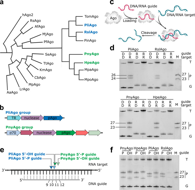
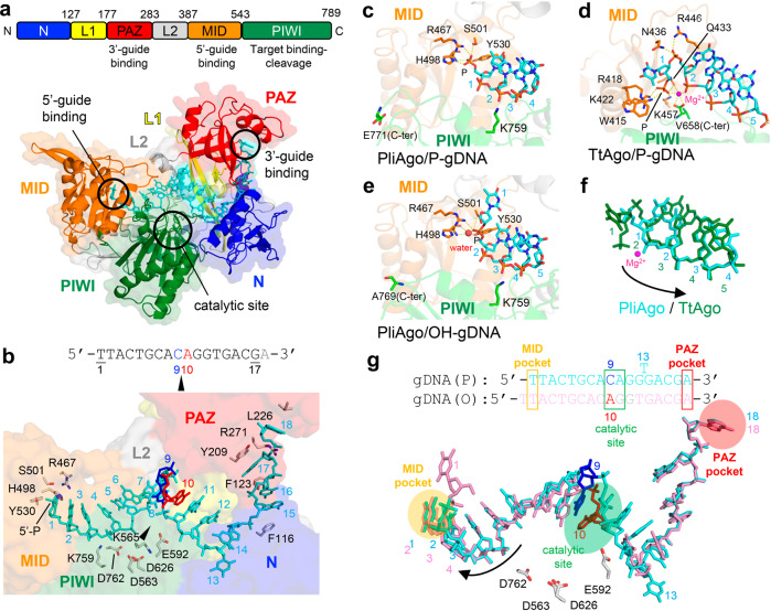
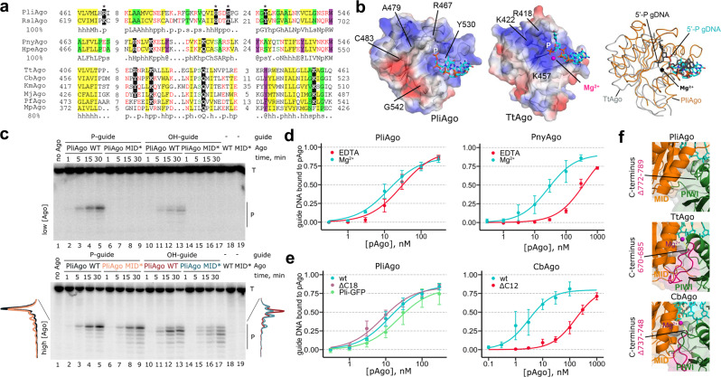
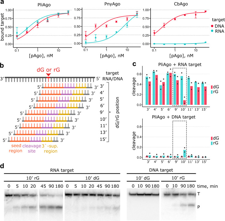
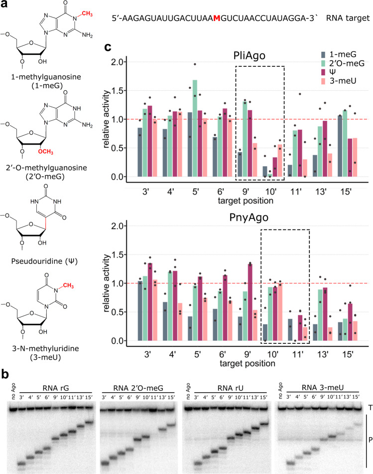
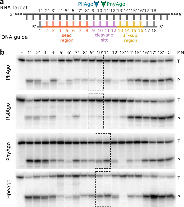
Argonaute proteins are programmable nucleases that have defense and regulatory functions in both eukaryotes and prokaryotes. All known prokaryotic Argonautes (pAgos) characterized so far act on DNA targets. Here, we describe a new class of pAgos that uniquely use DNA guides to process RNA targets. The biochemical and structural analysis of Pseudooceanicola lipolyticus pAgo (PliAgo) reveals an unusual organization of the guide binding pocket that does not rely on divalent cations and the canonical set of contacts for 5'-end interactions. Unconventional interactions of PliAgo with the 5'-phosphate of guide DNA define its new position within pAgo and shift the site of target RNA cleavage in comparison with known Argonautes. The specificity for RNA over DNA is defined by ribonucleotide residues at the cleavage site. The analysed pAgos sense mismatches and modifications in the RNA target. The results broaden our understanding of prokaryotic defense systems and extend the spectrum of programmable nucleases with potential use in RNA technology.


Conflict of interest statement

The authors declare no competing interests.

Figures

Fig. 1
Fig. 1. Two groups of pAgos prefer DNA guides and RNA targets.
a Phylogenetic tree of previously studied pAgos and of predicted D-R pAgos; pAgos studied in this work are shown in color (see Supplementary Fig. 1a for the full list of D-R pAgos and abbreviations). b Operon structure of D-R pAgos from the PliAgo (top) and PnyAgo (bottom) groups. pAgo-associated nuclease from the PD-(D/E)XK superfamily, putative transcription regulator (TR) and σ70 family factor are indicated. c Scheme of the cleavage assay. d Nucleic acid specificity of the two groups of pAgos. The reactions were performed with DNA (‘D’) or RNA (R’) guides and targets; positions of targets (‘T’), guides (‘G’) and reaction products (‘P’) are indicated (SYBR Gold staining). e Schematics of target RNA cleavage by the two groups of D-R pAgos in the presence (dark arrowheads) and in the absence (light arrowheads) of the 5’-phosphate in guide DNA. Nucleotide positions in guide DNA are indicated. f Analysis of RNA cleavage by D-R pAgos with 5′-P and 5′-OH guide DNAs. In panels d and f representative gels from 3 independent experiments with similar results are shown.
Fig. 2
Fig. 2. Structure of PliAgo and its interactions with guide DNA.
a The structure of the PliAgo and P-gDNA complex. (Top) The primary structure of PliAgo with amino acid numbering. Domains and their functions are colored and labeled. (Bottom) Overall structure of the PliAgo-P-gDNA complex. PliAgo and DNA are depicted as ribbon and stick models, respectively, with partially transparent surfaces. Domains are colored as in the top panel and labeled. b The PliAgo and P-gDNA interactions. (Top) The guide DNA sequence used for complex crystallization. Nucleotides that define the site of target RNA cleavage are colored. (Bottom) A magnified view of the PliAgo-P-gDNA interactions. PliAgo is depicted as a partially transparent surface. DNA nucleotides and amino acid residues responsible for guide positioning are shown as stick models and labeled. The catalytic site is indicated as a black triangle. The view is the same as a. ce The guide 5’-end binding in the PliAgo-P-gDNA complex c TtAgo-P-gDNA complex (PDB: 3DLH) d, and PliAgo-OH-gDNA complex e. PliAgo and TtAgo are depicted as partially transparent ribbon models. Amino acid residues responsible for 5’-end binding are shown as stick models and labeled. Salt bridges between amino acid residues and phosphate groups as well as coordination bonds of Mg2+/water are shown as yellow dashed lines. f Overlay of guide DNAs in the PliAgo-P-gDNA (cyan) and TtAgo-P-gDNA complexes (green). Positions of guide DNA nucleotides are labeled. Shifting the register of guide DNA from TtAgo to PliAgo is indicated by a black arrow. g 5’-phosphate dependent guide DNA positioning by PliAgo. (Top) The registers of 5’-P and 5’-OH guide DNAs in the PliAgo-gDNA complexes. (Bottom) Overlay of the guide DNA in the complexes of PliAgo with P-gDNA (cyan) and OH-gDNA (pink). Shifting the register of guide DNA without the 5’-phosphate group is indicated by a black arrow.
Fig. 3
Fig. 3. Role of the MID pocket in guide DNA binding and target RNA cleavage.
a Alignment of the MID pocket in various groups of pAgos. Conserved residues of the MID motif involved in 5’-nucleotide interactions are indicated in black. MpAgo has a hydrophobic MID pocket and binds 5′-hydroxylated guide RNAs but is more closely related to canonical pAgos than to PliAgo. The consensus for each group is shown underneath the alignment: ‘h’, hydrophobic residues (WFYMLIVACTH); ‘p’, polar (EDKRNQHTS); ‘s’, small (ACDGNPSTV); ‘o’, OH-containing (ST); ‘@‘, aromatic (YWFH). b Interactions of the MID domain with guide 5’-end (stick) in PliAgo (left) and TtAgo (PDB: 3DLH) (middle). Electrostatic potentials are shown as surfaces (blue, basic; red, acidic; white, neutral). (Right) Overlay of the MID pockets of PliAgo/P-gDNA (MID, orange; DNA, cyan) and TtAgo/gDNA complexes (MID, gray; DNA and Mg2+, black). c Kinetics of RNA cleavage by wild-type and MID* PliAgo (R467A/Y530A) loaded with 5’-P or 5’-OH gDNA, at low (50 nM) or high (500 nM) PliAgo concentrations (representative gel from two independent experiments). The reactions were performed with 5’-P32-labeled target RNA; positions of the target (‘T’) and the reactions products (‘P’) are indicated. The profiles of the cleavage products observed after 30 min with 500 nM PliAgo are shown on the left for P-gDNA (WT PliAgo, black; MID* PliAgo, orange) and on the right for OH-gDNA (WT, red; MID*, cyan). d Guide DNA binding by PliAgo and PnyAgo in the presence and in the absence of Mg2+. e Guide DNA binding by PliAgo, CbAgo and their variants PliAgo ΔC18 (deletion of the C-terminal 18 residues), PliAgo-GFP (C-teminal fusion with GFP), CbAgo ΔC12 (deletion of the C-terminal 12 residues). In d and e, means from 3 or 4 independent experiments are shown; error bars correspond to standard deviations. The amount of bound DNA is shown as a fraction of total 5’-P32-labeled DNA in the sample. f Comparison of the C-terminus positions in the structures of PliAgo (top), TtAgo (3DLH, middle) and CbAgo (6QZK, bottom). The C-terminal segment deleted in PliAgo (Δ772-789) is not solved on the structure. The corresponding segments in TtAgo (670-685) and CbAgo (737–748) are shown in magenta based on structural alignment.
Fig. 4
Fig. 4. Specificity of target recognition by D-R pAgos.
a Binding of target RNA and ssDNA of the same sequence by PliAgo, PnyAgo and CbAgo loaded with cognate guide DNA. Means and standard deviations from 3 independent experiments are shown. b Scheme of RNA/DNA targets containing a single dG/rG residue with differently positioned DNA guides (see Supplementary Table 1 for oligonucleotide sequences). Positions of the functional regions in guide DNA are indicated (seed, orange; central, magenta; 3’-supplementary region, ochre). c Effects of a single deoxyribonucleotide (dG) in the RNA target (top) and a single ribonucleotide (rG) in the DNA target (bottom) on target cleavage by PliAgo in comparison with control RNA and DNA targets. The reaction was performed at 37 °C for 30 min in the case of target RNA and for 3 h in the case of target DNA. Means from 2 independent experiments are shown. Target positions around the cleavage site are shown with dotted boxes. d Kinetics of target RNA (left) or target DNA (right) cleavage by PliAgo with containing ribonucleotide (rG) or deoxyribonucleotide (dG) residues at the position 10’. Representative gels from two independent experiments are shown.
Fig. 5
Fig. 5. D-R pAgos sense RNA modifications at specific target positions.
a Modified nucleotide residues with detected effects on the target RNA cleavage. The target RNA sequence is shown on the right (M, modified nucleotide), the guides are shown in Supplementary Fig. 8a. b Cleavage of modified RNAs containing 2’O-meG or 3-meU at the indicated positions by PnyAgo in comparison with control RNAs (rG or rU). Representative gels from 2 or 3 independent experiments. c Effects of RNA modifications at different positions relative to the guide 5’-end on target RNA cleavage by PliAgo (top) and PnyAgo (bottom). Target positions around the cleavage site are shown with dotted boxes. For each position, the efficiency of cleavage of modified RNA was normalized to the efficiency of cleavage of control RNA measured with the same guide DNA (‘relative activity’). Means from 2-3 independent experiments are shown.
Fig. 6
Fig. 6. D-R pAgos sense mismatches in the RNA target.
a Scheme of the guide-target duplex with indicated functional regions in guide DNA. The cleavage sites by PliAgo and PnyAgo are indicated. b Effects of single-nucleotide mismatches on the efficiency of RNA cleavage by D-R pAgos (see Supplementary Table 1 for guide DNA and target RNA sequences). The reactions were performed with 5’-P32-labeled target RNA at 37 °C for 60 min for PliAgo, RslAgo, HpeAgo and 10 min for PnyAgo; positions of the target (‘T’) and the reactions products (‘P’) are indicated. Representative gels from three independent experiments are shown (see Supplementary Fig. 9 for means and errors). The positions around the cleavage sites of corresponding pAgo proteins are shown with dotted boxes.

Similar articles

Cited by

References

    1. Koonin EV. Evolution of RNA- and DNA-guided antivirus defense systems in prokaryotes and eukaryotes: common ancestry vs convergence. Biol. Direct. 2017;12:5. - PMC - PubMed
    1. Makarova KS, Wolf YI, van der Oost J, Koonin EV. Prokaryotic homologs of Argonaute proteins are predicted to function as key components of a novel system of defense against mobile genetic elements. Biol. Direct. 2009;4:29. - PMC - PubMed
    1. Lisitskaya L, Aravin AA, Kulbachinskiy A. DNA interference and beyond: structure and functions of prokaryotic Argonaute proteins. Nat. Commun. 2018;9:5165. - PMC - PubMed
    1. Dimitriu T, Szczelkun MD, Westra ER. Evolutionary ecology and interplay of prokaryotic innate and adaptive immune systems. Curr. Biol. 2020;30:R1189–R1202. - PMC - PubMed
    1. Nussenzweig PM, Marraffini LA. Molecular mechanisms of CRISPR-Cas immunity in bacteria. Annu. Rev. Genet. 2020;54:93–120. - PubMed

Publication types

MeSH terms