. 2022 Aug 8;8(1):353.
doi: 10.1038/s41420-022-01146-7.

Nrf2 signaling activation by a small molecule activator compound 16 inhibits hydrogen peroxide-induced oxidative injury and death in osteoblasts

Affiliations

Nrf2 signaling activation by a small molecule activator compound 16 inhibits hydrogen peroxide-induced oxidative injury and death in osteoblasts

Jing-Wei Zhao et al. Cell Death Discov. .

Abstract

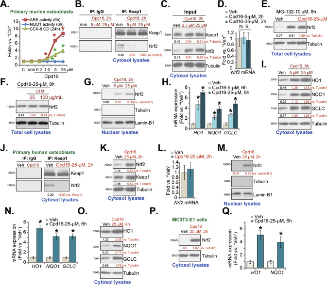
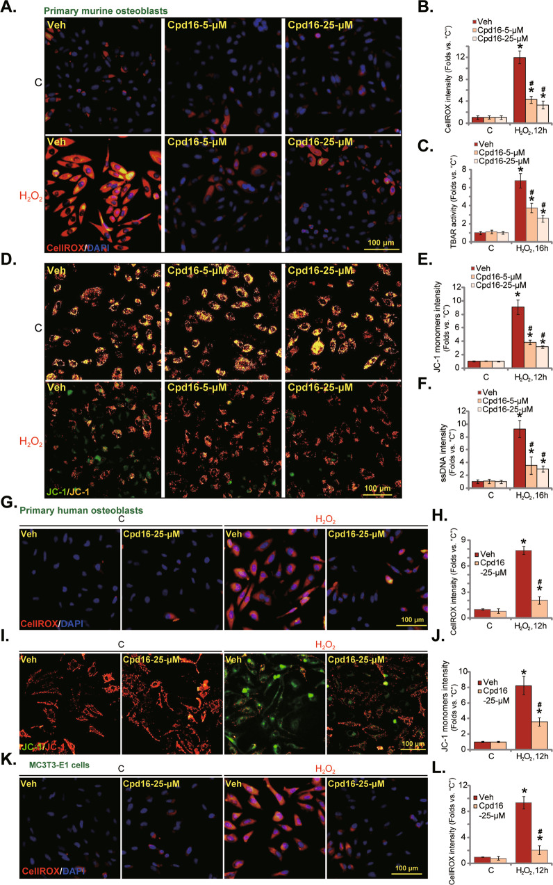
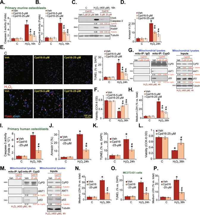
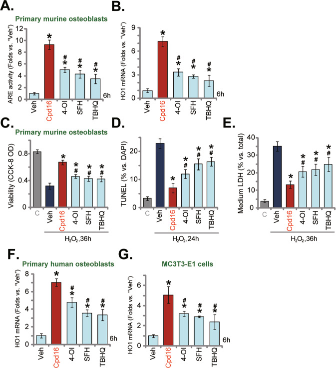
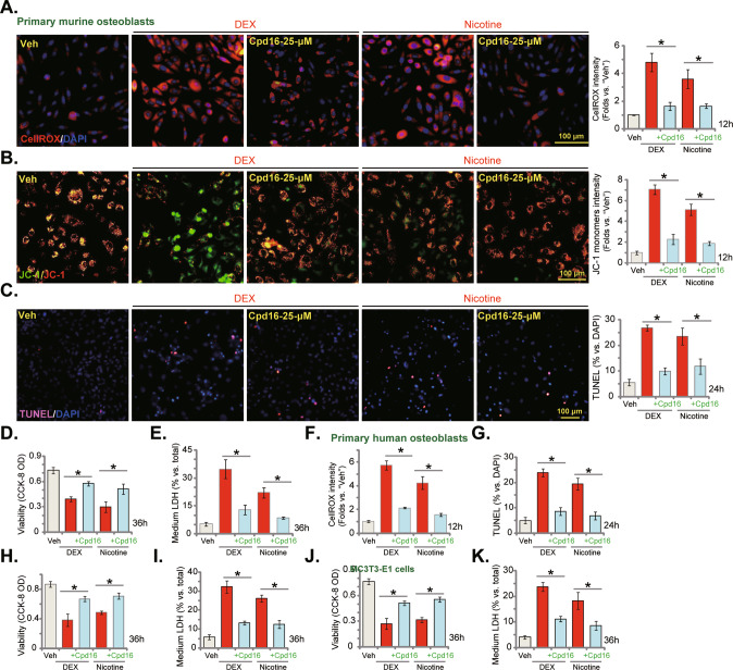
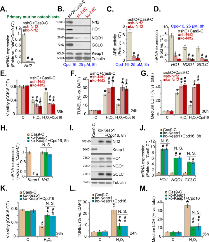
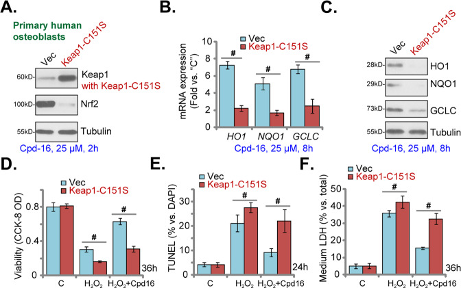
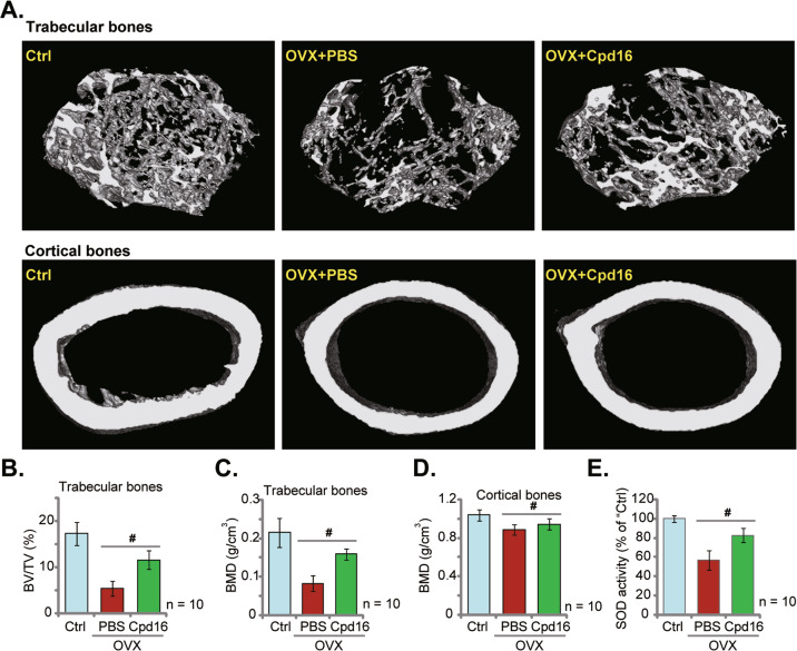
We explored the potential activity of compound 16 (Cpd16), a novel small molecule Nrf2 activator, in hydrogen peroxide (H2O2)-stimulated osteoblasts. In the primary murine/human osteoblasts and MC3T3-E1 murine osteoblastic cells, Cpd16 treatment at micro-molar concentrations caused disassociation of Keap1-Nrf2 and Nrf2 cascade activation. Cpd16 induced stabilization of Nrf2 protein and its nuclear translocation, thereby increasing the antioxidant response elements (ARE) reporter activity and Nrf2 response genes transcription in murine and human osteoblasts. Significantly, Cpd16 mitigated oxidative injury in H2O2-stimulited osteoblasts. H2O2-provoked apoptosis as well as programmed necrosis in osteoblasts were significantly alleviated by the novel Nrf2 activator. Cpd16-induced Nrf2 activation and osteoblasts protection were stronger than other known Nrf2 activators. Dexamethasone- and nicotine-caused oxidative stress and death in osteoblasts were attenuated by Cpd16 as well. Cpd16-induced osteoblast cytoprotection was abolished by Nrf2 short hairpin RNA or knockout, but was mimicked by Keap1 knockout. Keap1 Cys151S mutation abolished Cpd16-induced Nrf2 cascade activation and osteoblasts protection against H2O2. Importantly, weekly Cpd16 administration largely ameliorated trabecular bone loss in ovariectomy mice. Together, Cpd16 alleviates H2O2-induced oxidative stress and death in osteoblasts by activating Nrf2 cascade.


Conflict of interest statement

The authors declare no competing interests.

Figures

Fig. 1
Fig. 1. Cpd16 activates Nrf2 signaling cascade in osteoblasts.
Primary murine osteoblasts (AD, GI), human osteoblasts (JO), or the MC3T3-E1 murine osteoblastic cells (P, Q) were stimulated with Cpd16, ARE activity and NQO1 activity as well as cell viability were measured (A); Keap1-Nrf2 association was measured through co-immunoprecipitation (Co-IP) (B, J); Proteins in cytosol/nuclear fraction lysates were examined (C, G, I, K, M, O, P), with mRNAs measured by qRT-PCR (D, H, L, N, Q). The primary murine osteoblasts were treated with MG-132 (10 μM) or plus Cpd16 (25 μM) for 8 h, total protein lysates were tested (E). The murine osteoblasts were pretreated for 1 h with cycloheximide (CHX, 25/100 μg/mL), following by Cpd16 (25 μM) stimulation for another 8 h, listed proteins were shown (F). “C” is untreated control (same for all Figures). “Veh” is vehicle control (0.1% DMSO) (same for all Figures). *P < 0.05 versus “Veh” cells.
Fig. 2
Fig. 2. Cpd16 ameliorates H2O2-provoked oxidative injury in osteoblasts.
Primary murine osteoblasts (AF), human osteoblasts (GJ), or the MC3T3-E1 murine osteoblastic cells (K and L) were pretreated (for 2 h) with Cpd16 (5/25 μM), or plus H2O2 (400 μM) stimulation; ROS contents (the CellROX intensity assay, A, B, G, H, K, L), TBAR activity (C), JC-1 dye staining (D, E, I, J), and ssDNA contents (F) were measured. *P < 0.05 versus “C” cells. #P < 0.05 versus cells with H2O2 stimulation but “Veh” pretreatment. Scale bar = 100 μm.
Fig. 3
Fig. 3. Cpd16 ameliorates H2O2-induced apoptosis and programmed necrosis in osteoblasts.
Primary murine osteoblasts (AH), human osteoblasts (IN) or the MC3T3-E1 murine osteoblastic cells (O, P) were pretreated (for 2 h) with Cpd16 (5/25 μM), or plus H2O2 (400 μM) stimulation; the caspase-3/-9 activities (A, B, I) were measured; apoptosis-associated proteins were measured (C); cell apoptosis was examined by Annexin V flow cytometry (D, J, results were quantified) and the nuclear TUNEL staining (E, K, and O, results were quantified) assays, with cell viability measured through CCK-8 assays (F, L); CyPD-ANT1-p53 mitochondrial complexation and the expression were shown (G, M), and cell necrosis measured through measuring LDH releasing (H, N, P). *P < 0.05 versus “C” cells. #P < 0.05 versus cells with H2O2 stimulation but “Veh” pretreatment. Scale bar = 100 μm.
Fig. 4
Fig. 4. Cpd16-induced Nrf2 activation and osteoblasts protection against H2O2 were stronger than other known Nrf2 activators.
The primary murine osteoblasts (A, B), human osteoblasts (F), and MC3T3-E1 murine osteoblastic cells (G) were treated with 25 μM of Cpd16, Sulforaphane (SFH), 4-octyl itaconate (4-OI), tert-butylhydroquinone (TBHQ), and cultured for 6 h, the relative ARE activity (A) and HO1 mRNA (B, F, G) expression were tested. The primary murine osteoblasts were pretreated with 25 μM of Cpd16, SFH, 4-OI, or TBHQ for 2 h, followed by H2O2 (400 μM) stimulation, and viability, apoptosis, and necrosis were measured through CCK-8 (C), TUNEL-nuclei staining (D), and LDH releasing (E) assays, respectively. *P < 0.05 versus “Veh”. #P < 0.05. versus “Cpd16”.
Fig. 5
Fig. 5. Cpd16 inhibits dexamethasone- and nicotine-induced oxidative injury in osteoblasts.
The primary murine osteoblasts (AE), human osteoblasts (FI), or the MC3T3-E1 murine osteoblastic cells (J, K) were pretreated (for 2 h) with Cpd16 (25 μM), followed with or without dexamethasone (DEX, 2 μM) or nicotine (1 μM) treatments, ROS, depolarization of mitochondria, apoptosis, viability, and necrosis were tested by CellROX staining (A, F), JC-1 staining (B), TUNEL-nuclei staining (C, G), CCK-8 (D, H, J), and LDH releasing (E, I, K) assays, respectively. *P < 0.05. Scale bar = 100 μm (AC).
Fig. 6
Fig. 6. Nrf2 activation is indispensable for Cpd16-mediated osteoblast cytoprotection.
Nrf2 mRNA expression in the listed primary murine osteoblasts was measured (A); the osteoblasts were treated with Cpd16, and listed proteins (B), ARE activity (C), and listed mRNAs (D) were measured; the murine osteoblasts were pretreated with Cpd16 (25 μM) for 2 h, followed by H2O2 (400 μM) stimulation, with cell viability, apoptosis and necrosis tested by CCK-8 (E), TUNEL-nuclei staining (F), and LDH releasing G assays, respectively. Keap1-Nrf2 mRNA expression in the ko-Keap1 or Cas9-C murine osteoblasts was measured (H); the ko-Keap1 murine osteoblasts were also treated with or without Cpd16 (25 μM), expression of listed proteins and mRNAs (I, J) was measured; the ko-Keap1 osteoblasts were pretreated with Cpd16 (25 μM) for 2 h, followed by H2O2 (400 μM) stimulation; cell viability, apoptosis, and necrosis were measured by CCK-8 (K), TUNEL-nuclei (L), and LDH releasing M assays, respectively. *P < 0.05 versus “C” cells. #P < 0.05 versus “shC+Cas9-C”/“Cas9-C” osteoblasts. “N.S.” stands for the non-statistical difference (P > 0.05).
Fig. 7
Fig. 7. Keap1 Cys151S mutation abolishes Cpd16-induced Nrf2 cascade activation and osteoblasts cytoprotection.
Stable primary human osteoblasts with Cys151S Keap1 (“Keap1-C151S”) or Vector were treated with Cpd16 (25 μM), listed proteins and genes were measured (AC). Alternatively, the primary human osteoblasts were pretreated with Cpd16 (25 μM) for 2 h, followed by H2O2 (400 μM) stimulation, and cell viability, apoptosis, and necrosis were tested by CCK-8 (D), TUNEL-nuclei staining (E), and LDH releasing F assays, respectively. #P < 0.05.
Fig. 8
Fig. 8. Cpd16 administration largely ameliorates trabecular bone loss in OVX mice.
The female C57/BL6 mice were subject to bilateral ovariectomy (OVX) procedure. Afterward, Cpd16 (at 5 mg/kg) or PBS were intraperitoneally injected (i.p.) at the first day of each week, and mice were sacrificed after 8 weeks. The representative micro-CT images of trabecular bones and cortical bones were presented (A). BV/TV (%, B) and bone mineral density (BMD, g/cm3, C) of trabecular bones were calculated. BMD of cortical bones was recorded as well (D). The relative SOD activity in the left tibia bone tissues in different groups was shown (E). The control group mice were orally administered with PBS (“Ctrl”). 10 mice per group. #P < 0.05.

Similar articles

Cited by

References

    1. Lewiecki EM. New targets for intervention in the treatment of postmenopausal osteoporosis. Nat Rev Rheumatol. 2011;7:631–8. doi: 10.1038/nrrheum.2011.130. - DOI - PubMed
    1. Harvey N, Dennison E, Cooper C. Osteoporosis: impact on health and economics. Nat Rev Rheumatol. 2010;6:99–105. doi: 10.1038/nrrheum.2009.260. - DOI - PubMed
    1. Dempster DW. Bone histomorphometry in glucocorticoid-induced osteoporosis. J Bone Min Res. 1989;4:137–41. doi: 10.1002/jbmr.5650040202. - DOI - PubMed
    1. Zalavras CG, Lieberman JR. Osteonecrosis of the femoral head: evaluation and treatment. J Am Acad Orthop Surg. 2014;22:455–64. doi: 10.5435/JAAOS-22-07-455. - DOI - PubMed
    1. Lieberman JR, Engstrom SM, Meneghini RM, SooHoo NF. Which factors influence preservation of the osteonecrotic femoral head? Clin Orthop Relat Res. 2012;470:525–34. doi: 10.1007/s11999-011-2050-4. - DOI - PMC - PubMed