. 2022 Aug 8;12(1):13330.
doi: 10.1038/s41598-022-17629-7.

Characterization of extracellular vesicles from Lactiplantibacillus plantarum

Affiliations

Characterization of extracellular vesicles from Lactiplantibacillus plantarum

Atsushi Kurata et al. Sci Rep. .

Abstract

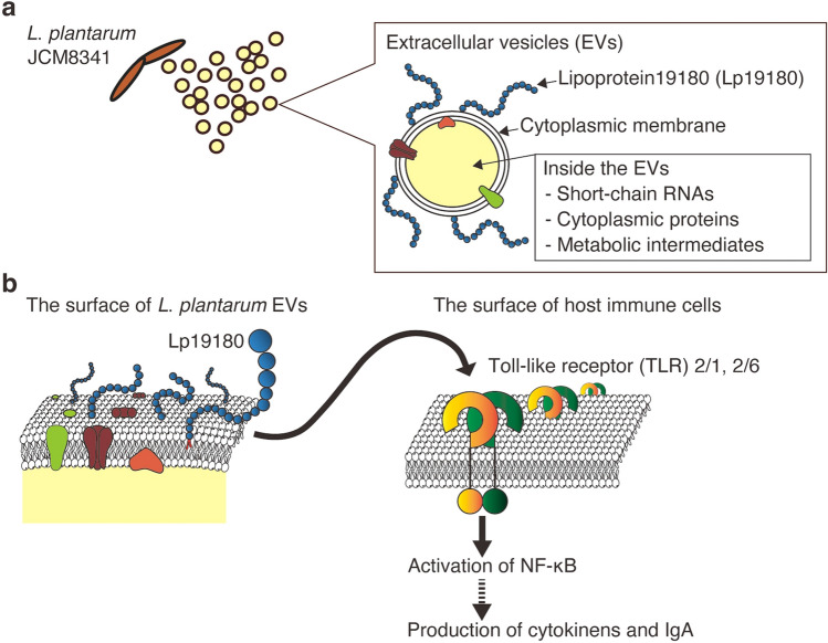
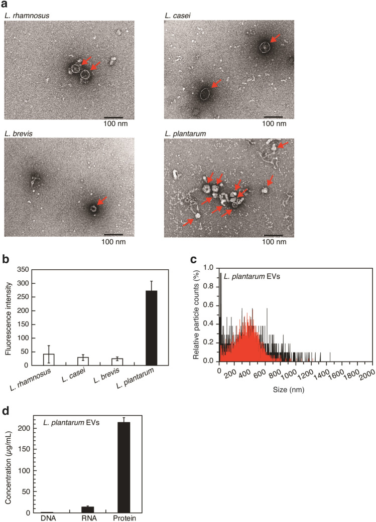
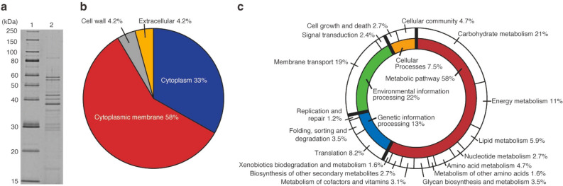
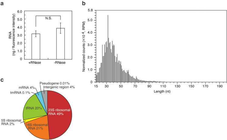
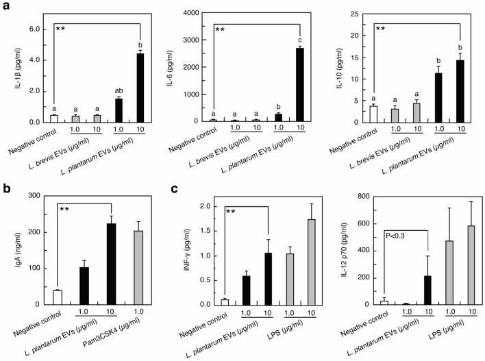
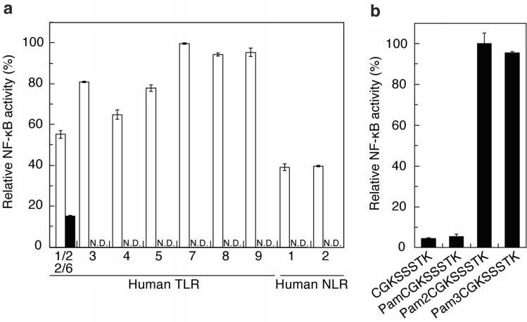
We investigated the characteristics and functionalities of extracellular vesicles (EVs) from Lactiplantibacillus plantarum (previously Lactobacillus plantarum) towards host immune cells. L. plantarum produces EVs that have a cytoplasmic membrane and contain cytoplasmic metabolites, membrane and cytoplasmic proteins, and small RNAs, but not bacterial cell wall components, namely, lipoteichoic acid and peptidoglycan. In the presence of L. plantarum EVs, Raw264 cells inducibly produced the pro-inflammatory cytokines IL-1β and IL-6, the anti-inflammatory cytokine IL-10, and IF-γ and IL-12, which are involved in the differentiation of naive T-helper cells into T-helper type 1 cells. IgA was produced by PP cells following the addition of EVs. Therefore, L. plantarum EVs activated innate and acquired immune responses. L. plantarum EVs are recognized by Toll-like receptor 2 (TLR2), which activates NF-κB, but not by other TLRs or NOD-like receptors. N-acylated peptides from lipoprotein19180 (Lp19180) in L. plantarum EVs were identified as novel TLR2 ligands. Therefore, L. plantarum induces an immunostimulation though the TLR2 recognition of the N-acylated amino acid moiety of Lp19180 in EVs. Additionally, we detected a large amount of EVs in the rat gastrointestinal tract for the first time, suggesting that EVs released by probiotics function as a modulator of intestinal immunity.


Conflict of interest statement

The authors declare no competing interests.

Figures

Figure 1
Figure 1
Properties of EVs produced by L. plantarum and their effects on host immune cells. (a) Biochemical components of L. plantarum EVs. (b) Schematic sketch of NF-κB activation by Lp19180 via TLR2 and the subsequent stimulation of innate and adaptive immunities.
Figure 2
Figure 2
Characterization of EVs produced by L. plantarum. (a) TEM images of lactobacillus EVs. Red arrows indicate EVs. Scale bar, 100 nm. (b) EVs production of lactobacilli. Means ± SD, n = 3. (c) Size distributions of EVs (black: all particles in the L. plantarum EVs fraction, red: EVs labeled with FM4-64). The total numbers of particles in L. plantarum EVs and those of particles labeled by FM4-64 in L. plantarum EVs are represented as 100%. (d) DNA, RNA, and protein in L. plantarum EVs. Means ± SD, n = 3.
Figure 3
Figure 3
Proteins in L. plantarum EVs. (a) SDS-PAGE profile of L. plantarum EVs. Lane 1: Marker, Lane 2: L. plantarum EVs (0.3 µg). Proteins were separated by SDS-PAGE using 12.5% gel (Gellex International, Tokyo, Japan) with constant voltage at 300 V and then detected using SYPRO Ruby staining (Lonza, Rockland, ME). The cropped gel was displayed, and original gel was indicated in Supplementary Fig. 3a_original_gel file. (b) Subcellular localization of proteins in L. plantarum EVs. (c) Biological processes involving proteins in L. plantarum EVs.
Figure 4
Figure 4
Small RNAs in L. plantarum EVs. (a) The amount of RNA per fluorescence intensity of EVs. The amount of RNA after the RNase treatment was compared with that without the treatment. Means ± SD, n = 3. Two-tailed unpaired Student’s t-test, N.S. not significant. (b) Strand length distribution of RNAs. (c) Classification of small RNAs in L. plantarum EVs.
Figure 5
Figure 5
Stimulation of immune cells by L. plantarum EVs. The production of cytokines from Raw264 cells (a,c) and IgA from PP cells (b) was indicated. Pam3CSK4 (b) and LPS (c) were used as positive controls. PBS (a–c) was used as the negative control. Means ± SD, n = 3. (a) Bars identified by the same letters are not significantly different from each other (P > 0.01) by the Tukey’s test. (a–c) the Dunnett’s test, **P < 0.01 compared with negative control.
Figure 6
Figure 6
Human cell receptors involved in the recognition of L. plantarum EVs and lipopeptides from Lp19180. The recognition abilities of (a) L. plantarum EVs (closed bar) and each positive control (open bar, described in the Materials and Methods) and (b) acylated N-terminal peptides derived from Lp19180 were evaluated based on NF-κB-inducible SEAP activity in duplicate wells. Means ± SD. N.D. not detected.

References

    1. Man SM. Inflammasomes in the gastrointestinal tract: Infection, cancer and gut microbiota homeostasis. Nat. Rev. Gastroenterol. Hepatol. 2018;15:721–737. doi: 10.1038/s41575-018-0054-1. - DOI - PMC - PubMed
    1. Dang AT, Marsland BJ. Microbes, metabolites, and the gut–lung axis. Mucosal. Immunol. 2019;12:843–850. doi: 10.1038/s41385-019-0160-6. - DOI - PubMed
    1. Philpott DJ, Girardin SE. The role of Toll-like receptors and Nod proteins in bacterial infection. Mol. Immunol. 2004;41:1099–1108. doi: 10.1016/j.molimm.2004.06.012. - DOI - PubMed
    1. Bunker JJ, Bendelac A. IgA responses to microbiota. Immunity. 2018;49:211–224. doi: 10.1016/j.immuni.2018.08.011. - DOI - PMC - PubMed
    1. Tezuka H, Ohteki T. Regulation of IgA production by intestinal dendritic cells and related cells. Front. Immunol. 2019;10:1891. doi: 10.3389/fimmu.2019.01891. - DOI - PMC - PubMed

Publication types

LinkOut - more resources