Review
. 2022 Aug;52(9):1601-1614.
doi: 10.1007/s00247-022-05396-6. Epub 2022 Aug 9.

Update on aneurysmal bone cyst: pathophysiology, histology, imaging and treatment

Affiliations
Review

Update on aneurysmal bone cyst: pathophysiology, histology, imaging and treatment

Ricardo Restrepo et al. Pediatr Radiol. 2022 Aug.

Abstract

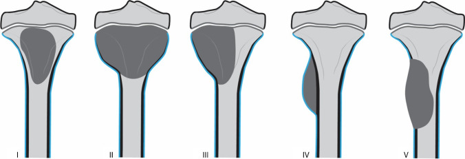
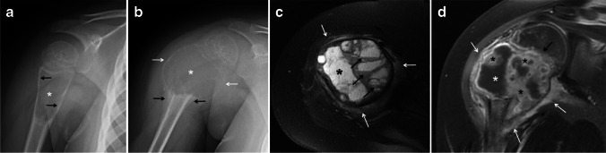
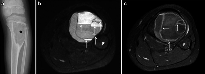
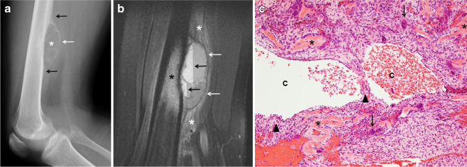
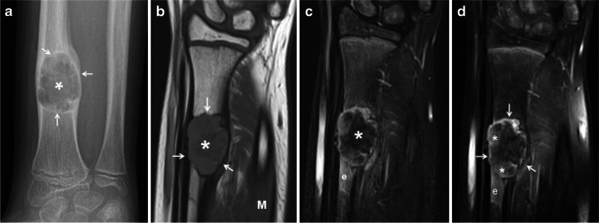
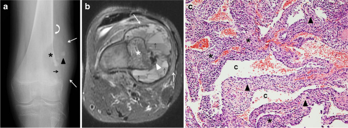
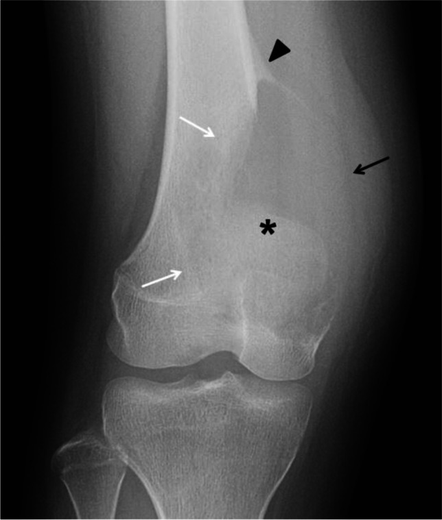
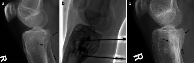
Aneurysmal bone cyst (ABC) is a benign but locally aggressive lesion that predominantly affects children and young adults. ABC, which accounts for approximately 70% of the cases, is now recognized to be a true neoplasm, whereas ABC-like changes associated to other bone neoplasms (also referred in the literature as secondary ABC) accounts for the remaining 30%. The solid variant of ABC is also considered a true neoplasm but is rare. ABC can involve any bone in the body, and although it has a metaphyseal preference, it can involve any part of a bone and soft tissues. As with any bone tumor, the initial evaluation of ABCs should be done with radiographs followed by magnetic resonance imaging or less frequently computed tomography for further characterization. The imaging appearance of ABC is variable; however, a lytic and expansile lesion with fluid-fluid levels is the most common presentation. The main differential diagnosis of an ABC in the pediatric population is unicameral bone cyst (UBC) and telangiectatic osteosarcoma, therefore a biopsy is recommended before treatment. The therapeutic options of ABC range from curettage with or without adjuncts such as phenol, liquid nitrogen, argon laser and bone grafting or bone substitutes to more recently employed alternatives such as image-guided sclerotherapy with various sclerosing agents and monoclonal antibodies (e.g., Denosumab).

Keywords: Aneurysmal bone cyst; Bone; Children; Computed tomography; Magnetic resonance imaging; Radiography; Sclerotherapy.


Conflict of interest statement

None

Figures

Fig. 1
Fig. 1
Aneurysmal bone cyst classification according to morphology. Reproduced with permission from Capanna et al. [23]. Type I: centrally located lesions that are well contained with no outline or slightly expanded outline. Type II: very expansile tumors with cortical thinning involving the entire bone segment. Type III: eccentric metaphyseal lesions that typically involve only one cortex. Type IV: subperiosteal lesions growing away from the bone. Type V: periosteal lesions expanding peripherally to ultimately penetrate the underlying cortex
Fig. 2
Fig. 2
Rapid progression of a proximal humeral aneurysmal bone cyst from a type I to a type II in a 5-year-old girl. a An initial anteroposterior radiograph of the shoulder shows a proximal metadiaphyseal, lytic lesion (asterisk) of the humerus. The lesion causes endosteal scalloping (arrows), is slightly expansile and has no internal septations or matrix. b An anteroposterior radiograph of the shoulder obtained 7 months later shows significant interval growth of the lesion (asterisk) with blurring of the physeal margin. The lesion is now expansile and has marked cortical thinning (white arrows) with no sclerotic border or areas of mineralization. Aggressive periosteal reaction (black arrows) is identified at the distal margin of the lesion. c An axial fat-suppressed T2-weighted magnetic resonance (MR) image of the humeral lesion shows the expansile, multiseptated humeral lesion with a dominant cystic cavity (asterisk), fluid-fluid levels (black arrows) and mild adjacent soft-tissue edema (white arrows). d A coronal fat-suppressed, contrast-enhanced T1-weighted MR image of the humeral lesion clearly shows the focal bone expansion and physeal involvement (black arrow). The lesion is multicystic; the dominant cyst (white asterisk) displays peripheral contrast enhancement and areas of enhancing solid tissue (black asterisks). A rim of peripheral enhancing edema is present (white arrows)
Fig. 3
Fig. 3
Type III aneurysmal bone cyst of the tibia in a 10-year-old girl. a An anteroposterior radiograph of the tibia shows an eccentric, well-defined, juxtaphyseal, lytic and expansile lesion (asterisk) involving the proximal tibial metadiaphysis. b An axial fat-suppressed T2-weighted magnetic resonance (MR) image of the leg shows fluid-fluid levels throughout the lesion (arrows). c A fat-suppressed, contrast-enhanced, T1-weighted MR image of the lesions shows thin, enhancing septations (arrows). F fibula
Fig. 4
Fig. 4
Type V aneurysmal bone cyst of the femur in a 16-year-old boy. a A lateral radiograph of the femur shows a superficial, parosteal, lytic and expansile lesion (asterisk) involving the distal femoral diaphysis. The lesion is exophytic extending into the adjacent soft tissues surrounded by a thin, calcified shell (white arrow). Periosteal reaction (black arrows) is identified at the proximal and distal margins of the lesion. b A sagittal fat-suppressed T2-weighted magnetic resonance image of the femur shows the exophytic mass involving the femoral cortex with a thin hypointense peripheral shell (white arrows) extending into the soft tissues. Fluid-fluid levels (black arrows) and adjacent bone marrow (black asterisk) as well as soft-tissue edema are present (white asterisks). c Hematoxylin and eosin staining, magnification 100X: Solid areas and cystic spaces (C) filled with blood, with cellular septa (arrowheads), benign osteoid (asterisks), stromal cells and giant cells (arrows)
Fig. 5
Fig. 5
Aneurysmal bone cyst (ABC) of the cervical spine involving two consecutive vertebrae in a 16-year-old boy before and after percutaneous sclerotherapy with doxycycline. a A coronal reconstruction of a contrast-enhanced computed tomography (CT) scan image of the cervical spine using bone window shows a large, expansile lytic lesion (asterisk) involving the transverse processes (white arrows), articular pillars and body of C5 and C6 (black arrows). b A coronal reconstruction of a non-enhanced CT scan image of the cervical spine using bone window 6 years later after sclerotherapy shows complete interval healing of the lesion (asterisk) with diffuse sclerosis, remodeling of the bone and localized fusion at the site of the treated ABC
Fig. 6
Fig. 6
Aneurysmal bone cyst (ABC) of the skull and petrous bone in a 10-year-old boy. a An axial T2-weighted magnetic resonance image of the head shows a large, elliptical, expansile mass (calipers) in the left side of the posterior fossa causing mass effect upon the left cerebellar hemisphere and partial effacement of the fourth ventricle (white arrow). The mass is multiseptated and contains fluid-fluid levels of different signal intensity distributed throughout the lesion (black arrows). b An axial computed tomography scan image using bone windows shows to better advantage the petrous bone and occipital bone involvement by the ABC (asterisk) as well as a thin peripheral calcified shell (white arrows)
Fig. 7
Fig. 7
Epiphyseal extension of an aneurysmal bone cyst of the left distal femur in a 14-year-old girl. An anteroposterior radiograph of the left femur shows a well-defined, lytic, expansile metadiaphyseal lesion (black asterisk) with multiple internal septations causing cortical thinning. There is clear epiphyseal extension (white asterisk) of the lesion across the physis (arrow)
Fig. 8
Fig. 8
Chondroblastoma containing aneurysmal bone cyst (ABC)-like changes in a 13-year-old boy. A sagittal fat-suppressed T2-weighted magnetic resonance image of the knee shows a well-defined, lobulated lesion involving the posterior aspect of the proximal tibial epiphysis (arrows) corresponding to the chondroblastoma. The lesion is cystic and multiseptated containing a fluid-fluid level (arrowhead). Note the extensive, surrounding bone marrow and soft-tissue edema (asterisks) characteristic of chondroblastomas. After resection, the diagnosis of chondroblastoma with ABC-like changes was confirmed
Fig. 9
Fig. 9
Solid variant aneurysmal bone cyst of the right radius in a 9-year-old girl. a An anteroposterior radiograph of the right wrist shows an expansile, lytic lesion (asterisk) involving the distal diaphysis of the radius. The lesion is geographical with endosteal scalloping and a sclerotic border (arrows). Minimal nonaggressive periosteal reaction is present proximally. b A coronal T1-weighted magnetic resonance (MR) image shows the lesion (asterisk) as solid and expansile with a slightly hyperintense parenchyma compared to muscle (M) and a sclerotic border (arrows). c A coronal fat-suppressed T2-weighted MR image shows the expansile lesion (asterisk) as heterogeneous in signal but predominantly hypointense. Note the adjacent bone marrow edema (e). d A coronal fat-suppressed, contrast-enhanced, T1-weighted MR image shows patchy, heterogeneous contrast enhancement (asterisks) by the lesion (arrows) and by the adjacent bone marrow edema (e)
Fig. 10
Fig. 10
Telangiectatic osteosarcoma in a 17-year-old boy. a An initial anteroposterior radiograph of the knee shows a distal femoral metadiaphyseal, geographical, eccentric, lytic lesion (asterisk) with no sclerotic border. There is focal destruction of the distal femoral cortex (black arrow), focal areas of mineralization (arrowhead) and an associated soft-tissue mass (white arrows). Codman’s triangle is present proximally (curved arrow). b An axial fat-suppressed T2-weighted magnetic resonance image of the femoral lesion shows the eccentric, osseous mass (asterisk) with multiple fluid-fluid levels located in the periphery (black arrows), hypointense areas of mineralization (arrowhead) and adjacent soft tissue edema (white arrows). c Hematoxylin and eosin staining, magnification 100X: solid areas and cystic spaces (C) filled with blood, fibrous cellular septa (arrowheads), malignant stromal cells (asterisks)
Fig. 11
Fig. 11
Radiographically aggressive, biopsy proven aneurysmal bone cyst (ABC) of the distal femur in a 17-year-old boy (compare the striking similarity with patient from Fig. 10). An anteroposterior radiograph of the distal left femur shows a lytic distal metadiaphyseal lesion (asterisk) of the left femur. The lesion extends into the epiphysis and adjacent soft tissues with no calcified peripheral shell (black arrow). The ABC has a wide zone of transition (white arrows) and aggressive periosteal reaction proximally (arrowhead). The lesion was biopsied twice and ABC was confirmed twice
Fig. 12
Fig. 12
Type III aneurysmal bone cyst in a 16-year-old boy before, during and after percutaneous sclerotherapy with doxycycline. a A lateral radiograph of the proximal tibia shows an eccentric, well-defined, lytic and expansile lesion (arrows) involving the proximal tibial metaphysis with a faint peripheral calcified shell. b A fluoroscopic image of the lesion during sclerotherapy shows the insertion of two spinal needles and injection of contrast accumulating in several intralesional locules. c A lateral radiograph of the proximal tibia 6 years later after several treatments with sclerotherapy shows complete interval healing of the lesion (arrows) with some sclerosis and remodeling of the bone but no significant residual deformity

References

    1. Cottalorda J, Kohler R, Sales de Gauzy J, et al. Epidemiology of aneurysmal bone cyst in children: a multicenter study and literature review. J Pediatr Orthop B. 2004;13:389–394. doi: 10.1097/01202412-200411000-00008. - DOI - PubMed
    1. Zehetgruber H, Bittner B, Gruber D, et al. Prevalence of aneurysmal and solitary bone cysts in young patients. Clin Orthop Relat Res. 2005;439:136–143. doi: 10.1097/01.blo.0000173256.85016.c4. - DOI - PubMed
    1. Leithner A, Windhager R, Lang S, et al. Aneurysmal bone cyst. A population based epidemiologic study and literature review. Clin Orthop Relat Res. 1999;363:176–179. doi: 10.1097/00003086-199906000-00023. - DOI - PubMed
    1. Vergel De Dios AM, Bond JR, Shives TC, et al. Aneurysmal bone cyst. A clinicopathologic study of 238 cases. Cancer. 1992;69:2921–2931. doi: 10.1002/1097-0142(19920615)69:12<2921::AID-CNCR2820691210>3.0.CO;2-E. - DOI - PubMed
    1. Burch S, Hu S, Berven S. Aneurysmal bone cysts of the spine. Neurosurg Clin N Am. 2008;19:41–47. doi: 10.1016/j.nec.2007.09.005. - DOI - PubMed