. 2022 Jul 29:2022:8923615.
doi: 10.1155/2022/8923615. eCollection 2022.

Protective Effect of Curcuma Extract in an Ex Vivo Model of Retinal Degeneration via Antioxidant Activity and Targeting the SUMOylation

Affiliations

Protective Effect of Curcuma Extract in an Ex Vivo Model of Retinal Degeneration via Antioxidant Activity and Targeting the SUMOylation

Kambiz Hassanzadeh et al. Oxid Med Cell Longev. .

Abstract

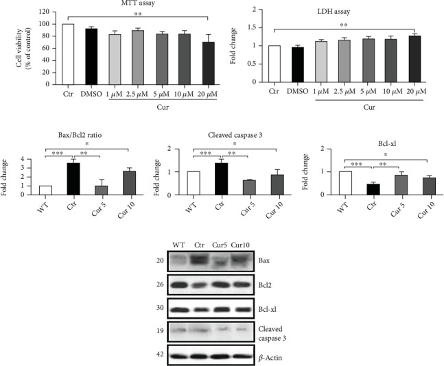
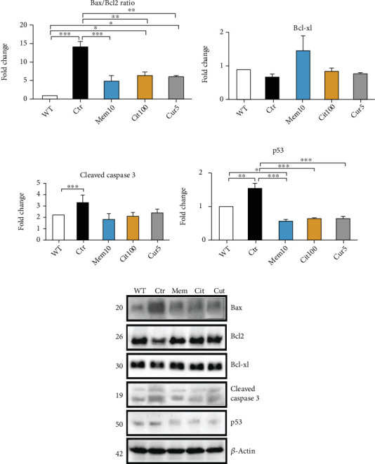
Retinal degeneration is the major and principal cause behind many incurable blindness diseases. Several studies indicated the neuroprotective effect of Curcuma longa in eye pathologies, specifically retinopathy. However, the molecular mechanism behind its effect has not been completely elucidated. Using an ex vivo model of retinal degeneration obtained from an ex vivo optic nerve cut (ONC), we demonstrated that Curcuma extract (Cur) exerted a neuroprotective effect. Importantly, Cur was able to modulate apoptosis and MAPK signaling pathway activation and prevent retinal ganglion cell (RGC) loss. Other well-known neuroprotective pharmacological tools, including memantine (Mem), citicoline (Cit), and ginkgolic acid (GA), were used to compare the potential mechanisms of Cur. The antioxidant activity of retinas treated with Cur following optic nerve cut was significantly higher than control, but Cur failed to change the retina glutamate content. Considering the antioxidant effect of Cur and taking advantage of our recent findings on the crosstalk between oxidative stress and post-translational protein modifiers, in particular, small ubiquitin-related modifier (SUMO), we were interested in exploring the effect of Cur on SUMOylation. We found that Cur significantly prevented the increase of protein SUMOylation, confirming our previous in vitro data indicating the cytoprotective effect of curcumin through modulating the oxidative stress and SUMO-JNK axis. Altogether, these results suggest that Curcuma protects the retina from degeneration via antioxidant activity and targets SUMOylation. Therefore, it might be considered for the combination therapy with other neuroprotective agents with different mechanisms in preclinical studies on retinal degeneration.


Conflict of interest statement

The authors declare that they have no conflicts of interest regarding the publication of this paper.

Figures

Figure 1
Figure 1
Effect of Curcuma extract on the cell viability assay and apoptotic signaling markers. (a) MTT assay has been done to check the cell viability in the presence of different concentrations of Curcuma extract. (b) LDH assay was performed to evaluate the cell death. (c–f) Representative western blots and relative quantifications in different treated groups. Data are shown as mean ± SEM. One-way ANOVA followed by Tukey's post hoc test was used to analyze the statistical differences among groups. A p value of less than 0.05 was considered significant in all analyses. ⁣p < 0.05, ⁣∗∗p < 0.01, and ⁣∗∗∗p < 0.001. WT: nontreated fresh retina; Ctr: control; Cur: Curcuma extract.
Figure 2
Figure 2
Effect of Curcuma extract on mitogen-activated protein kinase (MAPK) pathways. Representative western blots and relative quantifications showed a significant change in (a) p-JNK/JNK and (b) p-c-Jun/c-Jun but not (c) p-ERK/ERK in retina homogenates collected from different treatments. Data are shown as mean ± SEM. One-way ANOVA followed by Tukey's post hoc test was used to analyze the statistical differences among groups. A p value of less than 0.05 was considered significant in all analyses. ⁣p < 0.05 and ⁣∗∗p < 0.01. WT: nontreated fresh retina; Ctr: control; Cur: Curcuma extract.
Figure 3
Figure 3
Curcuma extract protected retinal damage in an ex vivo model of toxicity induction. (a–c) Representative western blots and relative quantifications for NeuN as the neuronal marker and BRN3a, a specific marker for RGCs, in retina homogenates collected from different treatments. (d and e) Representative histological (hematoxylin and eosin) staining and relative quantifications for retina thickness assay. Scale bar = 50 μm. Data are shown as mean ± SEM and were normalized to the nontreated group (WT). One-way ANOVA followed by Tukey's post hoc test was used to analyze the statistical differences among groups. A p value of less than 0.05 was considered significant in all analyses. ⁣p < 0.05, ⁣∗∗p < 0.01, and ⁣∗∗∗p < 0.001. WT: nontreated fresh retina; Ctr: control; Cur: Curcuma extract; Mem: memantine; Cit: citicoline.
Figure 4
Figure 4
Comparison of the effect of Curcuma extract with memantine and citicoline on apoptotic markers. Representative western blots and relative quantifications for apoptotic markers, including (a) Bax/Bcl2 ratio, (b) Bcl-xl, (c) caspase3, and (d) p53 in retina homogenates collected from different treatments. Data are shown as mean ± SEM and were normalized to the nontreated group in western blot (WT). One-way ANOVA followed by Tukey's post hoc test was used to analyze the statistical differences among groups. A p value of less than 0.05 was considered significant in all analyses. ⁣p < 0.05, ⁣∗∗p < 0.01, and ⁣∗∗∗p < 0.001. WT: nontreated fresh retina; Ctr: control; Cur: Curcuma extract; Mem: memantine; Cit: citicoline.
Figure 5
Figure 5
Total antioxidant capacity assay and retina glutamate content in different groups. (a) Trolox equivalent capacity measured in mouse retina lysates, showing quantity (nmol) per mg of retina tissue. (b) Comparison of the retina glutamate content between Curcuma extract treatment and memantine using HPLC. Data are shown as mean ± SEM. One-way ANOVA followed by Tukey's post hoc test was used to analyze the statistical differences among groups. A p value of less than 0.05 was considered significant in all analyses. ⁣p < 0.05 and ⁣∗∗p < 0.01. WT: nontreated fresh retina; Ctr: control; Cur: Curcuma extract; Mem: memantine; Cit: citicoline.
Figure 6
Figure 6
Effect of Curcuma extract on SUMOylation in the ex vivo model of retinal degeneration. Representative western blots and relative quantifications for (a) SUMO-1, (b) SUMO-2,3, and (c and d) Ubc9 in retina homogenates collected from different treatments. (e) Immunofluorescence analysis of retinal sections collected from different treatments. IF images indicated an increase of SUMO-1-positive cells (red dots) in samples collected from the Ctr, and Cur 5 and Cur 10 were able to prevent the SUMO-1 positive cell augmentation. Nuclei were stained with the nuclear marker DAPI (blue). Scale bar = 200 μm. In the western blot analysis, data are shown as mean ± SEM and were normalized to the nontreated group (WT). One-way ANOVA followed by Tukey's post hoc test was used to analyze the statistical differences among groups. A p value of less than 0.05 was considered significant in all analyses. ⁣p < 0.05 and ⁣∗∗p < 0.01. WT: nontreated fresh retina; Ctr: control; Cur: Curcuma extract; GA: ginkgolic acid.
Figure 7
Figure 7
Effect of Curcuma extract on ubiquitination and autophagy marker in the ex vivo model of retinal degeneration. Representative western blots and relative quantifications for (a) ubiquitin and (b) LC3B, the autophagy marker, in retina homogenates collected from different treatments. (c) A SUMO-1/ubiquitin ratio quantification. Data are shown as mean ± SEM and were normalized to the nontreated group (WT). One-way ANOVA followed by Tukey's post hoc test was used to analyze the statistical differences among groups. A p value of less than 0.05 was considered significant in all analyses. ⁣p < 0.05 and ⁣∗∗∗p < 0.001. WT: nontreated fresh retina; Ctr: control; Cur: Curcuma extract.
Figure 8
Figure 8
Summary cartoon of the obtained results. Representative flowchart cartoon of the obtained results showing how the retinal degeneration mouse model has been done and how the treatments were done for the 3 molecules used. The cartoon shows that three treatments have a different impact on the biological response in the retinal degeneration mouse model.

References

    1. Wert K. J., Lin J. H., Tsang S. H. General Pathophysiology in Retinal Degeneration. In: Casaroli-Marano R. P., editor. Cell-Based Therapy for Retinal Degenerative Disease . Basel: Karger; 2014. pp. 33–43. - DOI
    1. Morgan J. E. Retina ganglion cell degeneration in glaucoma: an opportunity missed? A review. Clinical & Experimental Ophthalmology . 2012;40(4):364–368. doi: 10.1111/J.1442-9071.2012.02789.X. - DOI - PubMed
    1. Tisi A., Feligioni M., Passacantando M., Ciancaglini M., Maccarone R. The impact of oxidative stress on blood-retinal barrier physiology in age-related macular degeneration. Cells . 2021;10:p. 64. doi: 10.3390/CELLS10010064. - DOI - PMC - PubMed
    1. Nucci C., Martucci A., Giannini C., Morrone L. A., Bagetta G., Mancino R. Neuroprotective agents in the management of glaucoma. Eye . 2018;32(5):938–945. doi: 10.1038/s41433-018-0050-2. - DOI - PMC - PubMed
    1. Thomas C. N., Berry M., Logan A., Blanch R. J., Ahmed Z. Caspases in retinal ganglion cell death and axon regeneration. Cell Death Discovery . 2017;3(1):1–13. doi: 10.1038/cddiscovery.2017.32. - DOI - PMC - PubMed

MeSH terms