. 2022 Jul 30:2022:2843990.
doi: 10.1155/2022/2843990. eCollection 2022.

MicroRNA-147a Targets SLC40A1 to Induce Ferroptosis in Human Glioblastoma

Affiliations

MicroRNA-147a Targets SLC40A1 to Induce Ferroptosis in Human Glioblastoma

Peng Xu et al. Anal Cell Pathol (Amst). .

Abstract

Objective: Glioblastoma is one of the most common malignant tumors in the brain, and these glioblastoma patients have very poor prognosis. Ferroptosis is involved in the progression of various tumors, including the glioblastoma. This study aims to determine the involvement of microRNA (miR)-147a in regulating ferroptosis of glioblastoma in vitro.

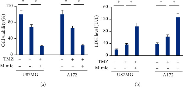
Methods: Human glioblastoma cell lines were transfected with the inhibitor, mimic and matched negative controls of miR-147a in the presence or absence of ferroptotic inducers. To knock down the endogenous solute carrier family 40 member 1 (SLC40A1), cells were transfected with the small interfering RNA against SLC40A1. In addition, cells with or without the miR-147a mimic treatment were also incubated with temozolomide (TMZ) to investigate whether miR-147a overexpression could sensitize human glioblastoma cells to TMZ chemotherapy in vitro.

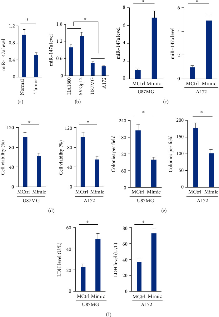
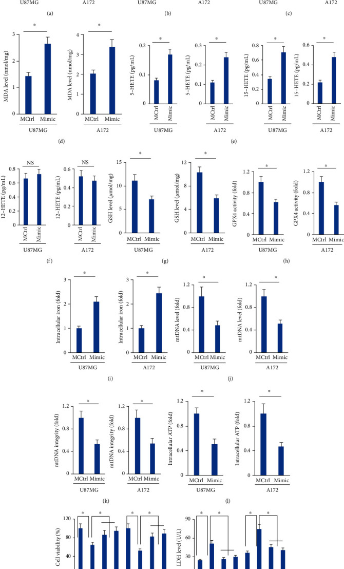
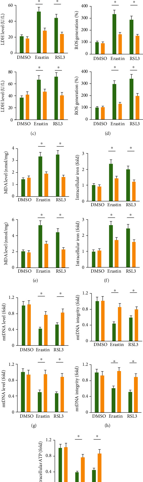
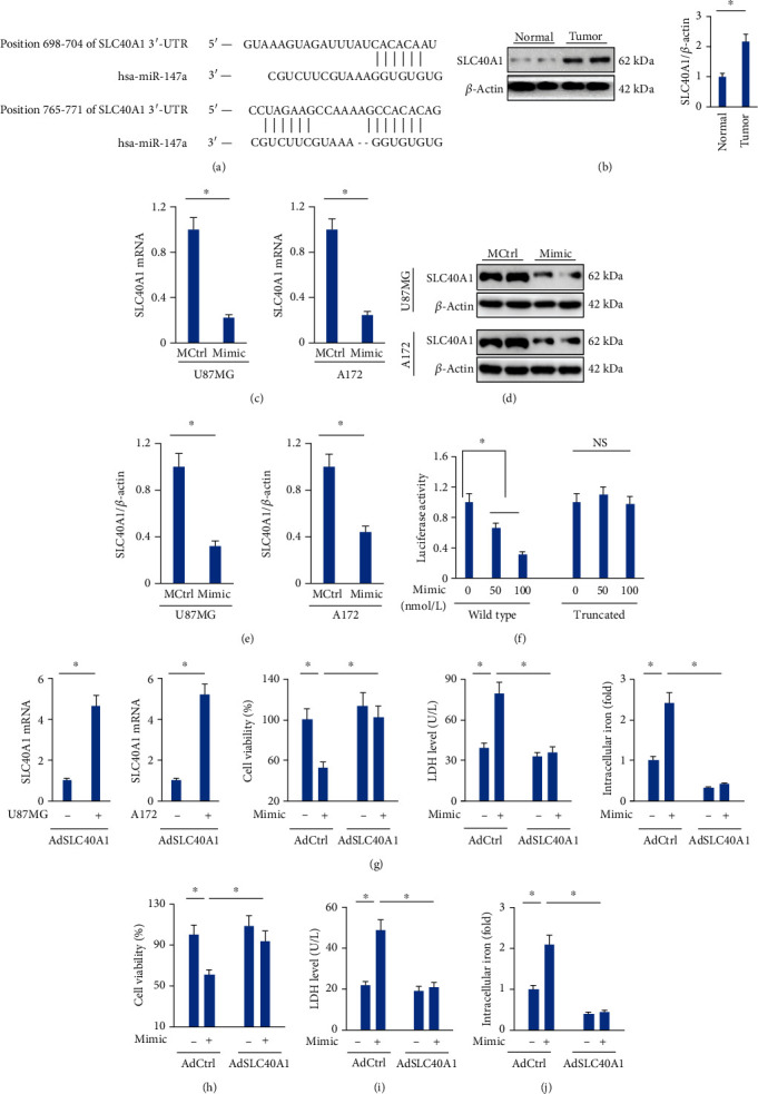
Results: We found that miR-147a level was decreased in human glioblastoma tissues and cell lines and that the miR-147a mimic significantly suppressed the growth of glioblastoma cells in vitro. In addition, miR-147a expression was elevated in human glioblastoma cells upon erastin or RSL3 stimulation. Treatment with the miR-147a mimic significantly induced ferroptosis of glioblastoma cells, and the ferroptotic inhibitors could block the miR-147a mimic-mediated tumor suppression in vitro. Conversely, the miR-147a inhibitor prevented erastin- or RSL3-induced ferroptosis and increased the viability of glioblastoma cells in vitro. Mechanistically, we determined that miR-147a directly bound to the 3'-untranslated region of SLC40A1 and inhibited SLC40A1-mediated iron export, thereby facilitating iron overload, lipid peroxidation, and ferroptosis. Furthermore, miR-147a mimic-treated human glioblastoma cells exhibited higher sensitivity to TMZ chemotherapy than those treated with the mimic control in vitro.

Conclusion: We for the first time determine that miR-147a targets SLC40A1 to induce ferroptosis in human glioblastoma in vitro.


Conflict of interest statement

The authors declare that they have no competing interests.

Figures

Figure 1
Figure 1
The miR-147a mimic suppresses cell survival and induces cell death of human glioblastoma cells. (a) miR-147a level in human glioblastoma tissues and normal brain tissues. (b) miR-147a level in human glioblastoma cells and normal brain astroglia cells. (c) miR-147a level in human glioblastoma cells treated with or without the miR-147a mimic. (d) Cell viability detected by CCK-8 assay. (e) Numbers of colonies. (f) LDH level in the medium from cells treated with or without the miR-147a mimic. N = 6 for each group. Data were expressed as the mean ± SD, and P < 0.05 was considered statistically significant.
Figure 2
Figure 2
The miR-147a mimic triggers ferroptosis of human glioblastoma cells. (a–b) miR-147a level in human glioblastoma cells treated with erastin or RSL3. (c) ROS level detected by DCFH-DA probe. (d) MDA level in cells treated with or without the miR-147a mimic. (e–f) 5-HETE, 12-HETE, and 15-HETE levels in the medium from cells treated with or without the miR-147a mimic. (g–h) GSH level and GPX4 activity in U87MG and A172 cells. (i) Relative level of intracellular iron. (j–k) Quantification of mtDNA content and integrity. (l) Quantification of intracellular ATP level. (m) Cell viability and LDH releases in the miR-147a mimic-treated human glioblastoma cells with Fer-1 or Lip-1 protection. N = 6 for each group. Data were expressed as the mean ± SD, and P < 0.05 was considered statistically significant.
Figure 3
Figure 3
The miR-147a inhibitor prevents ferroptosis of human glioblastoma cells. (a) miR-147a level in human glioblastoma cells treated with or without the miR-147a inhibitor. (b) Cell viability detected by CCK-8 assay. (c) LDH level in the medium from cells treated with or without the miR-147a inhibitor. (d) ROS level detected by DCFH-DA probe. (e) MDA level in cells treated with or without the miR-147a inhibitor. (f) Relative level of intracellular iron. (g–h) Quantification of mtDNA content and integrity. (i) Quantification of intracellular ATP level. N = 6 for each group. Data were expressed as the mean ± SD, and P < 0.05 was considered statistically significant.
Figure 4
Figure 4
The miR-147a mimic elicits ferroptosis of human glioblastoma cells through targeting SLC40A1. (a) Sequence alignment of miR-147a and the 3′-UTR of SLC40A1. (b) SLC40A1 protein level in human glioblastoma tissues and normal brain tissues. (c–e) The mRNA and protein levels of SLC40A1 in cells treated with or without the miR-147a mimic. (f) Relative luciferase activity. (g) The mRNA level SLC40A1 in cells infected with or without AdSCL40A1. (h) Cell viability detected by CCK-8 assay. (i) LDH level in the medium from the miR-147a mimic-treated cells with or without SLC40A1 overexpression. (j) Relative level of intracellular iron. N = 6 for each group. Data were expressed as the mean ± SD, and P < 0.05 was considered statistically significant. NS indicates no significance.
Figure 5
Figure 5
The miR-147a mimic sensitizes human glioblastoma cells to TMZ therapy. (a) Cell viability detected by CCK-8 assay. (b) LDH level in the medium from the miR-147a mimic-treated cells with or without TMZ stimulation. N = 6 for each group. Data were expressed as the mean ± SD, and P < 0.05 was considered statistically significant.

References

    1. Escoll M., Lastra D., Pajares M., et al. Transcription factor NRF2 uses the Hippo pathway effector TAZ to induce tumorigenesis in glioblastomas. Redox Biology . 2020;30, article 101425 doi: 10.1016/j.redox.2019.101425. - DOI - PMC - PubMed
    1. Tomar V. S., Patil V., Somasundaram K. Temozolomide induces activation of Wnt/β-catenin signaling in glioma cells via PI3K/Akt pathway: implications in glioma therapy. Cell Biology and Toxicology . 2020;36(3):273–278. doi: 10.1007/s10565-019-09502-7. - DOI - PubMed
    1. Wang B., Peng F., Huang W., et al. Rational drug design, synthesis, and biological evaluation of novel chiral tetrahydronaphthalene-fused spirooxindole as MDM2-CDK4 dual inhibitor against glioblastoma. Acta Pharmaceutica Sinica B . 2020;10(8):1492–1510. doi: 10.1016/j.apsb.2019.12.013. - DOI - PMC - PubMed
    1. Angelova P. R., Choi M. L., Berezhnov A. V., et al. Alpha synuclein aggregation drives ferroptosis: an interplay of iron, calcium and lipid peroxidation. Cell Death and Differentiation . 2020;27(10):2781–2796. doi: 10.1038/s41418-020-0542-z. - DOI - PMC - PubMed
    1. Li Y., Cao Y., Xiao J., et al. Inhibitor of apoptosis-stimulating protein of p53 inhibits ferroptosis and alleviates intestinal ischemia/reperfusion-induced acute lung injury. Cell Death and Differentiation . 2020;27(9):2635–2650. doi: 10.1038/s41418-020-0528-x. - DOI - PMC - PubMed

LinkOut - more resources