. 2022 Jul 30:2022:8440422.
doi: 10.1155/2022/8440422. eCollection 2022.

Transmembrane Protein ANTXR1 Regulates γ-Globin Expression by Targeting the Wnt/ β-Catenin Signaling Pathway

Affiliations

Transmembrane Protein ANTXR1 Regulates γ-Globin Expression by Targeting the Wnt/ β-Catenin Signaling Pathway

Tingting Jin et al. J Immunol Res. .

Abstract

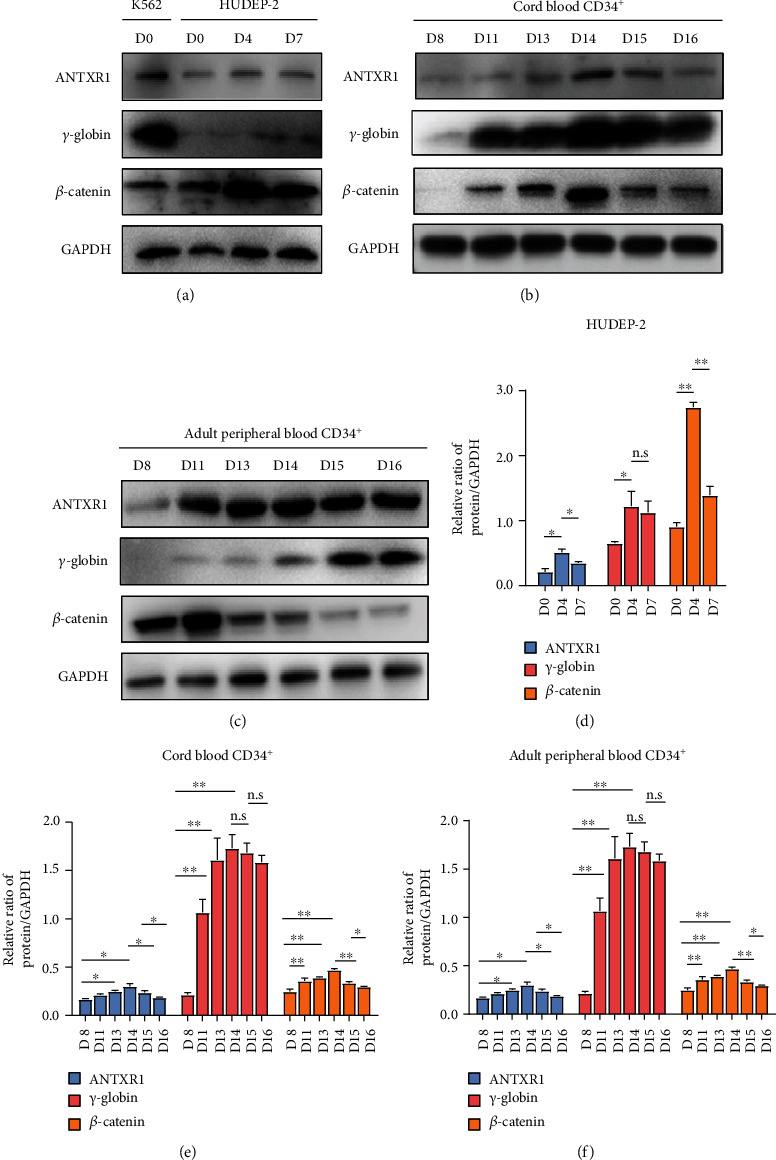
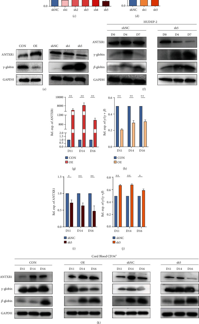
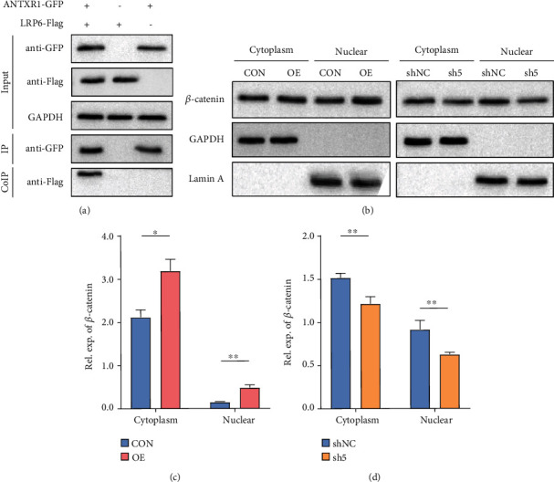
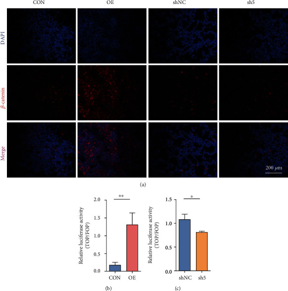
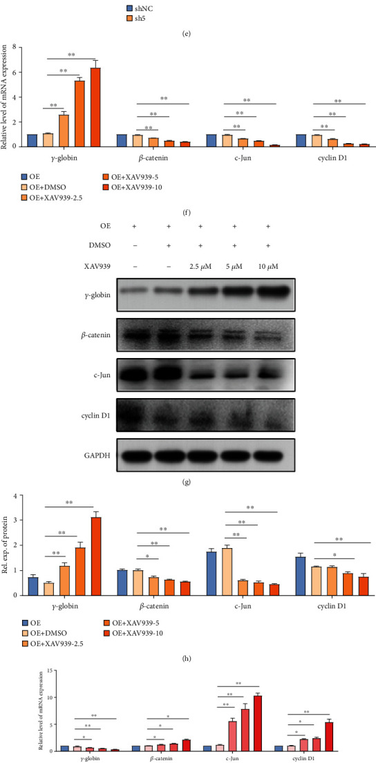
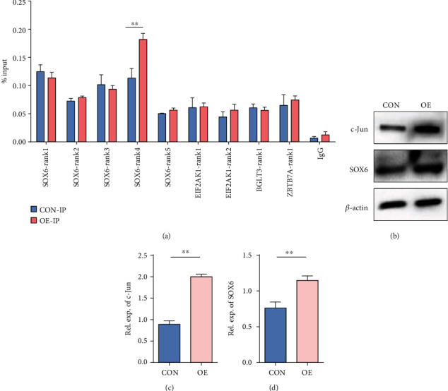
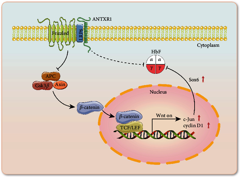
Reactivation of fetal hemoglobin (HbF, α2γ2) alleviates clinical symptoms in patients with β-thalassemia and sickle cell disease, although the regulatory mechanisms of γ-globin expression have not yet been fully elucidated. Recent studies found that interfering with the expression of the membrane protein ANTXR1 gene upregulated γ-globin levels. However, the exact mechanism by which ANTXR1 regulates γ-globin levels remains unclear. Our study showed that overexpression and knockdown of ANTXR1 in K562, cord blood CD34+, and HUDEP-2 cells decreased and increased γ-globin expression, respectively. ANTXR1 regulates the reactivation of fetal hemoglobin (HbF, α2γ2) in K562, cord blood CD34+, and adult peripheral blood CD34+ cells through interaction with LRP6 to promote the nuclear entry of β-catenin and activate the Wnt/β-catenin signaling pathway. The overexpression or knockdown of ANTXR1 on γ-globin and Wnt/β-catenin signaling in K562 cells was reversed by the inhibitor XAV939 and the activator LiCl, respectively, where XAV939 inhibits the transcription of β-catenin in the Wnt pathway, but LiCl inhibits GSK3-β. We also showed that the binding ability of the rank4 site in the transcriptional regulatory region of the SOX6 gene to c-Jun was significantly increased after overexpression of ANTXR1 in K562 cells. SOX6 protein expression was increased significantly after overexpression of the c-Jun gene, indicating that the transcription factor c-Jun initiated the transcription of SOX6, thereby silencing γ-globin. Our findings may provide a new intervention target for the treatment of β-hemoglobinopathies.


Conflict of interest statement

The authors declare no competing financial interests.

Figures

Figure 1
Figure 1
Expression of ANTXR1, γ-globin, and β-catenin during erythroid differentiation. (a) Western blot analysis was performed to detect the expression levels of ANTXR1, γ-globin, and β-catenin in K562 cells and HUDEP-2 cells at days 0, 4, and 7 of erythroid differentiation. (b) Western blot analysis for the expression of ANTXR1, γ-globin, and β-catenin in umbilical cord blood CD34+ cells during erythroid differentiation at days 8, 11, 13, 14, 15, and 16. (c) Western blot analysis for the expression of ANTXR1, γ-globin, and β-catenin in adult peripheral blood CD34+ cells during erythroid differentiation at days 8, 11, 13, 14, 15, and 16. (d) Protein quantification of the western blot in HUDEP-2 cells ANTXR1, γ-globin, and β-catenin at days 0, 4, and 7. (e) Quantification of western blot results of ANTXR1, γ-globin, and β-catenin in umbilical cord blood CD34+ cells at days 8, 11, 13, 14, 15, and 16. (f) Quantification of western blot results of ANTXR1, γ-globin, and β-catenin in adult peripheral blood CD34+ cells at days 8, 11, 13, 14, 15, and 16. These experiments n = 3. The error bar represents the SD. P < 0.05 and ∗∗P < 0.01.
Figure 2
Figure 2
Effects of ANTXR1 overexpression or knockdown on γ-globin mRNA and protein levels. (a, b) ANTXR1 was overexpressed using the PHAGE-fEF-1a-IRES-ZsGreen-2 vector in K562 cells, while cells transfected with the empty vector served as controls. ANTXR1 and γ-globin mRNA levels were measured by qRT-PCR. (c) In K562 cells, ANTXR1-sh1-5 was separately inserted into the pSIH1-H1-copGFPshRNA vector to test the knockdown efficiency of ANTXR1. Of the five shRNA vectors, ANTXR1-sh1 and ANTXR1-sh5 had the most significant knockdown efficiencies of 82% and 88%, respectively. (d) After the K562 cells were infected with ANTXR1-sh1, ANTXR1-sh5, and control shNC, respectively, the mRNA levels of ANTXR1 and γ-globin were determined using qRT-PCR. ANTXR1-sh5 had a greater effect on γ-globin expression than ANTXR1-sh1. (e) Western blot analysis was used to detect ANTXR1 and γ-globin levels after infection of K562 cells with ANTXR1 overexpression vectors and interference vectors (ANTXR1-sh1 and ANTXR1-sh5). (f) At days 0, 4, and 7 of erythroid differentiation after infection of HUDEP-2 cells with ANTXR1-sh5 and shNC vectors, ANTXR1,γ-globin, and β-globin expression was determined by western blotting. (g, h) After infection of umbilical cord blood CD34+ cells with overexpression of the ANTXR1 vector, the mRNA levels of ANTXR1 and γ/γ+β globin ratio at days 11, 14, and 16 of erythroid differentiation were determined by qRT-PCR. (i, j) After infection of umbilical cord blood CD34+ cells with ANTXR1-5 and shNC vectors, the mRNA levels of ANTXR1 and γ/γ+β globin ratio at days 11, 14, and 16 of erythroid differentiation were determined by qRT-PCR. (k) After infection of umbilical cord blood CD34+ cells with the ANTXR1 overexpression vector and interference vector, the protein levels of ANTXR1,γ-globin, and β-globin at days 11, 14, and 16 of erythroid differentiation were detected by western blotting. (l) Umbilical cord blood CD34+ cells interfered with ANTXR1 and were cultured under conditions that promoted erythroid maturation. The cells were collected at days 11, 14, and 16, respectively. Flow cytometry was used to detect HbF-immunostained cells (F cells). These experiments were repeated three times. The error bar represents the SD. P < 0.05 and ∗∗P < 0.01. NC designates the negative control.
Figure 3
Figure 3
ANTXR1 activates the Wnt/β-catenin signaling pathway by promoting β-catenin into the nucleus. (a) ANTXR1 interacted with LRP6 shown by the Co-IP assay that the lysates from the K562 cells transfected with the ANTXR1–GFP and LRP6-Flag plasmids by using GFP- and Flag-targeting antibodies. (b) β-Catenin expression in the cytoplasm and nucleus was detected by western blot analysis after K562 cells were transfected with the ANTXR1 vector, control vector, ANTXR1-shRNA vector, or NC-shRNA vector. (c, d) Quantification of western blots. The error bar represents the SD.P < 0.05 and ∗∗P < 0.01. NC designates the negative control.
Figure 4
Figure 4
ANTXR1 activates the Wnt/β-catenin signaling pathway by promoting β-catenin into the nucleus. (a) The expression of β-catenin in K562 cells after overexpression or knockdown of ANTXR1 was detected by immunofluorescence with a confocal laser scanning microscope. The color “red” represents β-catenin, and “blue” represents nuclei stained by DAPI. The merged images indicate the localization of β-catenin in both the nuclei and cytoplasm. (b, c) The TOP-/FOP-flash luciferase assay shows the activity of the Wnt/β-catenin signaling pathway in 293T cells overexpressing or knocking down the ANTXR1 gene. Experiments n = 3. The error bar represents the SD.P < 0.05 and ∗∗P < 0.01. NC designates the negative control.
Figure 5
Figure 5
Effect of ANTXR1 on the expression of key molecules of the Wnt/β-catenin pathway. (a) The mRNA expression levels of LRP6, β-catenin, c-Jun, and cyclin D1 were detected by qRT-PCR after overexpression of ANTXR1 in K562 cells. (b) The mRNA expression levels of LRP6, β-catenin, c-Jun, and cyclin D1 were detected by qRT-PCR, after K562 cells were transfected with ANTXR1-sh5. (c) The protein levels of LRP6, β-catenin, c-Jun, and cyclin D1 were measured by western blotting after K562 cells were overexpressed or knocked down by ANTXR1. (d, e) Quantification of the western blots is shown in (c). (f, g) The mRNA and protein expression levels of γ-globin, LRP6, β-catenin, c-Jun, and cyclin D1 were detected by qRT-PCR and western blotting, respectively, with K562 cells overexpressing ANTXR1 treated with 2.5, 5, and 10 μmol/L of XAV939 for 24 h. (h) Quantification of the western blots shown in (g). (i, j) The mRNA and protein expression levels of γ-globin, LRP6, β-catenin, c-Jun, and cyclin D1 were detected by qRT-PCR and western blotting, respectively, with K562 cells interfering with ANTXR1-sh5 which were treated with 5, 20, and 40 mmol/L of LiCl for 24 h. (k) Quantification of the western blots shown in (j). These experiments n = 3. The error bar represents the SD. P < 0.05, ∗∗P < 0.01. NC: negative control.
Figure 6
Figure 6
Overexpression of ANTXR1 promoted the combination of c-Jun and SOX6. (a) ChIP-qPCR assays were performed using K562 cells expressing ANTXR1, while an empty vector served as a control. The immunoprecipitated DNA of SOX6, EIF2AK, BGLT3, and ZBTB7A 4 binding site to c-Jun, as well as input DNA, were used as templates for quantitative fluorescence PCR. The enrichments were quantified by ChIP-qPCR and normalized by comparison to input DNA (% input). (b) After overexpression of c-Jun in K562 cells, the protein levels of c-Jun and SOX6 were measured by western blotting. (c, d) Quantification of the western blot. The experiments n = 3. The error bar represents the SD. ∗∗P < 0.01.
Figure 7
Figure 7
Molecular mechanisms by which ANTXR1 regulates γ-globin expression via the Wnt/β-catenin signaling pathway. The interaction of ANTXR1 with LRP6 may inhibit the activity of the β-catenin destructive complex consisting of Axin, APC, and Gsk3 via Frizzled. β-Catenin is then free to translocate to the nucleus where it binds to the TCF/LEF transcription factors, thereby promoting the transcription of target gene c-Jun, which, in turn, activates the expression of SOX6, a repressor of γ-globin genes, thereby inhibiting the expression of γ-globin genes.

Similar articles

Cited by

References

    1. Fard A. D., Kaviani S., Noruzinia M., Saki N., Mortaz E. Induction of fetal hemoglobin as a novel therapeutic strategy for β-hemoglobinopathy. Laboratory Hematology . 2014;20(1, article 12015) doi: 10.1532/lh96.12015. - DOI - PubMed
    1. Taher A. T., Weatherall D. J., Cappellini M. D. Thalassaemia. The Lancet . 2018;391(10116):155–167. doi: 10.1016/s0140-6736(17)31822-6. - DOI - PubMed
    1. Vinjamur D. S., Bauer D. E., Orkin S. H. Recent progress in understanding and manipulating haemoglobin switching for the haemoglobinopathies. British Journal of Haematology . 2018;180(5):630–643. doi: 10.1111/bjh.15038. - DOI - PubMed
    1. Ali H., Khan F., Musharraf S. G. Acyclovir induces fetal hemoglobin via downregulation of _γ -globin_ repressors, _BCL11A_ and _SOX6 trans_ -acting factors. Biochemical Pharmacology . 2021;190, article 114612 doi: 10.1016/j.bcp.2021.114612. - DOI - PubMed
    1. Sankaran V. G., Menne T. F., Xu J., et al. Human fetal hemoglobin expression is regulated by the developmental stage-specific repressor BCL11A. Science . 2008;322(5909):1839–1842. doi: 10.1126/science.1165409. - DOI - PubMed

LinkOut - more resources