. 2023 Feb 8;25(1):211-222.
doi: 10.1093/europace/euac116.

Personalized ablation vs. conventional ablation strategies to terminate atrial fibrillation and prevent recurrence

Affiliations

Personalized ablation vs. conventional ablation strategies to terminate atrial fibrillation and prevent recurrence

Luca Azzolin et al. Europace. .

Abstract

Aims: The long-term success rate of ablation therapy is still sub-optimal in patients with persistent atrial fibrillation (AF), mostly due to arrhythmia recurrence originating from arrhythmogenic sites outside the pulmonary veins. Computational modelling provides a framework to integrate and augment clinical data, potentially enabling the patient-specific identification of AF mechanisms and of the optimal ablation sites. We developed a technology to tailor ablations in anatomical and functional digital atrial twins of patients with persistent AF aiming to identify the most successful ablation strategy.

Methods and results: Twenty-nine patient-specific computational models integrating clinical information from tomographic imaging and electro-anatomical activation time and voltage maps were generated. Areas sustaining AF were identified by a personalized induction protocol at multiple locations. State-of-the-art anatomical and substrate ablation strategies were compared with our proposed Personalized Ablation Lines (PersonAL) plan, which consists of iteratively targeting emergent high dominant frequency (HDF) regions, to identify the optimal ablation strategy. Localized ablations were connected to the closest non-conductive barrier to prevent recurrence of AF or atrial tachycardia. The first application of the HDF strategy had a success of >98% and isolated only 5-6% of the left atrial myocardium. In contrast, conventional ablation strategies targeting anatomical or structural substrate resulted in isolation of up to 20% of left atrial myocardium. After a second iteration of the HDF strategy, no further arrhythmia episode could be induced in any of the patient-specific models.

Conclusion: The novel PersonAL in silico technology allows to unveil all AF-perpetuating areas and personalize ablation by leveraging atrial digital twins.

Keywords: Atrial fibrillation; Computational model; Digital twin; Electro-anatomical mapping; MRI; Personalized ablation.


Conflict of interest statement

Conflict of interest: The authors declare that the research was conducted in the absence of any commercial or financial relationships that could be construed as a potential conflict of interest.

Figures

Figure 1
Figure 1
Workflow to identify Personalized Ablation Lines (PersonAL) using atrial digital twins. Tomographic segmentations (CT or LGE-MRI) and/or electro-anatomical maps (EAM) are processed by a highly automated pipeline to generate a patient-specific Augmented Atria (AugmentA) computational model. In case both LGE-MRI and EAM are given, an augmented anatomical and functional digital twin integrating both non-invasive and invasive electrophysiological data is provided. Tailored simulations are performed to assess atrial fibrillation (AF) inducibility using the Pacing at the End of the Effective Refractory Period (PEERP) protocol and regions vulnerable to sustain AF are identified. Then, AF drivers are targeted for ablation and connecting lines to the closest non-conductive barrier (anatomical orifice or previous ablation) are included. The PEERP protocol is then repeated to localize post-ablation emergent AF episodes. If new drivers are identified, they are targeted for ablation. The latter two processes are repeated until no AF episode can be induced in the patient’s model. The PersonAL plan is finally exported to the clinical mapping system data format. SSM, statistical shape model; LAT, local activation time; IIR, image intensity ratio.
Figure 2
Figure 2
Anatomical, substrate, and functional ablation strategies. The ablation lesions are indicated by the black ablation lines. Ablation approaches targeting anatomical structures, shown in the green box, consist in PVI (i) and PVI + RL + ML (ii). Substrate ablations, shown in the blue box, include, e.g. approaches targeting regions with IIR > 1.2 (iv), LVAs (v), and regions with CV < 0.3 m/s (vii). Functional ablation strategies, shown in the red box, targeted HDF areas (viii).
Figure 3
Figure 3
Identification of HDF areas in Patient #20 with fibrotic tissue modelled according to low-voltage area <0.5 mV (LVA) in the clinical voltage map. (A) Atrial fibrillation episode perpetuated in LVAs close to the inferior left pulmonary vein and at the posterior septum; (B) DF map; (C) IIR map; (D) bipolar voltage map; (E) simulated activation (LAT) map.
Figure 4
Figure 4
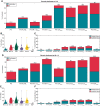
Success rate of each ablation strategy including connection lines to the closest non-conductive obstacle applied to the patient models cohort in which distribution of fibrotic tissue was modelled according to either voltage <0.5 mV (LVA) in clinical electrophysiological voltage maps (A) or LGE-MRI areas as identified by image intensity ratio (IIR) >1.2 (D). Ablation strategies that resulted in AF termination are shown in blue and conversion to atrial tachycardia (AT) in red. Percentage of atrial surface identified as substrate by the different substrate mapping methods before ablation in inducible patients with fibrosis adapted according to either LVA (B) or IIR >1.2 (E). Percentage of inactivated myocardium following each ablation strategy categorized in ablated tissue and isolated tissue in atrial models with fibrosis applied according to either LVA (C) or IIR >1.2 (F). IIR > 1.32, atrial regions with image intensity ratio (IIR) >1.32; IIR > 1.2, atrial regions with image intensity ratio (IIR) >1.2; LVA, low-voltage areas (bipolar voltage <0.5 mV); CV0.3, atrial regions with conduction velocity (CV) <0.3 m/s; CV0.4, atrial regions with CV <0.4 m/s; HDF, high dominant frequency areas; PVI, pulmonary vein isolation; PVI + ( )conn, PVI plus ablation targeting anatomical/structural/functional substrates and connecting localized ablation lesions to the closest non-conductive barrier (anatomical orifice or previous ablation). PVI + RL + ML, PVI plus roof line (RL) plus mitral isthmus line (ML).
Figure 5
Figure 5
Influence of inclusion of connecting lines to the closest non-conductive obstacle on the success rate for each ablation strategy applied to the patient models cohort in which distribution of fibrotic tissue was modelled according to either voltage <0.5 mV (LVA) in clinical electrophysiological voltage maps (A) or LGE-MRI areas as identified by image intensity ratio (IIR) >1.2 (B). Ablation strategies that resulted in AF termination are shown in blue and conversion to atrial tachycardia (AT) in red. IIR > 1.32, atrial regions with image intensity ratio (IIR) >1.32; IIR > 1.2, atrial regions with image intensity ratio (IIR) >1.2; LVA, low-voltage areas (bipolar voltage <0.5 mV); HDF, high dominant frequency (HDF) areas; PVI, pulmonary vein isolation; PVI + ( ), PVI plus ablation targeting anatomical/structural/functional substrates; PVI + ( )conn, PVI plus ablation targeting anatomical/structural/functional substrates and connecting localized ablation lesions to the closest non-conductive barrier (anatomical orifice or previous ablation).
Figure 6
Figure 6
First application of HDF ablation for Patient #18 with fibrosis modelled according to IIR > 1.2 from LGE-MRI. (A) Sustained AF episode in slow conduction area located on the anterior wall in proximity to the border zone of a low-voltage/IIR > 1.2 area; (B) DF map; (C) IIR map; (D) bipolar voltage map; (E) adapted electrophysiological LAT map.
Figure 7
Figure 7
PersonAL plan transferred to the original maps. The ablation lesions suggested by the PersonAL strategy are shown as black lines. (A) Sustained AF episode after PVI in Patient #18 colour coded by transmembrane voltage (TMV) with fibrosis distributed according to voltage <0.5 mV; (B) DF map; (C) original LGE-MRI segmentation colour coded by IIR; (D) original electro-anatomical map colour coded by clinical bipolar voltage; (E) atrial model colour coded by the in silico LAT map.

References

    1. Duytschaever M, Vijgen J, De Potter T, Scherr D, Van Herendael H, Knecht S, et al. . Standardized pulmonary vein isolation workflow to enclose veins with contiguous lesions: the multicentre VISTAX trial. EP Europace 2020;22:1645–52. - PubMed
    1. Verma A, Jiang C, Betts TR, Chen J, Deisenhofer I, Mantovan R, et al. . Approaches to catheter ablation for persistent atrial fibrillation. N Engl J Med 2015; 372:1812–22. - PubMed
    1. Rolf S, Kircher S, Arya A, Eitel C, Sommer P, Richter S, et al. . Tailored atrial substrate modification based on low-voltage areas in catheter ablation of atrial fibrillation. Circ Arrhythm Electrophysiol 2014;7:825–33. - PubMed
    1. Jadidi AS, Lehrmann H, Keyl C, Sorrel J, Markstein V, Minners J, et al. . Ablation of persistent atrial fibrillation targeting low-voltage areas with selective activation characteristics. Circ Arrhythm Electrophysiol 2016;9:e002962. - PubMed
    1. Kircher S, Arya A, Altmann D, Rolf S, Bollmann A, Sommer P, et al. . Individually tailored vs. standardized substrate modification during radiofrequency catheter ablation for atrial fibrillation: a randomized study. Europace 2018;20:1766–75. - PubMed