. 2022 Mar 9;11(3):S323-S333.
doi: 10.5152/eurjrheum.2021.20233. Online ahead of print.

Ultrasound-guided interventions in rheumatology

Affiliations

Ultrasound-guided interventions in rheumatology

Emili Gomez-Casanovas et al. Eur J Rheumatol. .

Abstract

Over the last few years, percutaneous procedures have undergone great advances, thanks to the ultrasound (US) guidance, due to the technical improvements in the US field, combined with the greater availability, good portability, and reduced cost of US devices. The direct target visualization and the real-time imaging performance enabled by US-guidance account for an improved accuracy in needle placement in several rheumatology interventions. So, ultrasound-guided procedures contribute to several diagnostic or therapeutic procedures such as fluid aspiration or treatment instillations of common joints, tendons, and bursas. In clinical practice, this fact is especially important in the case of depth areas like the hip, small anatomical structures as tendon sheaths or nerves in tenosynovitis or nervous blocks, or complex anatomical structures like the spine's facet joints. The US-guide is an essential tool for performing diagnostic procedures as synovial biopsy. US can also be combined with other imaging techniques, like the establishment portal arthroscopy for instance. Compared to older blind procedures, US-guided injections are more accurate and safer, and they result in better clinical outcomes in terms of joints improvement in function and decreased risk of damages caused by needle misplacement. With the ultrasound guided treatment, we can avoid the instillation of therapeutic products outside the predetermined target, which may be sometimes potentially harmful. The aim of this article is to describe the main generalities of ultrasound-guided procedures in rheumatology, their main advantages and disadvantages, and their particularities in joints where they are most frequently used, such as the shoulder, hip, and knee.


Figures

Figure 1.
Figure 1.
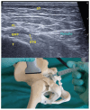
US-guided procedure in shoulder joint. ST, subcutaneous tissue; DM, deltoid muscle; ISM, infraspinatus muscle; SGN, spinoglenoid notch; GJ, glenohumeral joint.
Figure 2.
Figure 2.
US-guided procedure in subacromial-subdeltoid bursa. ST, subcutaneous tissue; DM, deltoid muscle; SBD, subacromial deltoid bursa; SST, supraspinatus tendon.
Figure 3.
Figure 3.
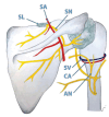
Anatomical representation of the partial shoulder innervation. SN, suprascapular nerve; SA, suprascapular artery; AN, axillar nerve; CA, circumflex artery; CV, circumflex vein; SL, suprascapular ligament. The original artwork was created by Tanit Gómez and published with her permission.
Figure 4.
Figure 4.
Ultrasonographic image for the nerve block or radiofrequency of the suprascapular nerve at the level of the suprascapular fossa. TRAP, trapezius muscle; SSM, supraspinatus muscle; SSA, suprascapular artery; SSTL, superior transverse scapular ligament; dotted line, recommended approach for in-plane puncture from medial to lateral; yellow dot, suprascapular nerve.
Figure 5.
Figure 5.
Ultrasonographic image for localization of the suprascapular nerve at the level of spinoglenoid fossa and its combination with an intra-articular technique. DEL, deltoid muscle; ISM, infraspinatus muscle; SGF, spino-glenoid fossa; GHJI, glenohumeral joint infiltration; SSNI, suprascapular nerve infiltration.
Figure 6.
Figure 6.
Ultrasonographic image for the localization of the posterior branch(es) of the AN at the humeral level. The image is somewhat prior to the insertion of the teres minor muscle, which, together with the circumflex artery, will serve as a guide for the localization of the AN and its branches.
Figure 7.
Figure 7.
US-guided procedure in hip joint. JC, joint capsule; FH, femoral head; JE, joint effusion; FAN, femoral anatomical neck.
Figure 8.
Figure 8.
US-guided procedure in hip joint using Doppler effect. F, femur; I, injectate.
Figure 9.
Figure 9.
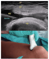
US-guided procedure in trochanter structure. ST, subcutaneal tissue; FL, fascia lata; GMT, gluteus medius tendon; T, trochanter; STB, superficial trochanter bursa.
Figure 10.
Figure 10.
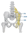
Anatomical representation of the partial hip innervation. FN, femoral nerve; ON, obturator nerve; AON, accessory obturator nerve. The original artwork was created by Tanit Gómez and published with her permission.
Figure 11.
Figure 11.
Ultrasonographic image for blocking or radiofrequency of the articular branch of the obturator nerve. Hip in slight abduction, external rotation and knee flexion of 20°, we will move the transducer distally from an initial position aligned with the iliopubic branch and inclination of about 30° caudo-cranial. We must find an image where we see, on the same plane, the ischium and the femoral head (or the acetabular notch). ONAB, articular branch of the obturator nerve; PEC, pectineus muscle; ON, obturator nerve; FH, femoral head; OE, external oblique muscle; FV, femoral vein; FA, femoral artery.
Figure 12.
Figure 12.
Ultrasonographic image for blocking or radiofrequency of the cranial articular branches of the femoral nerve. Transducer in a 20° oblique position at the level of the anterior inferior iliac spine. AIIS, anterior inferior iliac spine; ACE, acetabulumiliopubic eminence; ILP, iliopsoas muscle and its tendon; FN, femoral nerve; FA, femoral artery; Stars, points where we will find the superior articular terminal branches of the femoral nerve.
Figure 13.
Figure 13.
Ultrasonographic image for blocking or radiofrequency of the lower articular branches of the femoral nerve. Transducer in a 20° oblique position, which identifies the most cranial area of the femoral head. ILP, iliopsoas muscle; FN, femoral nerve; FA, femoral artery; HJC, hip joint capsule; RFT, rectus femoris muscle; Stars, points where we will find the superior articular terminal branches of the femoral nerve.
Figure 14.
Figure 14.
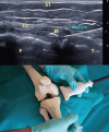
US-guided procedure in knee joint. Femoralpatellar lateral access. ST, subcutaneous tissue; QT, quadricipital tendon; IKE, intraarticular knee effusion; FD, femoral dyaphisis.
Figure 15.
Figure 15.
US-guided procedure in Baker's cyst. ST, subcutaneous tissue; CE, cyst effusion; SH, synovial hypertrophy.
Figure 16.
Figure 16.
US-guided procedure in medial interline of the knee. ST, subcutaneous tissue; CL, collateral ligament; M, meniscus; F, femur; T, tibia.
Figure 17.
Figure 17.
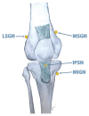
Anatomical localization of the puncture points used to blockade the genicular and infrapatellar branch of the saphenous (IPSN) nerves. MSGN, medial superior genicular nerve; MIGN, medial inferior genicular nerve; LSGN, lateral superior genicular nerve. The original artwork was created by Tanit Gómez and published with her permission.
Figure 18.
Figure 18.
Ultrasonographic image of the superior lateral genicular nerve. VL, vastus lateralis; VM, vastus medialis; SLGN, superior lateral genicular nerve; FS, femoral shaft; SLGA, superior lateral genicular artery; LFC, lateral femoral condyle.
Figure 19.
Figure 19.
Final position of the radio frequency cannulae for the three genicular nerves described.

Similar articles

References

    1. Hollander JL, Brown EM Jr, Jessar RA, Brown CY. Comparative effects of compound F (17-hy-droxycorticosterone) and cortisone injected locally into the rheumatoid arthritis joint. Ann Rheum Dis. 1951;10:473–476.. - PubMed
    1. Bianchi S, Zamorani P. US-guided interventional procedures. In Bianchi S, Martinolli C. (eds.): Ultrasound of the Musculoskeletal System. Berlin: Springer, 2007: 891–917..
    1. Bruyn GA, Iagnocco A, Naredo E. et al. OMERACT ultrasound working group. OMERACT definitions for ultrasonographic pathologies and elementary lesions of rheumatic disorders 15 years On. J Rheumatol. 2019;46:1388–1393.. (10.3899/jrheum.181095) - DOI - PubMed
    1. Gilliland CA, Salazar LD, Borchers JR. Ultrasound versus anatomic guidance for intraarticular and periarticular injection: A systematic review. Phys Sportsmed. 2011;39:121–131.. (10.3810/psm.2011.09.1928) - DOI - PubMed
    1. Park KD, Kim TK, Lee J, Lee WY, Ahn JK, Park Y. Palpation versus ultrasound-guided acromioclavicular joint intra-articular corticosteroid injections: A retrospective comparative clinical study. Pain Phys. 2015;18(4):333–341.. - PubMed

LinkOut - more resources