

T-cell receptor diversity in minimal change disease in the NEPTUNE study
- PMID: 35943576
- PMCID: PMC10037226
- DOI: 10.1007/s00467-022-05696-x
T-cell receptor diversity in minimal change disease in the NEPTUNE study
Abstract
Background: Minimal change disease (MCD) is the major cause of childhood idiopathic nephrotic syndrome, which is characterized by massive proteinuria and debilitating edema. Proteinuria in MCD is typically rapidly reversible with corticosteroid therapy, but relapses are common, and children often have many adverse events from the repeated courses of immunosuppressive therapy. The pathobiology of MCD remains poorly understood. Prior clinical observations suggest that abnormal T-cell function may play a central role in MCD pathogenesis. Based on these observations, we hypothesized that T-cell responses to specific exposures or antigens lead to a clonal expansion of T-cell subsets, a restriction in the T-cell repertoire, and an elaboration of specific circulating factors that trigger disease onset and relapses.
Methods: To test these hypotheses, we sequenced T-cell receptors in fourteen MCD, four focal segmental glomerulosclerosis (FSGS), and four membranous nephropathy (MN) patients with clinical data and blood samples drawn during active disease and during remission collected by the Nephrotic Syndrome Study Network (NEPTUNE). We calculated several T-cell receptor diversity metrics to assess possible differences between active disease and remission states in paired samples.
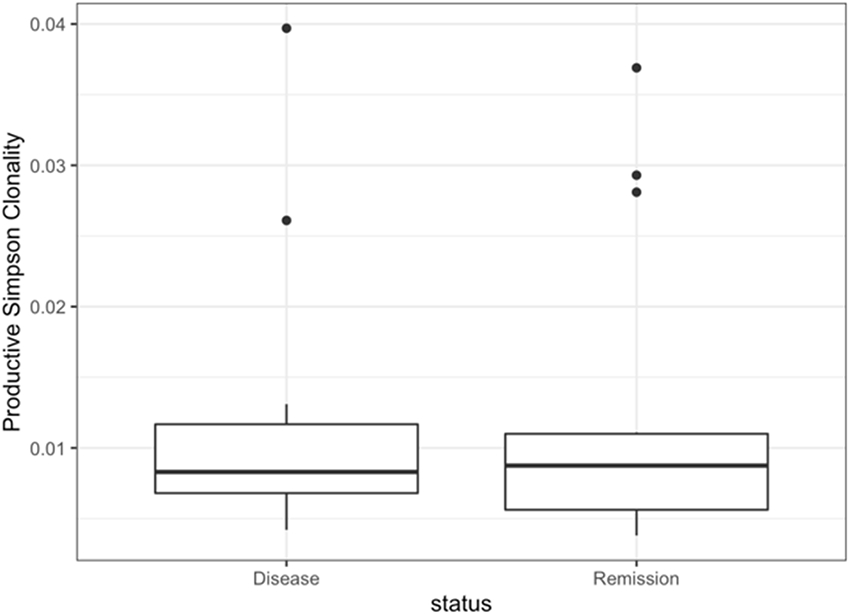
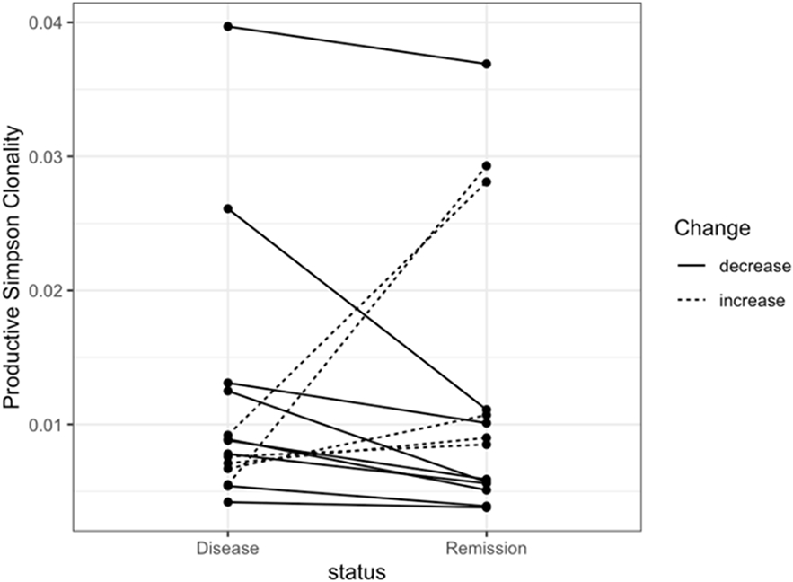
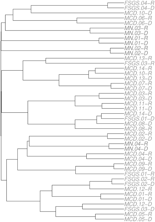
Results: Median productive clonality did not differ between MCD active disease (0.0083; range: 0.0042, 0.0397) and remission (0.0088; range: 0.0038, 0.0369). We did not identify dominant clonotypes in MCD active disease, and few clonotypes were shared with FSGS and MN patients.
Conclusions: While these data do not support an obvious role of the adaptive immune system T-cells in MCD pathogenesis, further study is warranted given the limited sample size. A higher resolution version of the Graphical abstract is available as Supplementary information.
Keywords: Adaptive immunity; Immunosequencing; Minimal change disease; NEPTUNE study; T-cell receptor.
© 2022. The Author(s), under exclusive licence to International Pediatric Nephrology Association.
Conflict of interest statement
Conflict of Interest Statement
J.R.S. has received research funding for randomized clinical trials in nephrotic syndrome from Goldfinch, Novartis, and Calliditas. He has also consulted for Maze, Goldfinch, Boehringer Ingelheim in the least three years and receives royalties from Sanofi for transgenic mice. The other authors have no conflicts of interest to declare.
Figures
References
-
- Niaudet P, Boyer O (2009) Idiopathic Nephrotic Syndrome in Children: Clinical Aspects. In: Avner E, Harmon W, Niaudet P, Yoshikawa N (eds) Pediatric Nephrology: Sixth Completely Revised, Updated and Enlarged Edition. Springer Berlin Heidelberg, Berlin, Heidelberg, pp 667–702
-
- Mak SK, Short CD, Mallick NP (1996) Long-term outcome of adult-onset minimal-change nephropathy. Nephrol Dial Transplant 11:2192–2201 - PubMed
-
- Eddy AA, Symons JM (2003) Nephrotic syndrome in childhood. The Lancet 362:629–639 - PubMed
-
- Garin EH, Laflam PF, Muffly K (2006) Proteinuria and fusion of podocyte foot processes in rats after infusion of cytokine from patients with idiopathic minimal lesion nephrotic syndrome. Nephron Exp Nephrol 102:e105–112 - PubMed
Publication types
MeSH terms
Substances
Grants and funding
LinkOut - more resources
Full Text Sources
Molecular Biology Databases
