. 2022 Nov:93:108-114.
doi: 10.1016/j.mri.2022.08.008. Epub 2022 Aug 6.

Reduced field-of-view and multi-shot DWI acquisition techniques: Prospective evaluation of image quality and distortion reduction in prostate cancer imaging

Affiliations

Reduced field-of-view and multi-shot DWI acquisition techniques: Prospective evaluation of image quality and distortion reduction in prostate cancer imaging

Edward M Lawrence et al. Magn Reson Imaging. 2022 Nov.

Abstract

Objectives: To prospectively compare image quality and apparent diffusion coefficient (ADC) quantification for reduced field-of-view (rFOV)- and multi-shot echo-planar imaging (msEPI)-based diffusion weighted imaging (DWI), using single-shot echo-planar-imaging (ssEPI) DWI as the reference.

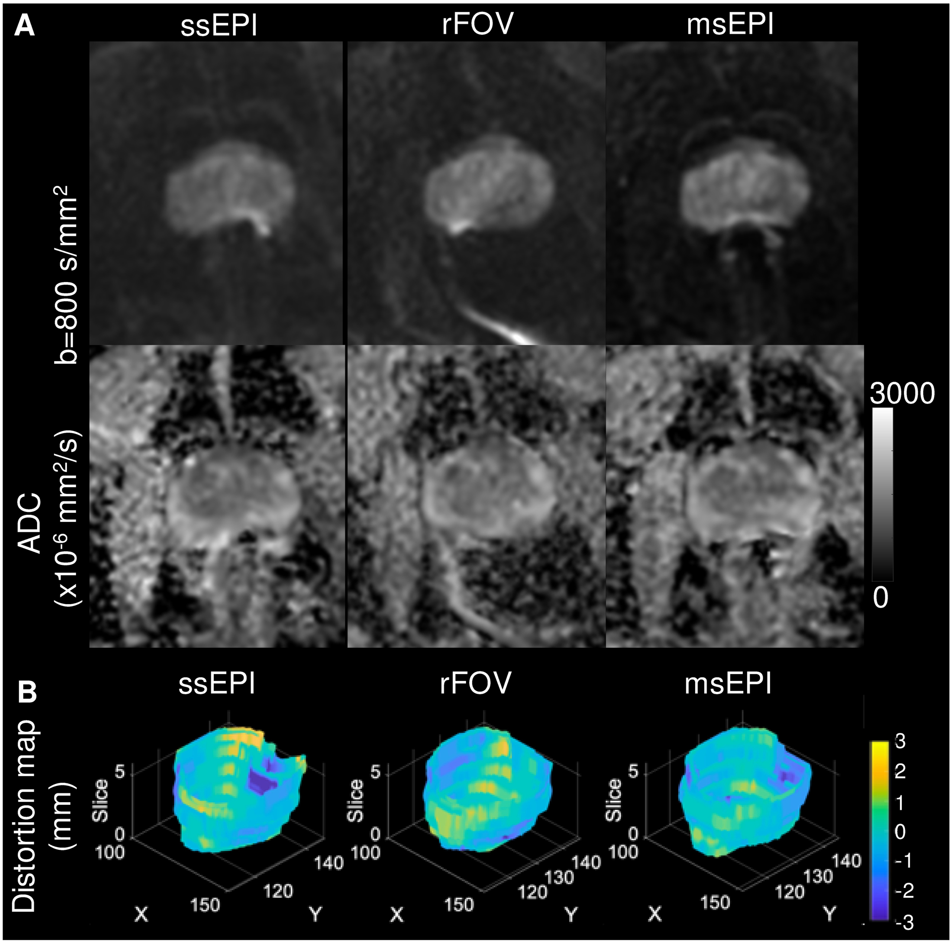
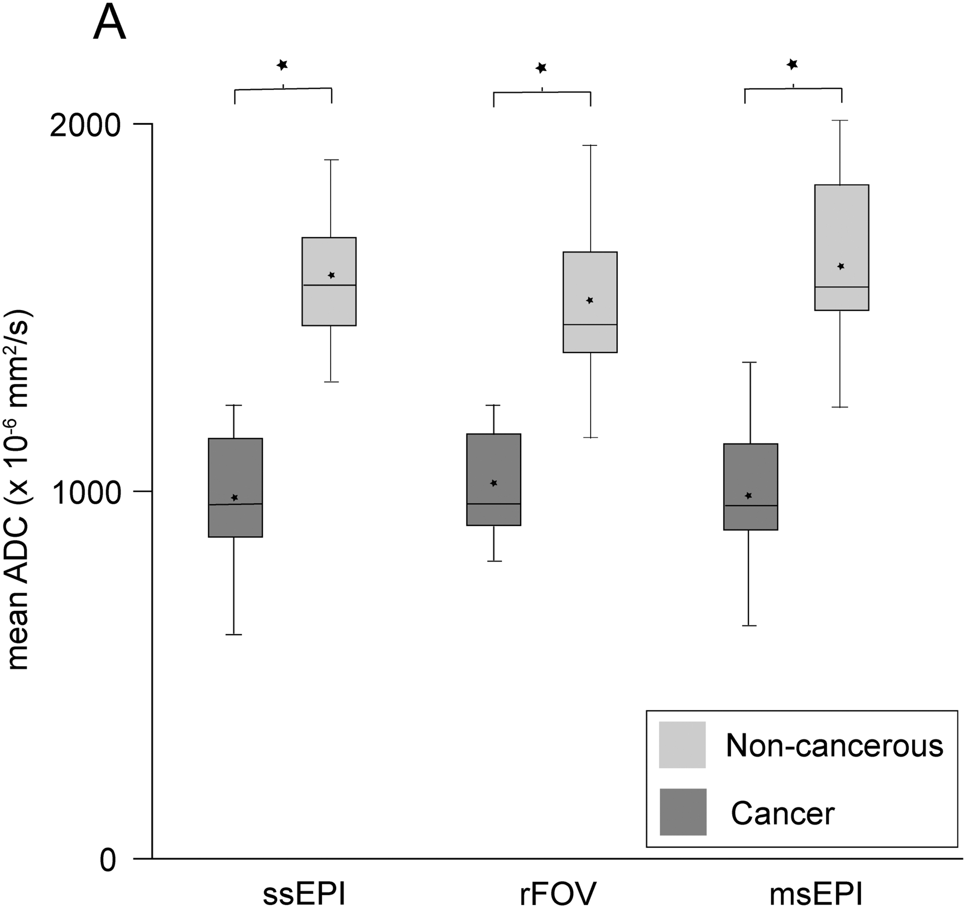
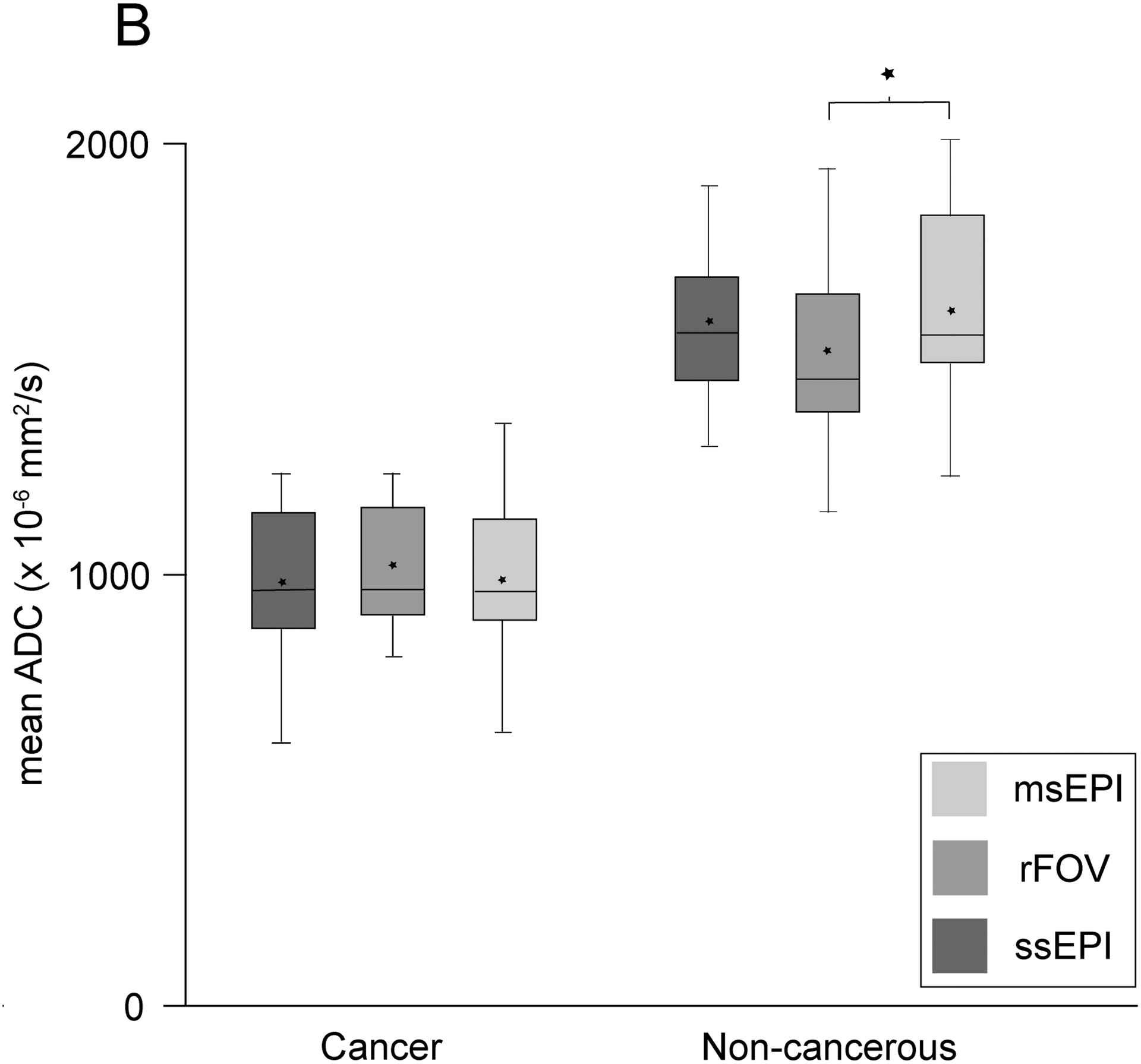
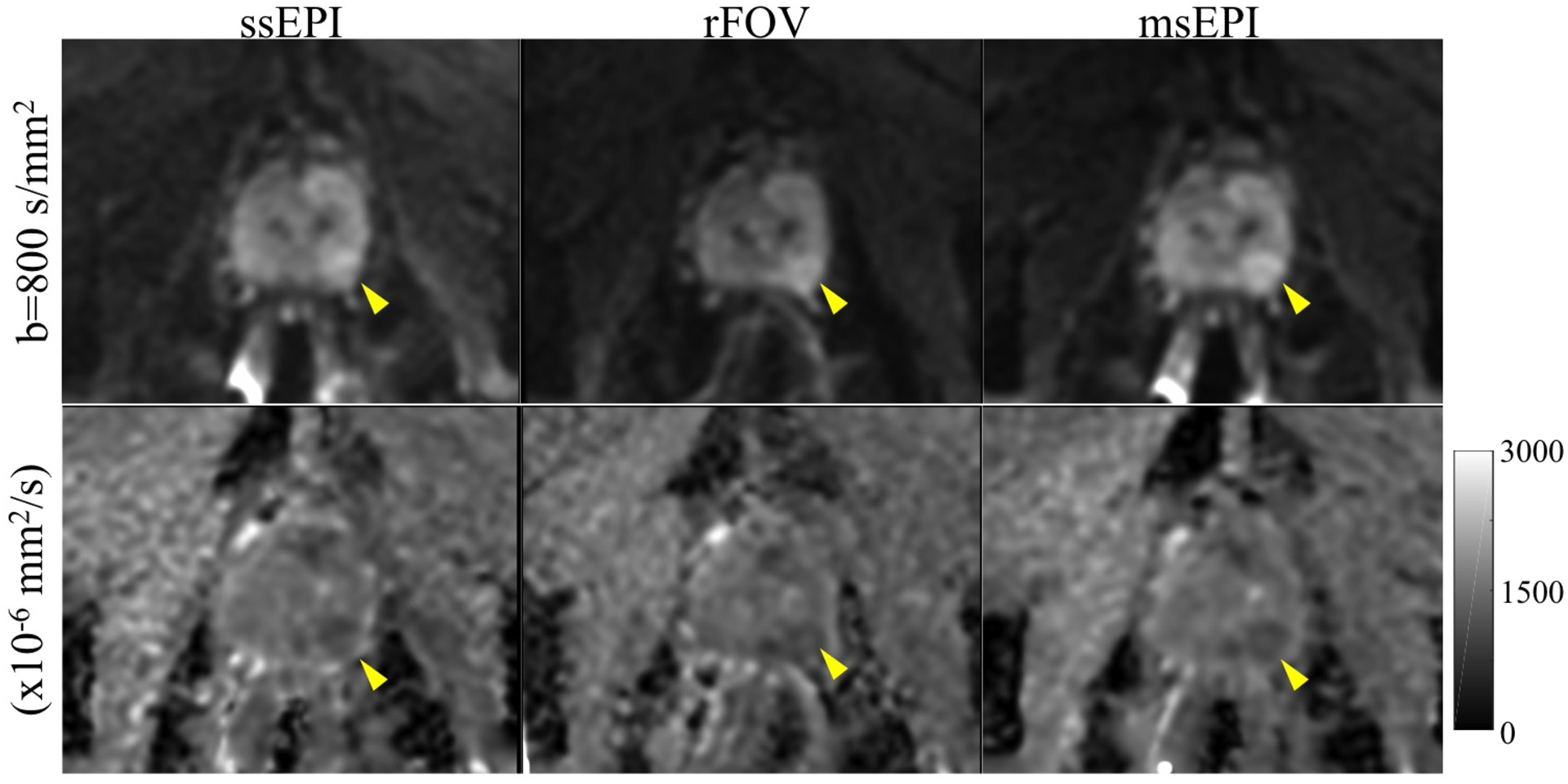
Methods: Under IRB approval and after informed consent, msEPI, rFOV, and ssEPI DWI acquisitions were prospectively added to clinical prostate MRI exams at 3.0 T. Image distortion was quantitatively evaluated by root-mean-squared displacement (dr.m.s.). Histogram-based quantitative ADC parameters were compared in a sub-set of patients for proven sites of prostate cancer and matched non-cancerous prostate. Three radiologists also independently evaluated the DWI sequences for subjective image quality and distortion/artifact on a 5-point Likert scale.

Results: Twenty-five patients were included (15 with proven sites of cancer). Average dr.m.s. demonstrated a small but statistically significant reduction in distortion for both rFOV and msEPI relative to ssEPI. Quantitative ADC parameters for prostate tumors demonstrated no significant difference across the 3 DWI acquisitions and each acquisition demonstrated a statistically significant decrease in mean ADC for tumor compared to normal prostate. Qualitative reader assessment demonstrated favorable image quality for rFOV and msEPI, more notable for msEPI.

Conclusions: rFOV and msEPI DWI techniques achieved reduction in image distortion, improvement in image quality, and maintained reproducible ADC quantification compared to the standard ssEPI.

Keywords: Diffusion weighted imaging; Echo planar imaging; Prostate cancer; image processing, computer-assisted; magnetic resonance imaging.


Figures

Figure 1.
Figure 1.. Reduced distortion using the rFOV and msEPI techniques.
(A) On the ssEPI images, rectal gas results in artifactual signal build-up (white arrow) and contour distortion (dashed arrow) of the posterior prostate. The artifact and distortion are reduced in both the rFOV and, to a greater extent, msEPI images. B. Distortion plots from the same patient demonstrate a small reduction in distortion for both rFOV and msEPI graphically and in a reduction in dr.m.s. (in mm) for rFOV (1.9) and msEPI (1.8) compared to ssEPI (2.2).
Figure 2.
Figure 2.. Comparison of mean apparent diffusion coefficient values for cancer and non-cancerous prostate regions.
2A) Boxplots grouped and compared according to diffusion acquisition sequence demonstrate that mean ADC was significantly lower for cancer regions compared to non-cancerous prostate for all DWI sequences (*p<0.0001). 2B) Boxplots grouped and compared according to tissue type demonstrate that mean ADC was not significantly different for all comparison except between the non-cancerous prostate values for rFOV compared to msEPI (*p<0.01). Box and whisker plots demonstrate the median (middle line), mean (star), upper and lower quartiles (top and bottom box margins), and minimum/maximum (whiskers). ADC, apparent diffusion coefficient; msEPI, multi-shot echo planar imaging; NC, non-cancerous; rFOV, reduced field-of-view; ssEPI, single-shot echo planar imaging;
Figure 3.
Figure 3.. Similar apparent diffusion coefficient (ADC) quantification in a 60-year old male with elevated serum prostate specific antigen (4.46 ng/mL).
Left peripheral zone PI-RADS 4 lesion (yellow arrowheads) is seen on all three DWI sequences, including the b=800 s/mm2 series (top row) and calculated ADC map (bottom row). The mean ADC value for the lesion was 1075.8, 1098.8, and 1067.5 ×10−6 mm2/s on ssEPI, rFOV, and msEPI, respectively. The patient subsequently underwent radical prostatectomy and was found to have Gleason 3+4 prostate cancer.

Similar articles

Cited by

References

    1. Stabile A, Giganti F, Rosenkrantz AB, Taneja SS, Villeirs G, Gill IS, et al. Multiparametric MRI for prostate cancer diagnosis: current status and future directions. Nat Rev Urol 2020;17:41–61. doi: 10.1038/s41585-019-0212-4. - DOI - PubMed
    1. Turkbey B, Rosenkrantz AB, Haider MA, Padhani AR, Villeirs G, Macura KJ, et al. Prostate Imaging Reporting and Data System Version 2.1: 2019 Update of Prostate Imaging Reporting and Data System Version 2. Eur Urol 2019;76:340–351. doi: 10.1016/j.eururo.2019.02.033 - DOI - PubMed
    1. Fütterer JJ, Briganti A, De Visschere P, Emberton M, Giannarini G, Kirkham A, et al. Can Clinically Significant Prostate Cancer Be Detected with Multiparametric Magnetic Resonance Imaging? A Systematic Review of the Literature. Eur Urol 2015;68:1045–1053. doi: 10.1016/j.eururo.2015.01.013. - DOI - PubMed
    1. Jung SI, Donati OF, Vargas HA, Goldman D, Hricak H, Akin O. Transition zone prostate cancer: incremental value of diffusion-weighted endorectal MR imaging in tumor detection and assessment of aggressiveness. Radiology 2013;269:493–503. doi: 10.1148/radiol.13130029. - DOI - PubMed
    1. Park SY, Oh YT, Jung DC, Cho NH, Choi YD, Rha KH, et al. Prediction of biochemical recurrence after radical prostatectomy with PI-RADS version 2 in prostate cancers: initial results. Eur Radiol 2016;26:2502–2509. doi: 10.1007/s00330-015-4077-5. - DOI - PubMed

Publication types

MeSH terms