

Kimura's disease of the left arm
- PMID: 35944940
- PMCID: PMC9367184
- DOI: 10.1136/bcr-2022-250758
Kimura's disease of the left arm
Abstract
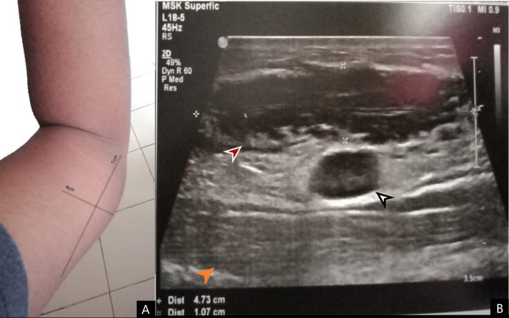
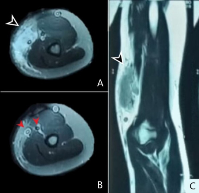
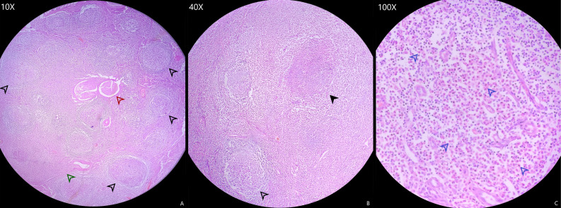
Kimura's disease is a rare, benign entity that causes subcutaneous angioblastic lymphoid hyperplasia with eosinophilia. It usually presents with subcutaneous lymphoid swellings with regional lymphadenopathy and salivary gland masses. Kimura's disease is frequently associated with renal involvement, which includes proteinuria and nephrotic syndrome as the most common presentations. This report presents a case of a man in his early 20s with a swelling in the medial aspect of the distal left arm with two previous episodes of nephrotic syndrome. Multiple enlarged axillary lymph nodes and epitrochlear nodes were noted. Ultrasonography revealed a mixed echogenic mass with enlarged lymph nodes. MRI showed a heterogeneous hyperintense lesion. Image-guided fine-needle aspiration showed multiple lymphocytes and eosinophils suggestive of Kimura's disease. The swelling was surgically excised under general anaesthesia. Histopathology of the excised mass confirmed the diagnosis of Kimura's disease.
Keywords: General surgery; Pathology; Screening (oncology); Surgery; Ultrasonography.
© BMJ Publishing Group Limited 2022. No commercial re-use. See rights and permissions. Published by BMJ.
Conflict of interest statement
Competing interests: None declared.
Figures
References
-
- Dhingra H, Nagpal R, Baliyan A. Kimura disease: case report and brief review of literature. Med Pharm Rep [Internet], 2018. Available: https://www.medpharmareports.com/index.php/mpr/article/view/1030 [Accessed 24 Jun 2022]. - PMC - PubMed
Publication types
MeSH terms
LinkOut - more resources
Full Text Sources