. 2022 Sep 21;84(10):1363-1367.
doi: 10.1292/jvms.22-0234. Epub 2022 Aug 10.

Sex differences in cervical spinal cord and spinal canal development in Thoroughbred horses

Affiliations

Sex differences in cervical spinal cord and spinal canal development in Thoroughbred horses

Taro Kondo et al. J Vet Med Sci. .

Abstract

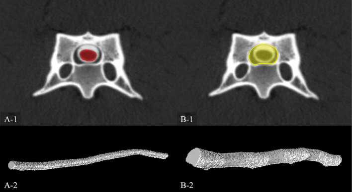
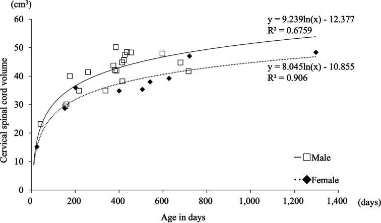
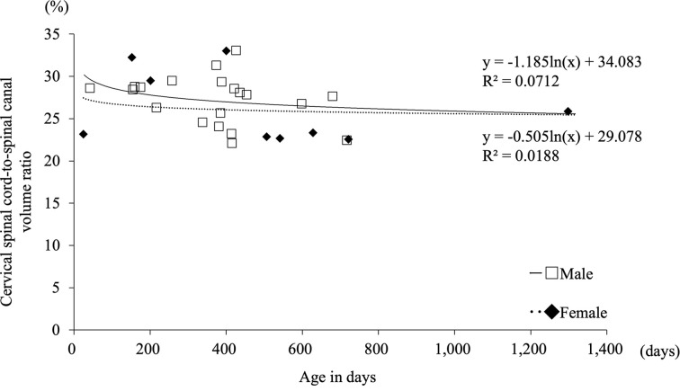
Cervical vertebral stenotic myelopathy (CVSM), a common cause of cervical spinal cord compression, is a neurological disease characterized by general proprioceptive ataxia and weakness of hindlimbs that tends to develop in young adult Thoroughbred horses. Although male horses seem to be at increased risk for CVSM, the mechanism for the occurrence of sex differences in the prevalence of CVSM is still poorly understood. Hence, we hypothesized that sex differences in the development of cervical spinal cord and spinal canal would affect the development of CVSM. This study aimed to evaluate sex differences in the development of cervical spinal cord and spinal canal in Thoroughbred horses. A total of 29 Thoroughbred horses underwent computed tomographic myelography. Thereafter, the volumes of cervical spinal cord and spinal canal were calculated. Accordingly, male horses had significantly lager cervical spinal cord volume and cervical spinal cord-to-spinal canal volume ratio than those of female horses (P<0.05). Sex differences in the cervical spinal cord-to-spinal canal volume ratio gradually decreased until around 1,400 days of age. Younger male horses have narrower interspace between the cervical spinal cord and spinal canal than younger female horses, suggesting that an imbalanced cervical spinal cord and spinal canal growth is one of the causes of CVSM.

Keywords: Thoroughbred; cervical spinal cord; computed tomographic myelography; spinal canal.


Figures

Fig. 1.
Fig. 1.
Areas of the cervical spinal cord (A-1) and spinal canal (B-1) were measured in the two-dimensional transverse plane. The measurement range spanned from the cranial top of the C3 to the caudal end of the C6. The three-dimensional images were reconstructed using the volume rendering technique. Thereafter, the three-dimensional images were used to calculate the volume of the cervical spinal cord (A-2) and spinal canal (B-2).
Fig. 2.
Fig. 2.
Relationship between cervical spinal cord volume and age in days. Male horses had a significantly larger cervical spinal cord volume than female horses (P<0.05). The cervical spinal cord volume dramatically increased until around 200 days of age, after which slow growth was observed.
Fig. 3.
Fig. 3.
Relationship between spinal canal volume and age in days. There was no significant difference in the spinal canal volume between the male and female horses (P=0.19). The spinal canal volume dramatically increased until around 200 days of age, after which slow growth was observed.
Fig. 4.
Fig. 4.
Relationship between the cervical spinal cord-to-spinal canal volume ratio and age in days. Male horses had a significantly higher cervical spinal cord-to-spinal canal volume ratio than female horses (P<0.05). Sex differences in the cervical spinal cord-to-spinal canal volume ratio gradually decreased until around 1,400 days of age.

Similar articles

Cited by

References

    1. Bahar S, Bolat D, Selcuk ML. 2013. The segmental morphometric properties of the horse cervical spinal cord: a study of cadaver. Scientific World Journal 2013: 734923. doi: 10.1155/2013/734923 - DOI - PMC - PubMed
    1. Claridge HAH, Piercy RJ, Parry A, Weller R. 2010. The 3D anatomy of the cervical articular process joints in the horse and their topographical relationship to the spinal cord. Equine Vet J 42: 726–731. doi: 10.1111/j.2042-3306.2010.00114.x - DOI - PubMed
    1. Craig LE, Dittmer KE, Thompson KG. 2015. Bones and joints. pp. 16–163. In: Jubb, Kennedy & Palmer’s Pathology of Domestic Animals Vol. 1, 6th ed. (Maxie, M. G. ed.), Saunders Elsevier, Amsterdam.
    1. Dimock WW. 1950. “Wobbles” an hereditary disease in horses. J Hered 41: 319–323. doi: 10.1093/oxfordjournals.jhered.a106074 - DOI - PubMed
    1. Falco MJ, Whitwell K, Palmer AC. 1976. An investigation into the genetics of ‘wobbler disease’ in thoroughbred horses in Britain. Equine Vet J 8: 165–169. doi: 10.1111/j.2042-3306.1976.tb03331.x - DOI - PubMed