. 2022 Sep 14;42(37):7016-7030.
doi: 10.1523/JNEUROSCI.0442-22.2022. Epub 2022 Aug 9.

Drosophila Homolog of the Human Carpenter Syndrome Linked Gene, MEGF8, Is Required for Synapse Development and Function

Affiliations

Drosophila Homolog of the Human Carpenter Syndrome Linked Gene, MEGF8, Is Required for Synapse Development and Function

Shuting Chen et al. J Neurosci. .

Abstract

Drosophila multiple epidermal growth factor-like domains 8 (dMegf8) is a homolog of human MEGF8 MEGF8 encodes a multidomain transmembrane protein which is highly conserved across species. In humans, MEGF8 mutations cause a rare genetic disorder called Carpenter syndrome, which is frequently associated with abnormal left-right patterning, cardiac defects, and learning disabilities. MEGF8 is also associated with psychiatric disorders. Despite its clinical relevance, MEGF8 remains poorly characterized; and although it is highly conserved, studies on animal models of Megf8 are also very limited. The presence of intellectual disabilities in Carpenter syndrome patients and association of MEGF8 with psychiatric disorders indicate that mutations in MEGF8 cause underlying defects in synaptic structure and functions. In this study, we investigated the role of Drosophila dMegf8 in glutamatergic synapses of the larval neuromuscular junctions (NMJ) in both males and females. We show that dMegf8 localizes to NMJ synapses and is required for proper synaptic growth. dMegf8 mutant larvae and adults show severe motor coordination deficits. At the NMJ, dMegf8 mutants show altered localization of presynaptic and postsynaptic proteins, defects in synaptic ultrastructure, and neurotransmission. Interestingly, dMegf8 mutants have reduced levels of the Type II BMP receptor Wishful thinking (Wit). dMegf8 displays genetic interactions with neurexin-1 (dnrx) and wit, and in association with Dnrx and Wit plays an essential role in synapse organization. Our studies provide insights into human MEGF8 functions and potentially into mechanisms that may underlie intellectual disabilities observed in Carpenter syndrome as well as MEGF8-related synaptic structural and/or functional deficits in psychiatric disorders.SIGNIFICANCE STATEMENT Carpenter syndrome, known for over a century now, is a genetic disorder linked to mutations in Multiple Epidermal Growth Factor-like Domains 8 (MEGF8) gene and associated with intellectual disabilities among other symptoms. MEGF8 is also associated with psychiatric disorders. Despite the high genetic conservation and clinical relevance, the functions of MEGF8 remain largely uncharacterized. Patients with intellectual disabilities and psychiatric diseases often have an underlying defect in synaptic structure and function. This work defines the role of the fly homolog of human MEGF8, dMegf8, in glutamatergic synapse growth, organization, and function and provide insights into potential functions of MEGF8 in human central synapses and synaptic mechanisms that may underlie psychiatric disorders and intellectual disabilities seen in Carpenter syndrome.

Keywords: BMP signaling; Carpenter syndrome; Drosophila larval NMJ; MEGF8; Neurexin-1; synapses.


Figures

Figure 1.
Figure 1.
Generation of dMegf8 mutants. A, Protein domain structure of human MEGF8, mouse Megf8, and Drosophila dMegf8. Green bar represents the antibody region in dMegf8. B, Genomic structure of dMegf8 showing exons 1-5. The targeting construct using CRISPR/Cas9 for recombination and the final targeted allele of dMegf8HSC is shown. Blue boxes next to loxP sites on both sides represent phage C31 integration sites referred to as attP sites in the targeting vector. Red arrows with numbers indicate location of the primers for genotyping that differentiate the WT and the dMegf8HSC mutants. C, D, PCR confirmation of the targeted deletion using primer combinations 1 + 2 for WT and 1 + 3 for dMegf8HSC mutants in B. E, Immunoblot analysis of dMegf8 showing presence of dMegf8 in WT, loss of dMeg8 in dMegf8HSC mutants, and overexpression of dMegf8 in elav>dMegf8. The blot was probed for actin as loading control. F–I, WT (+/+) and dMegf8HSC larval locomotor behaviors assayed by measuring the number of 0.5 cm2 grids crossed in 30 s (F), time taken in seconds for larvae to exit a circle of 1.5 cm in diameter (G), number of full body peristaltic contractions in 1 min (H), and time taken in seconds for larvae to right themselves when turned on their dorsal surface (I). n = 50 larvae. Data are mean ± SEM. t(46.49) = 18.22, ****p < 0.0001 (Welch's Student's t test) (F), t(78) = 4.833, ****p < 0.0001 (unpaired Student's t test) (G), t(78) = 5.753, ****p < 0.0001 (unpaired Student's t test) (H), and t(78) = 6.863, ****p < 0.0001 (unpaired Student's t test) (I). J, Adult locomotion assay using climbing ability of WT (+/+) (black) and dMegf8HSC mutants (red) flies. n = 50 flies. Data are mean ± SEM. Interaction: F(9,80) = 4.579, ****p < 0.0001 (two-way ANOVA test).
Figure 2.
Figure 2.
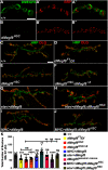
dMegf8 is expressed in synaptic terminals and required for synaptic bouton growth. A–B′′, Confocal images in (A) +/+ and (B) dMegf8HSC mutant third instar larvae NMJ Type Ib boutons at muscles 6/7 labeled with anti-dMegf8 (green) and anti-Brp (red). C–J, Confocal images in (C) dMegf8HSC, (D) dMegf8 point mutant dMegf8F1/Df, (E) dMegf8 null mutant (dMegf8HSC), (F) dMegf8HSC/dMegf8Δ8 trans-allelic combination, (G) presynaptic overexpression of dMegf8 (elav>dMegf8), (H) presynaptic rescue (elav>dMegf8;dMegf8HSC), (I) postsynaptic overexpression of dMegf8 (MHC>dMegf8), and (J) postsynaptic rescue (MHC>dMegf8;dMegf8HSC) third instar larvae NMJ at muscles 6/7 labeled with the presynaptic marker Hrp (green) and the postsynaptic marker Dlg (red). K, Quantification of total bouton numbers in indicated genotypes. K, Data are mean ± SEM (one-way ANOVA test with Tukey's multiple comparisons): F(8,160) = 22.3064, +/+ versus F1/Df: ***p = 0.0001, +/+ versus dMegf8HSC: ****p < 0.0001, +/+ versus dMegf8HSC/Δ8: ****p < 0.0001, +/+ versus MHC>dMegf8: p = 0.7496, +/+ versus elav>dMegf8: **p = 0.0014, +/+ versus Genomic Rescue: p = 0.9467, +/+ versus MHC>dMegf8; dMegf8HSC: ****p < 0.0001, +/+ versus elav>dMegf8; dMegf8HSC: p = 0.9992, dMegf8HSC versus F1/Df: p = 0.9915, dMegf8HSC versus dMegf8HSC/Δ8: p = 0.9989, dMegf8HSC versus genomic Rescue: **p = 0.0027, dMegf8HSC versus MHC>dMegf8; dMegf8HSC: p > 0.9999, dMegf8HSC versus elav>dMegf8; dMegf8HSC: ****p < 0.0001. Scale bars: A–B′′, 5 μm; C–J, 20 μm.
Figure 3.
Figure 3.
dMegf8HSC mutants show altered localization of presynaptic/postsynaptic proteins. A–B′′, Confocal images in (A) +/+ and (B) dMegf8HSC mutant third instar larval NMJ Type Ib boutons at muscles 6/7 labeled with the presynaptic protein Brp (green) and the postsynaptic protein GluRIIA (red). C, Quantification of Brp puncta/bouton area (μm2) in WT and dMegf8HSC mutants. D–E′, Confocal images in (D) +/+ and (E) dMegf8HSC mutant third instar larvae NMJ labeled with the presynaptic marker Hrp (green) and the postsynaptic protein Dlg (red). C, Quantification data are mean ± SEM; t(62) = 11.14. ****p < 0.0001 (unpaired Student's t test). F–G′, Dlg localization in dMegf8HSC mutants that have either presynaptic expression (elav>dMegf8) (F,F′) or postsynaptic expression (MHC>dMegf8) (G–G′). Scale bars: A–B′, F, G, 5 μm; D–E′, F′, G′, 20 μm.
Figure 4.
Figure 4.
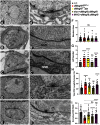
Loss of dMegf8 causes synaptic ultrastructural defects. A–E′, TEM images of cross sections through Type Ib boutons in (A) +/+, (B) dMegf8HSC mutant, (C) dMegf8 point mutant dMegf8F1/Df, (D) presynaptic rescue (elav>dMegf8;dMegf8HSC), and (E) postsynaptic rescue (MHC>dMegf8;dMegf8HSC) at low magnification (A–E) and high magnification (A′–E′). Arrows indicate the active zones (AZs). Arrowheads indicate the PSDs in A–E. F–I, Quantification in (F) total bouton area, (G) number of AZs, (H) total PSD length/perimeter (%), and (I) normalized SSR width in represented genotypes. F–I, Data are mean ± SEM (one-way ANOVA test with Tukey's multiple comparisons). F, F(4,183) = 0.4529, +/+ versus dMegf8HSC: p = 0.7990, +/+ versus F1/Df: p > 0.9999, +/+ versus elav>dMegf8; dMegf8HSC: p = 0.9994, +/+ versus MHC>dMegf8; dMegf8HSC: p > 0.9999. G, F(4.000,159.4) = 25.97, +/+ versus dMegf8HSC: ****p < 0.0001, +/+ versus F1/Df: ****p < 0.0001, +/+ versus elav>dMegf8; dMegf8HSC: p = 0.9999, +/+ versus MHC>dMegf8; dMegf8HSC: ****p < 0.0001 (Brown-Forsythe and Welch ANOVA with Games-Howell's multiple comparisons specifically). H, F(4,179) = 18.18, +/+ versus dMegf8HSC: ****p < 0.0001, +/+ versus F1/Df: *p = 0.0292, +/+ versus elav>dMegf8; dMegf8HSC: p > 0.9999, +/+ versus MHC>dMegf8; dMegf8HSC: ****p < 0.0001. I, F(4,169) = 16.03, +/+ versus dMegf8HSC: ****p < 0.0001, +/+ versus F1/Df: *p = 0.0136, +/+ versus elav>dMegf8; dMegf8HSC: ****p < 0.0001, +/+ versus MHC>dMegf8; dMegf8HSC: p = 0.1136, dMegf8HSC versus elav>dMegf8; dMegf8HSC: p = 0.2102, dMegf8HSC versus MHC>dMegf8; dMegf8HSC: ****p < 0.0001. Scale bars: A–E, 600 nm; A′–E′, 200 nm.
Figure 5.
Figure 5.
dMegf8 is required for proper synaptic transmission. A–D, Representative electrophysiological traces showing EJPs from (A) WT (wCS), (B) dMegf8HSC mutant, (C) presynaptic rescue (elav>dMegf8;dMegf8HSC), and (D) postsynaptic rescue (MHC>dMegf8;dMegf8HSC). E–I, Quantification of (E) EJP amplitude, (F) mEJP amplitude, (G) quantal contents, (H) paired pulse ratio, and (I) mEJP frequency in respective genotypes. E–I, Data are mean ± SEM (one-way ANOVA test with Tukey's multiple comparisons). E, F(4,58) = 11.53, +/+ versus dMegf8HSC: ****p < 0.0001, +/+ versus elav>dMegf8; dMegf8HSC: p = 0.9980, +/+ versus MHC>dMegf8; dMegf8HSC: **p = 0.0026. F, F(4,58) = 4.311, +/+ versus dMegf8HSC: p = 0.9658, +/+ versus elav>dMegf8; dMegf8HSC: p = 0.9936, +/+ versus MHC>dMegf8; dMegf8HSC: p = 0.0861. G, F(4,43) = 13.95, +/+ versus dMegf8HSC: ****p < 0.0001, +/+ versus elav>dMegf8; dMegf8HSC: p = 0.9503, +/+ versus MHC>dMegf8; dMegf8HSC: ****p < 0.0001. H, F(4,58) = 3.195, +/+ versus dMegf8HSC: p = 0.1143, +/+ versus elav>dMegf8; dMegf8HSC: p = 0.2908, +/+ versus MHC>dMegf8; dMegf8HSC: p = 0.0800. I, F(4,58) = 6.865, +/+ versus dMegf8HSC: p = 0.9998, +/+ versus elav>dMegf8; dMegf8HSC: p = 0.9088, +/+ versus MHC>dMegf8; dMegf8HSC: p = 0.2920.
Figure 6.
Figure 6.
Interdependency of dMegf8, Dnrx, and Wit in their localization and stability. A–D′′, Confocal images in (A) +/+, (B) dMegf8HSC mutant, (C) dnrx mutant, and (D) wit mutant third instar larval NMJ labeled with anti-dMegf8 (green) and anti-Brp (red). E–G′, Confocal images in (E) +/+, (F) dnrx mutant, and (G) dMegf8HSC mutant third instar larval NMJ labeled with anti-Dnrx (green) and anti-Brp (red). H–I′, Confocal images of (H) elav>wit and (I) elav>wit; dMegf8HSC larval NMJ labeled with anti-Hrp (green) and anti-Wit (red). J, Quantification of dMegf8 fluorescence intensity/bouton area in WT (+/+), dMegf8HSC, dnrx, and wit mutants. Data are mean ± SEM; F(7,37) = 18.25, +/+ versus dMegf8HSC: ****p < 0.0001, +/+ versus dnrx−/−: ***p = 0.0005, +/+ versus wit−/−: ****p < 0.0001 (one-way ANOVA test with Tukey's multiple comparisons). K, Quantification of Dnrx fluorescence intensity/bouton area in WT (+/+), dnrx, and dMegf8HSC mutants. Data are mean ± SEM; F(2,21) = 46.54, +/+ versus dnrx−/−: ****p < 0.0001, +/+ versus dMegf8HSC: ****p < 0.0001 (one-way ANOVA test with Tukey's multiple comparisons). L, Quantification of Wit/Hrp fluorescence intensity ratio in elav>wit and elav>wit; dMegf8HSC mutants. Data are mean ± SEM; t(24) = 11.02, ****p < 0.0001 (unpaired Student's t test). Scale bars: A–D′′, 10 μm; E–G′, 5 μm; H–I′, 5 μm.
Figure 7.
Figure 7.
BMP downstream effectors in dMegf8 mutants and genetic interactions between dMegf8, dnrx, and wit. A–B′, Confocal images in (A) +/+ and (B) dMegf8HSC mutant third instar larval NMJ labeled with anti-PS-1 (red) and anti-Hrp (green). C, D, Confocal images of (C) +/+ and (D) dMegf8HSC mutant larval VNC labeled with anti-Smad (red). E, Quantification of PS-1 fluorescence intensity/bouton area in the specified genotypes in A, B. Data are mean ± SEM; t(34) = 0.1063, p = 0.9159 (unpaired Student's t test). F, Quantification of Smad fluorescence intensity/bouton area in the specified genotypes in C, D. Data are mean ± SEM; t(34) = 1.063, p = 0.2951 (unpaired Student's t test). G–J, Representative immunoblots showing total levels of Smad (G) and Trio (H), and corresponding quantification of ratio of band intensities of Smad (I) and Trio (J). I, Data are mean ± SEM; F(2,6) = 31.42, +/+ versus dMegf8HSC: p = 0.7653, +/+ versus wit−/−: **p= 0.0011 (one-way ANOVA with Tukey's multiple comparisons). J, Data are mean ± SEM; t(4) = 0.5412, p = 0.6171 (unpaired Student's t test). K–R, Confocal images in (K) dMegf8 heterozygote (dMegf8HSC+/−), (L) dMegf8, dnrx trans-heterozygote (dMegf8HSC+/−;dnrx+/−), (M) dnrx−/− mutant, (N) dMegf8, dnrx double mutant (dMegf8HSC−/−;dnrx−/−), (O) wit heterozygote (wit+/−), (P) dMegf8, wit trans-heterozygote (dMegf8HSC+/−;wit+/−), (Q) wit−/− mutant, and (R) dMegf8, wit double mutant (dMegf8HSC−/−;wit−/−) third instar larval NMJ labeled with anti-Hrp (green) and anti-Dlg (red). S, T, Quantification of total bouton numbers in indicated genotypes. S, Data are mean ± SEM; F(6,107) = 21.47, +/+ versus dMegf8+/−: p > 0.9999, +/+ versus dnrx+/−: p = 0.7589, +/+ versus dMegf8+/−;dnrx+/−: ****p < 0.0001, +/+ versus dnrx−/−: ****p < 0.0001, +/+ versus dMegf8−/−: ****p < 0.0001, +/+ versus dMegf8−/−; dnrx−/−: ****p < 0.0001, dMegf8+/−; dnrx+/− versus dnrx−/−: p = 0.5614, dnrx−/− versus dMegf8−/−: p = 0.9379, dnrx−/− versus dMegf8−/−; dnrx−/−: p = 0.8967 (one-way ANOVA test with Tukey's multiple comparisons). T, Data are mean ± SEM; F(7,120) = 21.32, +/+ versus dMegf8+/−: p = 0.9211, +/+ versus witA12/-: p = 0.9985, +/+ versus dMegf8+/−; witA12/-: ***p = 0.0009, +/+ versus dMegf8+/−; witB11/-: ***p = 0.0007, +/+ versus dMegf8−/−: ****p < 0.0001, +/+ versus witA12/B11: ****p < 0.0001, +/+ versus dMegf8−/−; witA12/B11: ****p < 0.0001, dMegf8−/− versus witA12/B11: p = 0.9612, dMegf8−/− versus dMegf8−/−; witA12/B11: p = 0.9994 (one-way ANOVA test with Tukey's multiple comparisons). Scale bars: A–H, 20 μm.
Figure 8.
Figure 8.
Biochemical interactions between dMegf8, Dnrx, and Wit. A–C, Representative immunoblots from adult head lysates showing total levels of dMegf8 in WT (+/+), dMegf8F1/Df (A) and in dnrx mutant (B), and quantification of the normalized ratio of protein band intensities (C). +/+ versus dMegf8F1/Df: t(4) = 6.815, **p = 0.0024 (unpaired Student's t test); +/+ versus dnrx−/−: t(6) = 0.1961, p = 0.8510 (unpaired Student's t test). D, E, Representative immunoblots from adult head lysates showing total levels of Dnrx in WT (+/+), dMegf8HSC, and dnrx mutants (as a negative control), and quantification of the normalized ratio of protein band intensities (E). +/+ versus dMegf8HSC: t(6) = 0.3921, p = 0.7085 (unpaired Student's t test). F, G, Representative immunoblots from larval musculature lysates showing levels of Wit in specified genotypes, and quantification of the normalized ratio of protein band intensities (G). F(2,11) = 45.91, +/+ versus dMegf8HSC: *p = 0.0107, +/+ versus elav>dMegf8: ***p = 0.0003 (one-way ANOVA test with Tukey's multiple comparisons). H, I, Representative immunoblots from larval musculature lysates showing levels of Wit in specified genotypes, and quantification of the normalized ratio of protein band intensities (I). F(2,12) = 37.98, +/+ versus elav>wit;dMegf8HSC: *p = 0.0396, +/+ versus elav>wit: ****p < 0.0001 (one-way ANOVA test with Tukey's multiple comparisons). Actin was used as the loading control in A, B, D, F, H. J, K, dMegf8 coimmunoprecipitated with anti-Dnrx (J) and anti-Wit (K) antibodies, respectively, and probed with anti-dMegf8. C, E, G, I, Data are mean ± SEM; *p < 0.05; **p < 0.01; ***p < 0.001; ****p < 0.0001; unpaired Student's t test (C,E) and one-way ANOVA test with Tukey's multiple comparisons (G,I).
Figure 9.
Figure 9.
Schematic model of dMegf8 function at the NMJ synapse. A schematic model depicting dMegf8 and other known presynaptic and postsynaptic proteins at the NMJ. Based on our immunohistochemical, ultrastructural, electrophysiological, biochemical, and genetic rescue analyses, we propose that dMegf8 functions presynaptically to coordinate BMP signaling for the synaptic bouton growth and postsynaptically to organize the SSR and other synaptic structures that are necessary for proper synaptic function. While dMegf8 interactions with Dnrx and Wit are established, uncovering potential molecular interactions between dMegf8 and Dnlg1, Tkv, and Sax, or other synaptic proteins would shed further light on the functions of dMegf8 at the synapse.

Similar articles

Cited by

References

    1. Aberle H, Haghighi AP, Fetter RD, McCabe BD, Magalhaes TR, Goodman CS (2002) wishful thinking encodes a BMP Type II receptor that regulates synaptic growth in Drosophila. Neuron 33:545–558. 10.1016/S0896-6273(02)00589-5 - DOI - PubMed
    1. Alessandri JL, Dagoneau N, Laville JM, Baruteau J, Hebert JC, Cormier-Daire V (2010) RAB23 mutation in a large family from Comoros Islands with Carpenter syndrome. Am J Med Genet A 152A:982–986. 10.1002/ajmg.a.33327 - DOI - PubMed
    1. Awasaki T, Saito M, Sone M, Suzuki E, Sakai R, Ito K, Hama C (2000) The Drosophila trio plays an essential role in patterning of axons by regulating their directional extension. Neuron 26:119–131. 10.1016/S0896-6273(00)81143-5 - DOI - PubMed
    1. Ball RW, Warren-Paquin M, Tsurudome K, Liao EH, Elazzouzi F, Cavanagh C, An BS, Wang TT, White JH, Haghighi AP (2010) Retrograde BMP signaling controls synaptic growth at the NMJ by regulating trio expression in motor neurons. Neuron 66:536–549. 10.1016/j.neuron.2010.04.011 - DOI - PubMed
    1. Banerjee S, Riordan M (2018) Coordinated regulation of axonal microtubule organization and transport by Drosophila neurexin and BMP pathway. Sci Rep 8:17337. 10.1038/s41598-018-35618-7 - DOI - PMC - PubMed

Publication types

Supplementary concepts

LinkOut - more resources