

Risk factors of radiographic severity of massive rotator cuff tear
- PMID: 35945235
- PMCID: PMC9363414
- DOI: 10.1038/s41598-022-17624-y
Risk factors of radiographic severity of massive rotator cuff tear
Abstract
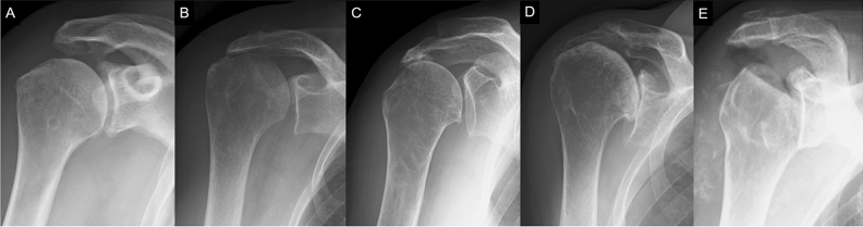
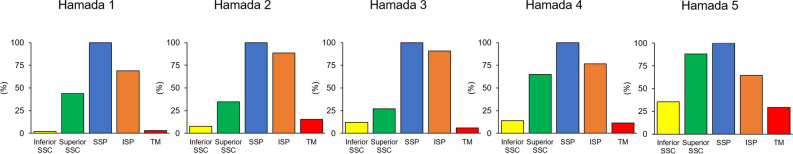
As massive rotator cuff tears progress, various radiographic changes occur; however, the factors associated with radiographic changes remain largely unknown. This study aimed to determine the factors that affect radiographic severity in massive rotator cuff tears using multivariate analyses. We retrospectively reviewed 210 shoulders with chronic massive rotator cuff tears. The dependent variables were superior migration of the humeral head (Hamada grades 2-3), narrowing of the glenohumeral joint (grade 4), and humeral head collapse (grade 5). Baseline variables that were significant in univariate analyses were included in multivariate models. There were 91, 59, 43, and 17 shoulders classified as Hamada grades 1, 2-3, 4, and 5, respectively. Multivariate analysis showed that infraspinatus tear (P = 0.015) and long head of biceps (LHB) tendon rupture (P = 0.007) were associated with superior migration of humeral head. Superior subscapularis tear (P = 0.003) and LHB tendon rupture (P < 0.001) were associated with narrowing of glenohumeral joint. Female sex (P = 0.006) and superior subscapularis tear (P = 0.006) were associated with humeral head collapse. This study identified the rupture of infraspinatus and LHB as risk factors of superior migration of humeral head, and the rupture of subscapularis and LHB and female sex as risk factors of cuff tear arthropathy.
© 2022. The Author(s).
Conflict of interest statement
The authors declare no competing interests
Figures
Similar articles
-
Relationship between humeral head translation, spur formations, and the locations of tendon tears in massive rotator cuff tears.BMC Res Notes. 2025 Jan 27;18(1):39. doi: 10.1186/s13104-024-07079-x. BMC Res Notes. 2025. PMID: 39871361 Free PMC article.
-
[Coraco-humeral space and rotator cuff tears].Rev Chir Orthop Reparatrice Appar Mot. 1999 Nov;85(7):677-83. Rev Chir Orthop Reparatrice Appar Mot. 1999. PMID: 10612131 French.
-
Relationship between the Hamada Grade and underlying pathological conditions in the rotator cuff and long head of biceps in symptomatic patients with rotator cuff tears.JSES Int. 2022 Feb 18;6(3):488-494. doi: 10.1016/j.jseint.2022.01.012. eCollection 2022 May. JSES Int. 2022. PMID: 35572420 Free PMC article.
-
Pectoralis major and pectoralis minor transfer for irreparable subscapularis tendon tears.Oper Orthop Traumatol. 2022 Feb;34(1):45-54. doi: 10.1007/s00064-021-00760-5. Epub 2022 Feb 3. Oper Orthop Traumatol. 2022. PMID: 35113176 Review. English.
-
Repair of tears of the subscapularis. Surgical technique.J Bone Joint Surg Am. 2006 Mar;88 Suppl 1 Pt 1:1-10. doi: 10.2106/JBJS.E.00842. J Bone Joint Surg Am. 2006. PMID: 16510795 Review.
Cited by
-
Collagen and platelet-rich plasma in partial-thickness rotator cuff injuries. Friends or only indifferent neighbours? Randomised controlled trial.BMC Musculoskelet Disord. 2022 Dec 20;23(1):1109. doi: 10.1186/s12891-022-06089-9. BMC Musculoskelet Disord. 2022. PMID: 36536333 Free PMC article. Clinical Trial.
-
The Relationship Among Probable SARCopenia, Osteoporosis and SuprasPinatus Tendon Tears in Postmenopausal Women: The SARCOSP Study.Calcif Tissue Int. 2024 Apr;114(4):340-347. doi: 10.1007/s00223-024-01183-7. Epub 2024 Feb 12. Calcif Tissue Int. 2024. PMID: 38342790 Free PMC article.
-
Development of a novel animal model of rotator cuff tear arthropathy replicating clinical features of progressive osteoarthritis with subchondral bone collapse.Osteoarthr Cartil Open. 2023 Jul 27;5(3):100389. doi: 10.1016/j.ocarto.2023.100389. eCollection 2023 Sep. Osteoarthr Cartil Open. 2023. PMID: 37560389 Free PMC article.
-
Parsonage-Turner Syndrome Following AstraZeneca COVID-19 Vaccination.Curr Drug Saf. 2025;20(4):526-531. doi: 10.2174/0115748863322806240808095118. Curr Drug Saf. 2025. PMID: 39248062
-
Improved clinical outcomes after arthroscopic partial rotator cuff repair with rotator interval shift for the treatment of massive irreparable anterior L-shaped rotator cuff tears.J Orthop Surg Res. 2025 Jun 3;20(1):562. doi: 10.1186/s13018-025-05975-x. J Orthop Surg Res. 2025. PMID: 40462137 Free PMC article.
References
Publication types
MeSH terms
LinkOut - more resources
Full Text Sources
Medical
Research Materials
