. 2023 May;48(6):877-886.
doi: 10.1038/s41386-022-01412-3. Epub 2022 Aug 9.

mGluR5 PAMs rescue cortical and behavioural defects in a mouse model of CDKL5 deficiency disorder

Affiliations

mGluR5 PAMs rescue cortical and behavioural defects in a mouse model of CDKL5 deficiency disorder

Antonia Gurgone et al. Neuropsychopharmacology. 2023 May.

Abstract

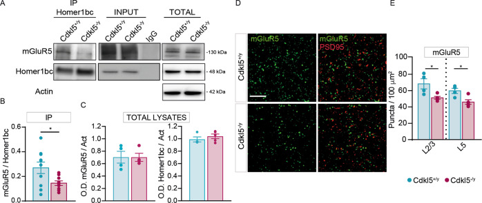
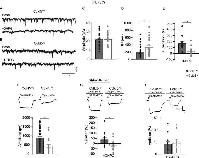
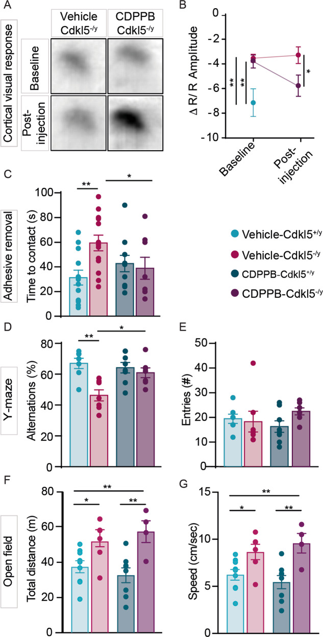
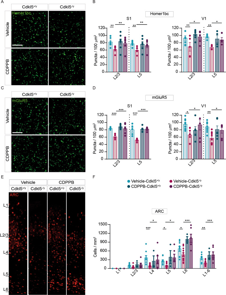
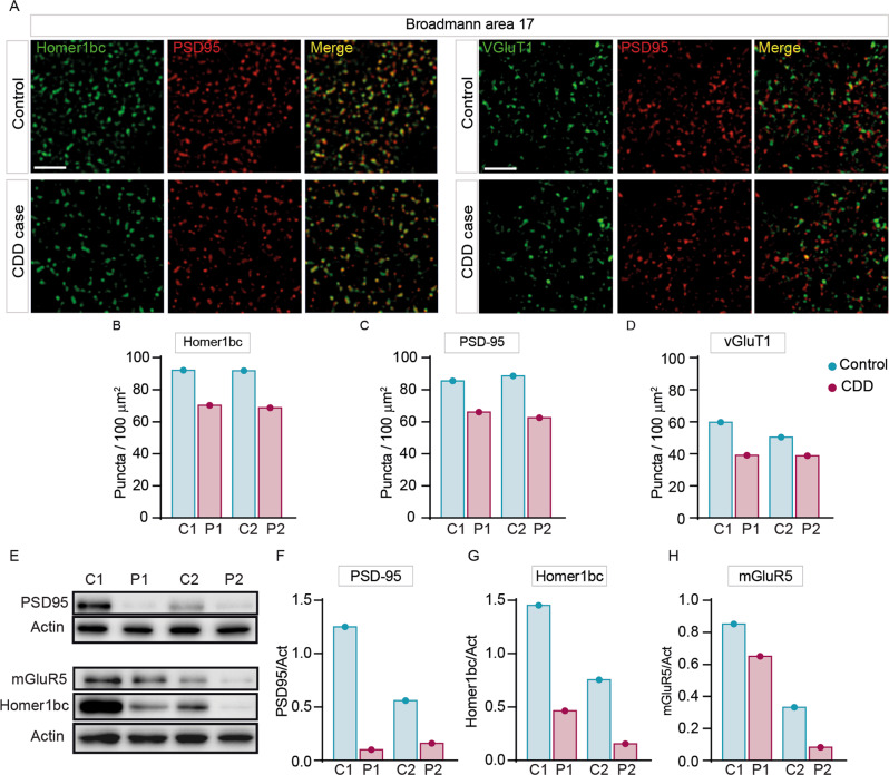
Cyclin-dependent kinase-like 5 (CDKL5) deficiency disorder (CDD) is a devastating rare neurodevelopmental disease without a cure, caused by mutations of the serine/threonine kinase CDKL5 highly expressed in the forebrain. CDD is characterized by early-onset seizures, severe intellectual disabilities, autistic-like traits, sensorimotor and cortical visual impairments (CVI). The lack of an effective therapeutic strategy for CDD urgently demands the identification of novel druggable targets potentially relevant for CDD pathophysiology. To this aim, we studied Class I metabotropic glutamate receptors 5 (mGluR5) because of their important role in the neuropathological signs produced by the lack of CDKL5 in-vivo, such as defective synaptogenesis, dendritic spines formation/maturation, synaptic transmission and plasticity. Importantly, mGluR5 function strictly depends on the correct expression of the postsynaptic protein Homer1bc that we previously found atypical in the cerebral cortex of Cdkl5-/y mice. In this study, we reveal that CDKL5 loss tampers with (i) the binding strength of Homer1bc-mGluR5 complexes, (ii) the synaptic localization of mGluR5 and (iii) the mGluR5-mediated enhancement of NMDA-induced neuronal responses. Importantly, we showed that the stimulation of mGluR5 activity by administering in mice specific positive-allosteric-modulators (PAMs), i.e., 3-Cyano-N-(1,3-diphenyl-1H-pyrazol-5-yl)benzamide (CDPPB) or RO6807794, corrected the synaptic, functional and behavioral defects shown by Cdkl5-/y mice. Notably, in the visual cortex of 2 CDD patients we found changes in synaptic organization that recapitulate those of mutant CDKL5 mice, including the reduced expression of mGluR5, suggesting that these receptors represent a promising therapeutic target for CDD.


Conflict of interest statement

The authors declare no competing interests.

Figures

Fig. 1
Fig. 1. CDKL5 loss is responsible for both the disruption of mGluR5-Homer1bc interaction and the reduction of mGluR5 localization in the cortical neuropil.
A Co-IP of cortical synaptosomal fraction (P2) from P56 mice by using anti-Homer1bc. IgG: control lane in the absence of antibodies. Immunoprecipitates, inputs (P2) and total cortical lysates were analyzed by immunoblotting for mGluR5 and Homer1bc. B, C Bar graphs showing Co-IP (B) and total cortical lysates (C) quantitation expressed as optical density (O.D.). D Confocal microscopy images showing mGluR5+ (green) and PSD-95+ (red) immunopuncta in layers II/III of S1 cortex (scale bar: 5 µm). E Bar graphs displaying the density of mGluR5+ puncta. Student T test *p < 0.05 (Co-IP: n = 8; WB: n = 4 IFL: n = 4).
Fig. 2
Fig. 2. CDKL5 loss tampers with both mEPSCs and NMDA current.
A Sample traces of miniature excitatory postsynaptic current (mEPSC) recorded from Cdkl5+/y neurons (A, upper part) and Cdkl5-/y neurons (B, upper part) and after the application of DHPG (A, B lower part). C, D Bar graphs showing the mean average amplitude (C) and the inter-event interval (IEI) of mEPSCs (D). E Bar graphs displaying the % of IEI variation IEI after the application of DHPG (100 µM). F Representative traces of currents obtained with patch-clamp recordings on S1 neurons cultures from Cdkl5+/y and Cdkl5-/y embryos after NMDA (50 µM) application (upper part), bar graphs showing differences of INMDA current between genotypes (lower part). G Representative traces of NMDA currents on S1 neurons after 2-min application of DHPG (100 µM-upper part); bar graphs showing the % change of INMDA after the application of DHPG (lower part). H Representative traces of NMDA after 2-min CDPPB + NMDA application (upper part), bar graphs showing the % change of INMDA current after the application of CDPPB (lower part). Student’s t-test, chi-square, two-way ANOVA followed by Fisher’s multiple comparison test, *p < 0.05, **p < 0.01, ***p < 0.001 (mEPSC Cdkl5+/y n = 22 cells, Cdkl5-/y n = 28; minis+DHPG Cdkl5+/y: n = 12 cells; minis+DHPG Cdkl5-/y: n = 13 cells. NMDA: Cdkl5+/y n = 36 cells, Cdkl5-/y n = 23 cells; NMDA + DHPG Cdkl5+/y n = 15 cells and NMDA + DHPG Cdkl5-/y n = 14 cells; NMDA + CDPPB Cdkl5+/y n = 12 cells; NMDA + CDPPB Cdkl5-/y n = 9 cells).
Fig. 3
Fig. 3. Acute CDPPB treatment rescues visual response, sensorimotor and memory deficits in Cdkl5-/y mice.
A Samples images showing differences of IOS evoked responses in vehicle- and CDPPB-treated Cdkl5-/y mice. B Trajectory of the IOS amplitude in vehicle-Cdkl5+/y, vehicle-Cdkl5+/y and CDPPB-Cdkl5-/y treated mice. C Bar graphs showing contact latency with the tape placed under mice’s forepaw. D, E Bar graphs showing the percentage of the correct alternations (D) and the number of entries (E) made by Cdkl5+/y and Cdkl5-/y mice, treated with either vehicle or CDPPB, in the Y-maze. F, G Bar graphs showing the total distance traveled (F) and the mean speed (G) in the open field arena of mice treated with either vehicle or CDPPB. One-way ANOVA followed by Tukey’s multiple comparison; two-way ANOVA followed by Sidak or Bonferroni’s multiple comparison test, *p < 0.05, **p < 0.01 (IOS: vehicle-Cdkl5+/y n = 3, vehicle-Cdkl5-/y n = 8, CDPPB-Cdkl5-/y n = 6; behavioural tests: vehicle-Cdkl5+/y n = 12, vehicle-Cdkl5-/y n = 13, CDPPB-Cdkl5+/y n = 8, CDPPB-Cdkl5-/y n = 7).
Fig. 4
Fig. 4. Structural defects exhibited by Cdkl5-/y mice cortices are rescued by an acute CDPPB injection.
A, C Representative confocal images showing Homer1bc+ and mGluR5+ puncta in layer II-III of S1 cortex from either vehicle- or CDPPB-treated mice (scale bar: 5 µm). B, D Bar graphs showing both Homer1bc+ (B) and mGluR5+ (D) immunopuncta density in layers II-III and V of both S1 and V1 cortices in either vehicle- or CDPPB-treated mice. E Confocal images of ARC immunostaining on coronal sections of the V1 cortex from mice treated with vehicle or CDPPB (scale bar: 25 µm), and relative ARC+ cells density quantitation (F) throughout the cortical layers. Two-way ANOVA followed by Fisher’s multiple comparison test, *p < 0.05, **p < 0.01, ***p < 0.001; (n = 6 animals for each genotype).
Fig. 5
Fig. 5. Aberrant expression of excitatory synaptic proteins in the BA17 cortex of CDD patients.
A Illustrative confocal images taken from layers II-III of the BA17 cortex. (A) PSD-95+(red), Homer1bc+(green), VGluT1+(green) immunofluorescence puncta. Note the virtually complete overlapping of PSD-95 and Homer1bc immunofluorescence (scale bar: 5 µm). BD Bar graphs showing the analysis of puncta density in layers II-III of BA17 cortices. E Western blotting showing the expression of PSD-95, Homer1bc and mGluR5 in lysates from BA17 cortices. FH Bar graphs displaying the optical density (O.D.) analysis of PSD-95 (F), Homer1bc (G) and mGluR5 (H) expression. Student’s t-test, *p < 0.05, **p < 0.01 (C1 = F, 4 years old; P1 = F, 5.7 years old; C2 = F, 29 years old; P2 = F, 30 years old).

References

    1. Rusconi L, Salvatoni L, Giudici L, Bertani I, Kilstrup-Nielsen C, Broccoli V, et al. CDKL5 expression is modulated during neuronal development and its subcellular distribution is tightly regulated by the C-terminal tail. J Biol Chem. 2008;283:30101–11. doi: 10.1074/jbc.M804613200. - DOI - PMC - PubMed
    1. Baltussen LL, Negraes PD, Silvestre M, Claxton S, Moeskops M, Christodoulou E, et al. Chemical genetic identification of CDKL5 substrates reveals its role in neuronal microtubule dynamics. EMBO J. 2018;37:e99763. doi: 10.15252/embj.201899763. - DOI - PMC - PubMed
    1. Muñoz IM, Morgan ME, Peltier J, Weiland F, Gregorczyk M, Cm Brown F, et al. Phosphoproteomic screening identifies physiological substrates of the CDKL5 kinase. EMBO J. 2018;37:e99559. doi: 10.15252/embj.201899559. - DOI - PMC - PubMed
    1. Nawaz MS, Giarda E, Bedogni F, La Montanara P, Ricciardi S, Ciceri D, et al. CDKL5 and Shootin1 interact and concur in regulating neuronal polarization. PLoS One. 2016;11:e0148634. doi: 10.1371/journal.pone.0148634. - DOI - PMC - PubMed
    1. Trazzi S, De Franceschi M, Fuchs C, Bastianini S, Viggiano R, Lupori L, et al. CDKL5 protein substitution therapy rescues neurological phenotypes of a mouse model of CDKL5 disorder. Hum Mol Genet. 2018;27:1572–92. doi: 10.1093/hmg/ddy064. - DOI - PubMed

Publication types

MeSH terms

Substances

Supplementary concepts