. 2023 Feb;44(2):434-445.
doi: 10.1038/s41401-022-00958-8. Epub 2022 Aug 9.

14-Deoxygarcinol improves insulin sensitivity in high-fat diet-induced obese mice via mitigating NF-κB/Sirtuin 2-NLRP3-mediated adipose tissue remodeling

Affiliations

14-Deoxygarcinol improves insulin sensitivity in high-fat diet-induced obese mice via mitigating NF-κB/Sirtuin 2-NLRP3-mediated adipose tissue remodeling

Jia-Li Chen et al. Acta Pharmacol Sin. 2023 Feb.

Abstract

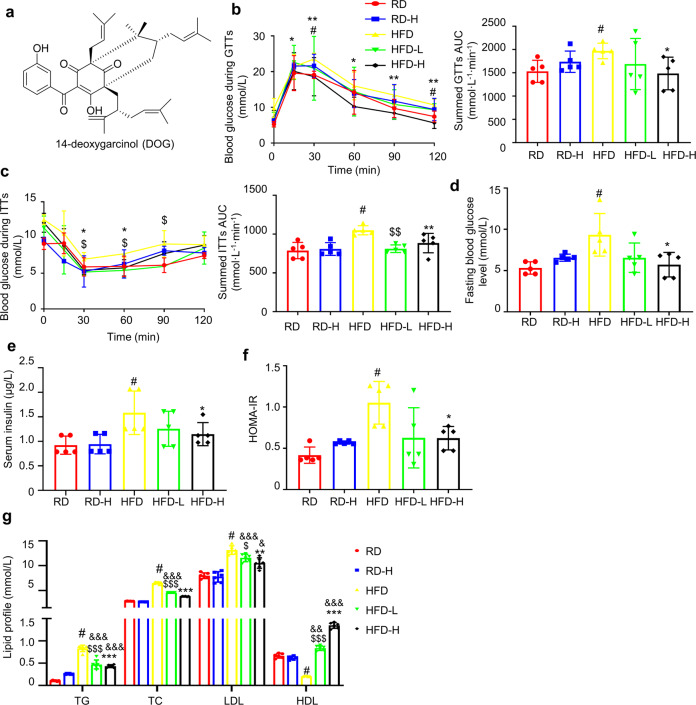
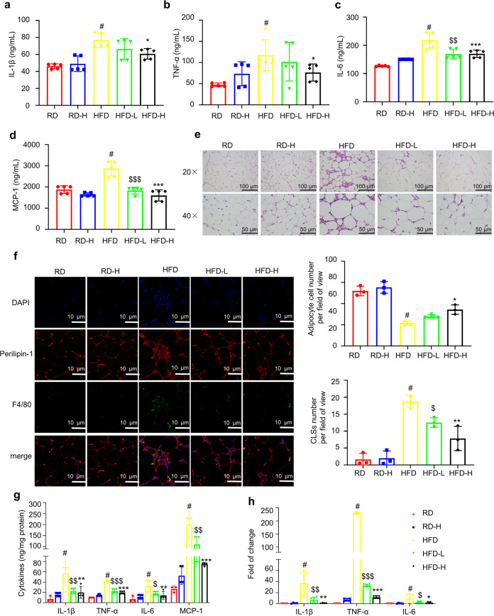
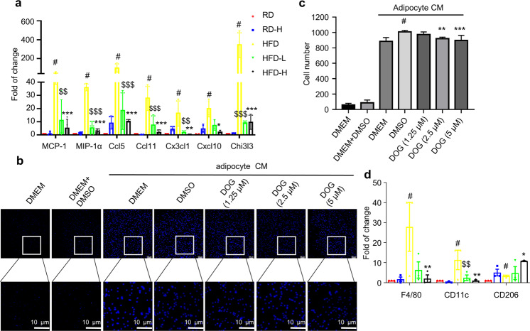
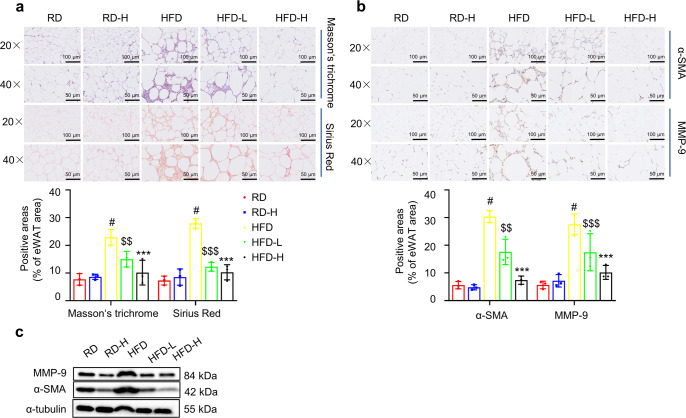
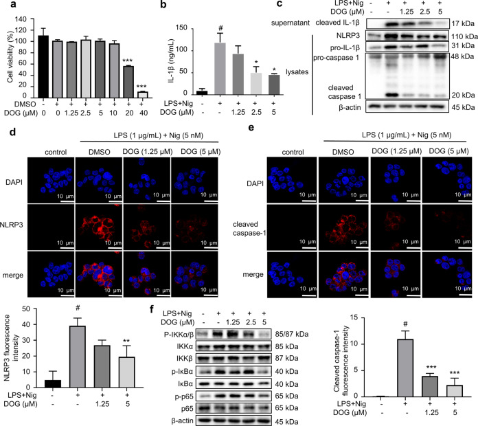
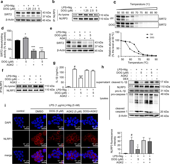
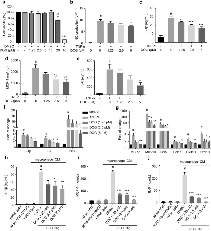
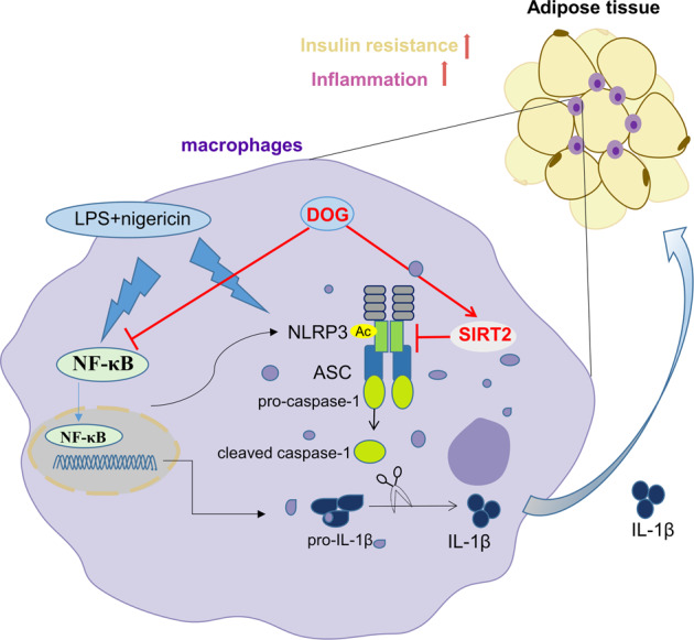
Interleukin (IL)-1β is a culprit of adipose tissue inflammation, which in turn causes systematic inflammation and insulin resistance in obese individuals. IL-1β is mainly produced in monocytes and macrophages and marginally in adipocytes, through cleavage of the inactive pro-IL-1β precursor by caspase-1, which is activated via the NLRP3 inflammasome complex. The nuclear factor-κB (NF-κB) transcription factor is the master regulator of inflammatory responses. Brindle berry (Garcinia cambogia) has been widely used as health products for treating obesity and related metabolic disorders, but its active principles remain unclear. We previously found a series of polyisoprenylated benzophenones from brindle berry with anti-inflammatory activities. In this study we investigated whether 14-deoxygarcinol (DOG), a major polyisoprenylated benzophenone from brindle berry, alleviated adipose tissue inflammation and insulin sensitivity in high-fat diet fed mice. The mice were administered DOG (2.5, 5 mg · kg-1 · d-1, i.p.) for 4 weeks. We showed that DOG injection dose-dependently improved insulin resistance and hyperlipidemia, but not adiposity in high-fat diet-fed mice. We found that DOG injection significantly alleviated adipose tissue inflammation via preventing macrophage infiltration and pro-inflammatory polarization of macrophages, and adipose tissue fibrosis via reducing the abnormal deposition of extracellular matrix. In LPS plus nigericin-stimulated THP-1 macrophages, DOG (1.25, 2.5, 5 μM) dose-dependently suppressed the activation of NLRP3 inflammasome and NF-κB signaling pathway. We demonstrated that DOG bound to and activated the deacetylase Sirtuin 2, which in turn deacetylated and inactivated NLRP3 inflammasome to reduce IL-1β secretion. Moreover, DOG (1.25, 2.5, 5 μM) dose-dependently mitigated inflammatory responses in macrophage conditioned media-treated adipocytes and suppressed macrophage migration toward adipocytes. Taken together, DOG might be a drug candidate to treat metabolic disorders through modulation of adipose tissue remodeling.

Keywords: 14-deoxygarcinol; NLRP3 inflammasome; Sirtuin 2; adipose tissue inflammation; insulin resistance; interleukin-1β.


Conflict of interest statement

The authors declare no competing interests.

Figures

Fig. 1
Fig. 1. DOG alleviates insulin resistance and hyperlipidemia in HFD-challenged mice.
a Chemical structure of DOG. b Glucose tolerance test was performed after 2-week DOG treatment. AUC (area under curve) of each group was calculated. c Insulin tolerance test was performed after 3-week DOG treatment. AUC of each group was calculated. The blood glucose levels (d) and the serum insulin levels (e) of mice were determined after 16 h fasting. f The HOMA-IR index. g The lipid parameters in serum were measured. Data are expressed as means ± SEM. n = 5. #P < 0.05, HFD vs. RD; &P < 0.05, &&P < 0.01, &&&P < 0.001, HFD-L, HFD-H vs. RD; $P < 0.05, $$P < 0.01, $$$P < 0.001, HFD vs. HFD-L; *P < 0.05, **P < 0.01, ***P < 0.001, HFD vs. HFD-H.
Fig. 2
Fig. 2. DOG ameliorates HFD-induced adipose tissue inflammation in mice.
The serum levels of IL-1β (a), TNF-α (b), IL-6 (c) and MCP-1 (d) were determined by ELISA kits. e Representative H&E staining images of eWAT. The number of adipocytes per field under 40× view in H&E-stained eWAT. f F4/80-positive staining in eWAT, scale bar = 10 μm. The number of CLSs per field of view was calculated using ImageJ software. g The levels of IL-1β, TNF-α, IL-6 and MCP-1 in eWAT were determined by ELISA kits. h The mRNA expression of IL-1β, TNF-α and IL-6 in eWAT. Data are expressed as means ± SEM. n = 5. #P < 0.05, HFD vs. RD; $P < 0.05, $$P < 0.01, $$$P < 0.001, HFD vs. HFD-L; *P < 0.05, **P < 0.01, ***P < 0.001, HFD vs. HFD-H.
Fig. 3
Fig. 3. DOG inhibits macrophage infiltration and pro-inflammatory polarization of macrophage in eWAT of obese mice.
a The mRNA expression of MCP-1, MIP-1α, Ccl5, Ccl11, Cx3cl1, Cxcl10 and Chi3l3 in eWAT. n = 5. Fully differentiated 3T3-L1 adipocytes were treated with various concentrations of DOG for 12 h. Then, the cells were changed to fresh medium. After 24 h, the medium supernatants were collected as adipocyte CM. THP-1 macrophages were cultured in adipocyte CM for 4 h and the migrated THP-1 macrophages were visualized by DAPI staining (b) and quantified (c). Data are expressed as means ± SEM. n = 3. #P < 0.05, DMEM + DMSO vs. adipocyte CM + DMSO; **P < 0.01, ***P < 0.001, adipocyte CM + DOG vs. adipocyte CM + DMSO. d The mRNA expression of F4/80, CD11c and CD206 in eWAT. Data are expressed as means ± SEM. n = 5. #P < 0.05, HFD vs. RD; $$P < 0.01, $$$P < 0.001, HFD vs. HFD-L; *P < 0.05, **P < 0.01, ***P < 0.001, HFD vs. HFD-H.
Fig. 4
Fig. 4. DOG ameliorates abnormal ECM components in eWAT of HFD-challenged mice.
a Representative images of Masson’s trichrome staining (collagenous connective tissue fibers, blue-purple) and Sirius Red staining (collagen I/III fibers, pale pink) of eWAT. Original magnification, ×20 (top) and ×40 (bottom). The proportion of positive-stained areas to total areas in Masson’s trichrome staining and Sirius Red staining of eWAT were estimated by ImageJ software. b Immunohistochemical staining of α-SMA and MMP-9 in eWAT. c The expression of MMP-9 and α-SMA in eWAT was detected by Western blotting. α-Tubulin was chosen as an internal loading control. Data are expressed as means ± SEM. n = 3. #P < 0.05, HFD vs. RD; $$P < 0.01, $$$P < 0.001, HFD vs. HFD-L; ***P < 0.001, HFD vs. HFD-H.
Fig. 5
Fig. 5. DOG suppresses LPS plus nigericin-stimulated IL-1β secretion in THP-1 cells.
a THP-1 cells were treated with different concentrations of DOG (from 1.25 to 40 μM) for 24 h. Cell viability was assessed by MTT assay (n = 6). THP-1 cells were cultured in the presence or absence of DOG for 12 h and stimulated with LPS for 4 h and then nigericin for 1 h. b The levels of IL-1β in the culture medium (n = 3). c The expression of NLRP3, pro-caspase 1, cleaved caspase 1, pro-IL-1β in the lysates, and cleaved IL-1β in the supernatant of THP-1 cells was detected by Western blotting. β-Actin was chosen as an internal loading control (n = 3). Immunofluorescence staining of NLRP3 (d) and cleaved caspase 1 (e). Scale bar = 10 μm. n = 3. f The protein levels of phospho-IKKα/β, IKKα, IKKβ, phospho-IκBα, IκBα, phospho-p65 and p65 were detected by Western blotting analyses. β-Actin was used as an internal loading control. n = 3. Data are expressed as means ± SEM. #P < 0.05, control vs. LPS + nigericin; *P < 0.05, **P < 0.01, ***P < 0.001 DOG vs. LPS + nigericin.
Fig. 6
Fig. 6. DOG inhibits the activation of NLRP3 inflammasome through activating SIRT2.
THP-1 cells were treated with various concentrations of DOG for 12 h, and then stimulated with LPS for 4 h and nigericin for 1 h. a The expression level of SIRT2 was detected by Western blotting (n = 3). β-Actin was chosen as an internal loading control. b The acetylated and total NLRP3 protein levels (n = 3). c CETSA was performed on THP-1 cells treated with or without DOG (5 μM) for 12 h (n = 3). d The effect of DOG or AGK2 on the deacetylating activity of SIRT2 (n = 6). THP-1 cells were treated with 5 µM DOG with or without 5 µM AGK2 for 12 h, and then stimulated with LPS for 4 h and nigericin for 1 h. e The expression level of SIRT2 was detected by Western blotting (n = 3). β-Actin was chosen as an internal loading control. f The acetylated and total NLRP3 protein levels (n = 3). g The levels of IL-1β in the culture medium from THP-1 cells (n = 6). h The expression of NLRP3, pro-caspase 1, cleaved caspase 1, and pro-IL-1β in the lysates, and cleaved IL-1β in the supernatant of THP-1 cells was detected by Western blotting (n = 3). β-Actin was chosen as an internal loading control. i Immunofluorescence staining of NLRP3. Scale bar = 10 μm. Data are expressed as means ± SEM. #P < 0.05, control vs. LPS + nigericin; *P < 0.05, ***P < 0.001, DOG vs. LPS + nigericin.
Fig. 7
Fig. 7. DOG attenuates macrophage CM or TNF-α-induced inflammatory responses in adipocytes.
a 3T3-L1 adipocytes were treated with different concentrations of DOG (from 1.25 to 40 μM) for 24 h. Cell viability was assessed by MTT assay. NO production (b) and the levels of IL-1β (c), MCP-1 (d) and IL-6 (e) in the culture medium from 3T3-L1 adipocytes were determined. f The mRNA expression of IL-1β, IL-6 and iNOS in 3T3-L1 adipocytes. g The mRNA expression of MCP-1, MIP-1α, Ccl5, Ccl11, Cx3cl1, and Cxcl10 in 3T3-L1 adipocytes. Data are expressed as means ± SEM. n = 6. #P < 0.05, control vs. TNF-α, *P < 0.05, **P < 0.01, ***P < 0.001, DOG vs. TNF-α. THP-1 cells were treated with 1.25, 2.5 or 5 μM DOG for 12 h. Subsequently, the cells were stimulated with LPS for 4 h and then nigericin for 1 h. Then, the cells were changed to fresh medium. After 24 h, the medium supernatants were collected as macrophage CM. The fully differentiated 3T3-L1 adipocytes were incubated in macrophage CM for 24 h. The levels of IL-1β (h), MCP-1 (i) and IL-6 (j) in 3T3-L1 adipocytes were determined. Data are expressed as means ± SEM. n = 6. #P < 0.05, RPMI-1640 + DMSO vs. macrophage CM + DMSO; *P < 0.05, **P < 0.01, ***P < 0.001, macrophage CM + DOG vs. macrophage CM + DMSO.
Fig. 8
Fig. 8. Schematic models of DOG in attenuating adipose tissue inflammation.
Therapeutically, DOG activates SIRT2 to suppress NF-κB/NLRP3 inflammasome-mediated IL-1β secretion, which in turn alleviates adipose tissue inflammation and improves insulin sensitivity.

Similar articles

Cited by

References

    1. World Health Organization. Obesity and overweight. 2021. https://www.who.int/news-room/fact-sheets/detail/obesity-and-overweight.
    1. Chu DT, Minh Nguyet NT, Dinh TC, Thai Lien NV, Nguyen KH, Nhu Ngoc VT, et al. An update on physical health and economic consequences of overweight and obesity. Diabetes Metab Syndr. 2018;12:1095–100. doi: 10.1016/j.dsx.2018.05.004. - DOI - PubMed
    1. Smith GI, Mittendorfer B, Klein S. Metabolically healthy obesity: facts and fantasies. J Clin Invest. 2019;129:3978–89. doi: 10.1172/JCI129186. - DOI - PMC - PubMed
    1. Lin K, Yang N, Luo W, Qian JF, Zhu WW, Ye SJ, et al. Direct cardio-protection of Dapagliflozin against obesity-related cardiomyopathy via NHE1/MAPK signaling. Acta Pharmacol Sin. 2022;43. 10.1038/s41401-022-00885-8. (In press). - PMC - PubMed
    1. Li D, Zhang T, Lu J, Peng C, Lin L. Natural constituents from food sources as therapeutic agents for obesity and metabolic diseases targeting adipose tissue inflammation. Crit Rev Food Sci Nutr. 2021;61:1947–65. doi: 10.1080/10408398.2020.1768044. - DOI - PubMed