

Early diagnosis of spinal tuberculosis by magnetic resonance: perfusion weighted imaging in a rabbit model
- PMID: 35945512
- PMCID: PMC9361608
- DOI: 10.1186/s12880-022-00870-x
Early diagnosis of spinal tuberculosis by magnetic resonance: perfusion weighted imaging in a rabbit model
Abstract
Background: This study aimed to analyze the application value of magnetic resonance (MR)-perfusion weighted imaging (PWI) in the early imaging diagnosis of rabbit spinal tuberculosis.
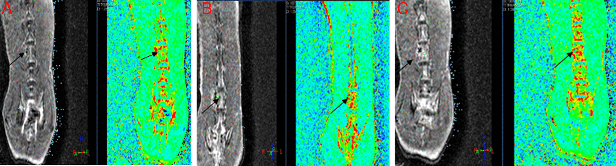
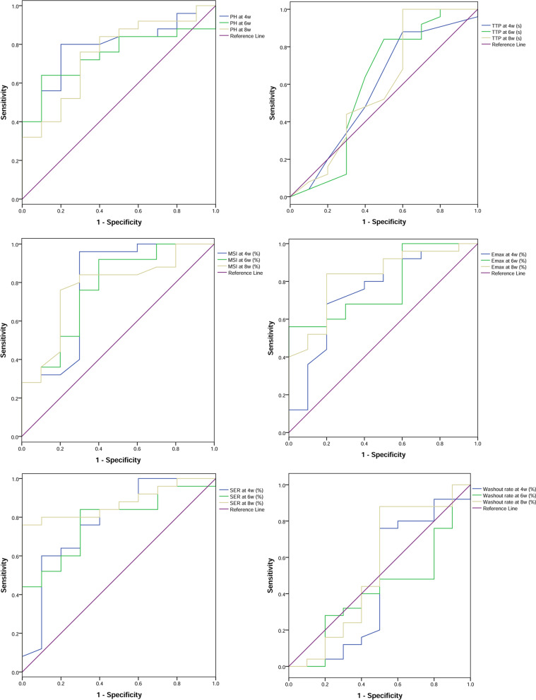
Methods: Spinal tuberculosis model was established using ATCC25177 Mycobacterium tuberculosis strain in the lumbar spine of rabbits. Forty rabbits were divided into 2 groups: rabbits in the experiment group were injected with 0.2 ml of 5.0 mg/ml tuberculosis suspension (n = 30) and those in the control group were injected with 0.2 ml of normal saline (n = 10) after vertebrae drilling surgery. Routine MRI and MR-PWI were performed at 4, 6, and 8 weeks after surgery. The statistical difference in terms of perfusion parameter values in the early MR-PWI scan of spinal tuberculosis between two groups was analyzed. The receiver operating characteristic (ROC) curve analysis was conducted for the accuracy of MR-PWI parameters in the early diagnosis of spinal tuberculosis.
Results: Except time to peak, the other perfusion parameters in the experiment group were all increased with time. In addition, the difference between the two groups, as well as the differences at each time point was statistically significant (all P < 0.05). First-pass enhancement rate (Efirst), early enhancement rate (Ee), peak height (PH), maximum slope of increase (MSI), maximum signal enhancement rate (Emax) and signal enhancement rate (SER) showed high values in early diagnosing spinal tuberculosis.
Conclusion: The parameters including Efirst, Ee, PH, MSI, Emax and SER may provide valuable imaging evidence for the early diagnosis of spinal tuberculosis in clinical application.
Keywords: Hemodynamics; Perfusion parameters; Perfusion weighted imaging; Spinal tuberculosis.
© 2022. The Author(s).
Conflict of interest statement
The authors declare that they have no conflict of interest.
Figures


Similar articles
-
Role of MR-DWI and MR-PWI in the radiotherapy of implanted pulmonary VX-2 carcinoma in rabbits.Chin J Cancer Res. 2014 Oct;26(5):532-42. doi: 10.3978/j.issn.1000-9604.2014.08.23. Chin J Cancer Res. 2014. PMID: 25400418 Free PMC article.
-
[Establishment of liver fibrosis in rabbit model and quantitative study on hepatic perfusion with dynamic whole-liver 3D MR imaging].Zhonghua Gan Zang Bing Za Zhi. 2009 May;17(5):350-3. Zhonghua Gan Zang Bing Za Zhi. 2009. PMID: 19497200 Chinese.
-
Establishment of a New Zealand rabbit model of spinal tuberculosis.J Spinal Disord Tech. 2015 Apr;28(3):E140-5. doi: 10.1097/BSD.0000000000000191. J Spinal Disord Tech. 2015. PMID: 25325713
-
Differentiation Between True Tumor Progression of Glioblastoma and Pseudoprogression Using Diffusion-Weighted Imaging and Perfusion-Weighted Imaging: Systematic Review and Meta-analysis.World Neurosurg. 2020 Dec;144:e100-e109. doi: 10.1016/j.wneu.2020.07.218. Epub 2020 Aug 7. World Neurosurg. 2020. PMID: 32777397
-
Advanced Neuroimaging in the Evaluation of Spinal Cord Tumors and Tumor Mimics: Diffusion Tensor and Perfusion-Weighted Imaging.Semin Ultrasound CT MR. 2017 Apr;38(2):163-175. doi: 10.1053/j.sult.2016.07.006. Epub 2016 Jul 12. Semin Ultrasound CT MR. 2017. PMID: 28347419 Review.
References
-
- Singh S, Dawar H, Das K, Mohapatra B, Prasad S. Functional and radiological outcomes of anterior decompression and posterior stabilization via posterior transpedicular approach in thoracic and thoracolumbar Pott's Disease: a retrospective study. Asian Spine J. 2017;11(4):618. doi: 10.4184/asj.2017.11.4.618. - DOI - PMC - PubMed
MeSH terms
LinkOut - more resources
Full Text Sources
