

Dynamics and structural changes of calmodulin upon interaction with the antagonist calmidazolium
- PMID: 35945584
- PMCID: PMC9361521
- DOI: 10.1186/s12915-022-01381-5
Dynamics and structural changes of calmodulin upon interaction with the antagonist calmidazolium
Abstract
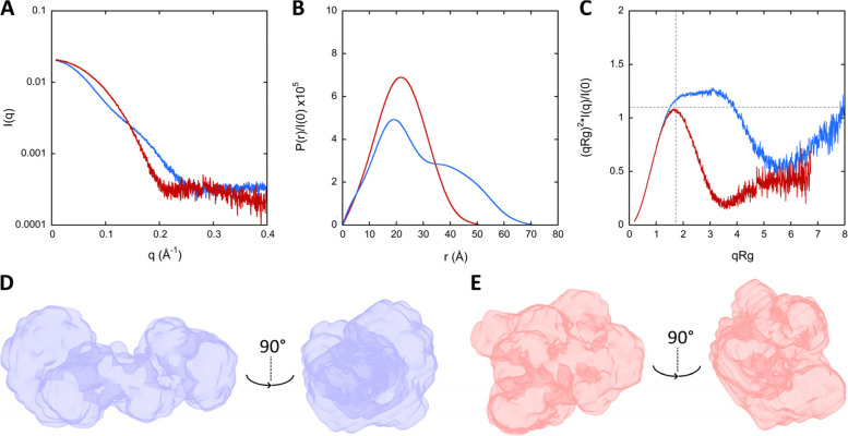
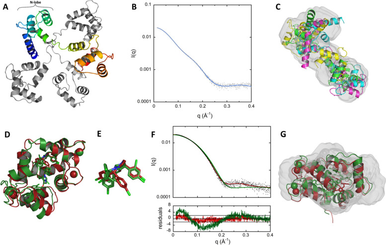
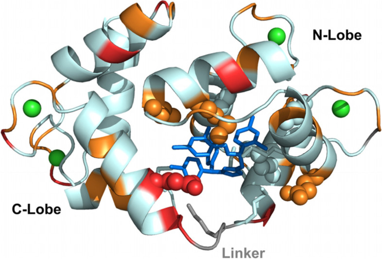
Background: Calmodulin (CaM) is an evolutionarily conserved eukaryotic multifunctional protein that functions as the major sensor of intracellular calcium signaling. Its calcium-modulated function regulates the activity of numerous effector proteins involved in a variety of physiological processes in diverse organs, from proliferation and apoptosis, to memory and immune responses. Due to the pleiotropic roles of CaM in normal and pathological cell functions, CaM antagonists are needed for fundamental studies as well as for potential therapeutic applications. Calmidazolium (CDZ) is a potent small molecule antagonist of CaM and one the most widely used inhibitors of CaM in cell biology. Yet, CDZ, as all other CaM antagonists described thus far, also affects additional cellular targets and its lack of selectivity hinders its application for dissecting calcium/CaM signaling. A better understanding of CaM:CDZ interaction is key to design analogs with improved selectivity. Here, we report a molecular characterization of CaM:CDZ complexes using an integrative structural biology approach combining SEC-SAXS, X-ray crystallography, HDX-MS, and NMR.
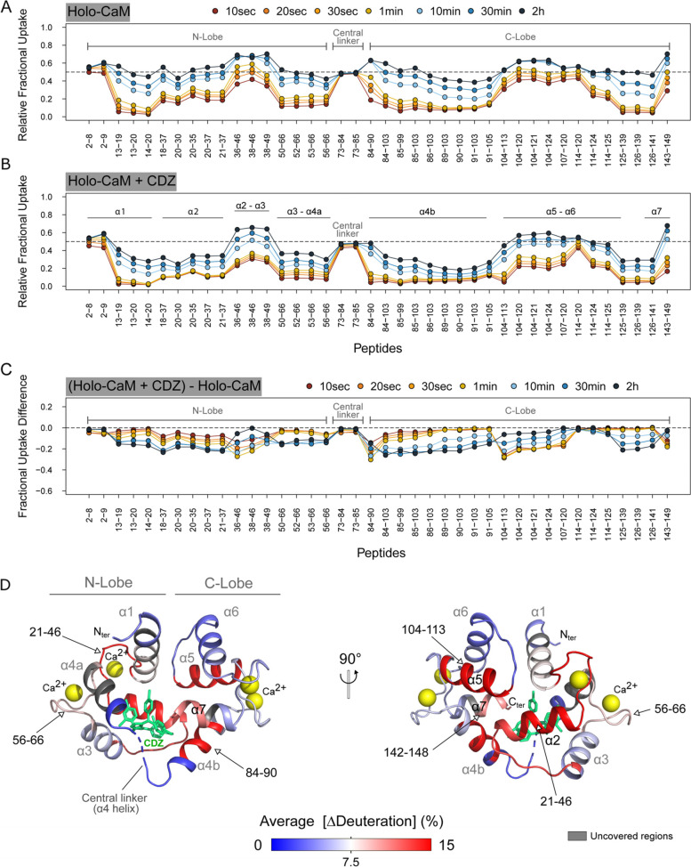
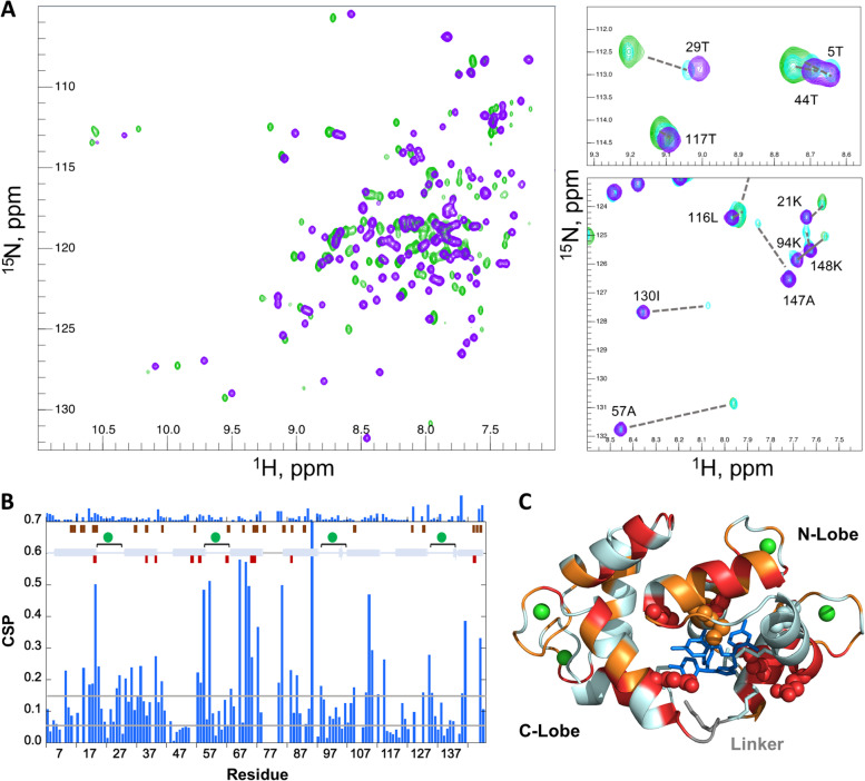
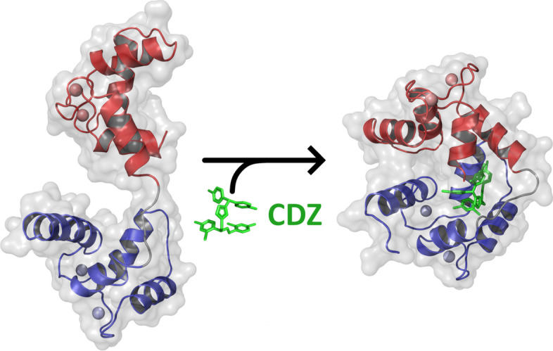
Results: We provide evidence that binding of a single molecule of CDZ induces an open-to-closed conformational reorientation of the two domains of CaM and results in a strong stabilization of its structural elements associated with a reduction of protein dynamics over a large time range. These CDZ-triggered CaM changes mimic those induced by CaM-binding peptides derived from physiological protein targets, despite their distinct chemical natures. CaM residues in close contact with CDZ and involved in the stabilization of the CaM:CDZ complex have been identified.
Conclusion: Our results provide molecular insights into CDZ-induced dynamics and structural changes of CaM leading to its inhibition and open the way to the rational design of more selective CaM antagonists. Calmidazolium is a potent and widely used inhibitor of calmodulin, a major mediator of calcium-signaling in eukaryotic cells. Structural characterization of calmidazolium-binding to calmodulin reveals that it triggers open-to-closed conformational changes similar to those induced by calmodulin-binding peptides derived from enzyme targets. These results provide molecular insights into CDZ-induced dynamics and structural changes of CaM leading to its inhibition and open the way to the rational design of more selective CaM antagonists.
Keywords: CDZ; CaM; Calmidazolium; Calmodulin; Calmodulin antagonist; Protein dynamics; Structure.
© 2022. The Author(s).
Conflict of interest statement
The authors declare that they have no competing interests.
Figures
Similar articles
-
Calmidazolium, a calmodulin antagonist, stimulates calcium-troponin C and calcium-calmodulin-dependent activation of striated muscle myofilaments.J Biol Chem. 1987 Dec 15;262(35):17240-6. J Biol Chem. 1987. PMID: 2960681
-
Calcium transport by sarcoplasmic reticulum of vascular smooth muscle: II. Effects of calmodulin and calmodulin inhibitors.J Cell Physiol. 1992 Oct;153(1):169-75. doi: 10.1002/jcp.1041530121. J Cell Physiol. 1992. PMID: 1522130
-
A SOX9 defect of calmodulin-dependent nuclear import in campomelic dysplasia/autosomal sex reversal.J Biol Chem. 2003 Sep 5;278(36):33839-47. doi: 10.1074/jbc.M302078200. Epub 2003 Jun 16. J Biol Chem. 2003. PMID: 12810722
-
Structure, dynamics and interaction with kinase targets: computer simulations of calmodulin.Biochim Biophys Acta. 2004 Mar 11;1697(1-2):289-300. doi: 10.1016/j.bbapap.2003.11.032. Biochim Biophys Acta. 2004. PMID: 15023369 Review.
-
Structural Diversity in Calmodulin - Peptide Interactions.Curr Protein Pept Sci. 2019;20(11):1102-1111. doi: 10.2174/1389203720666190925101937. Curr Protein Pept Sci. 2019. PMID: 31553290 Review.
Cited by
-
Identification and validation of autophagy-related genes in primary open-angle glaucoma.BMC Med Genomics. 2023 Nov 15;16(1):287. doi: 10.1186/s12920-023-01722-5. BMC Med Genomics. 2023. PMID: 37968618 Free PMC article.
-
Phytochemical Interactions with Calmodulin and Critical Calmodulin Binding Proteins Involved in Amyloidogenesis in Alzheimer's Disease.Biomolecules. 2023 Apr 15;13(4):678. doi: 10.3390/biom13040678. Biomolecules. 2023. PMID: 37189425 Free PMC article. Review.
-
Marine Polysaccharides Carrageenans Enhance Eryptosis and Alter Lipid Order of Cell Membranes in Erythrocytes.Cell Biochem Biophys. 2024 Jun;82(2):747-766. doi: 10.1007/s12013-024-01225-9. Epub 2024 Feb 9. Cell Biochem Biophys. 2024. PMID: 38334853
-
Small-angle X-ray scattering studies of enzymes.Curr Opin Chem Biol. 2023 Feb;72:102232. doi: 10.1016/j.cbpa.2022.102232. Epub 2022 Nov 30. Curr Opin Chem Biol. 2023. PMID: 36462455 Free PMC article. Review.
-
New Insights into the Regulation of mTOR Signaling via Ca2+-Binding Proteins.Int J Mol Sci. 2023 Feb 15;24(4):3923. doi: 10.3390/ijms24043923. Int J Mol Sci. 2023. PMID: 36835331 Free PMC article. Review.
References
Publication types
MeSH terms
Substances
LinkOut - more resources
Full Text Sources
