. 2022 Dec;42(12):2216-2229.
doi: 10.1177/0271678X221119288. Epub 2022 Aug 9.

Persistent neuroinflammation and behavioural deficits after single mild traumatic brain injury

Affiliations

Persistent neuroinflammation and behavioural deficits after single mild traumatic brain injury

Antoine Drieu et al. J Cereb Blood Flow Metab. 2022 Dec.

Abstract

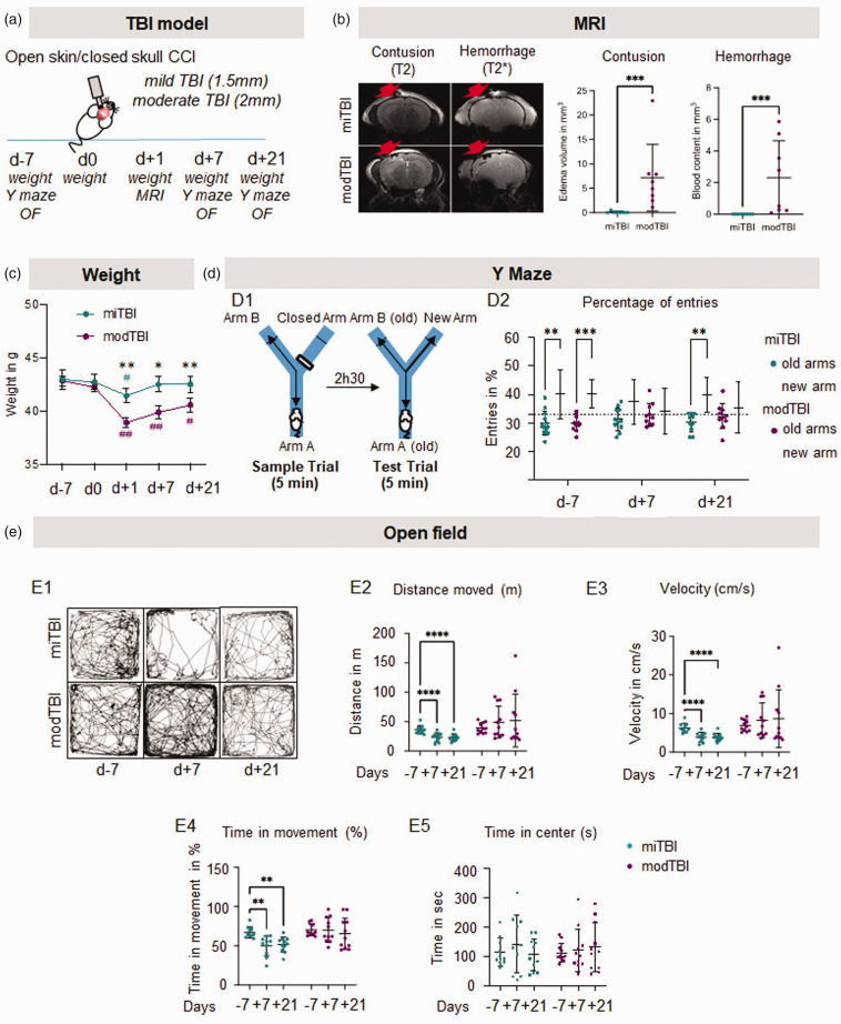
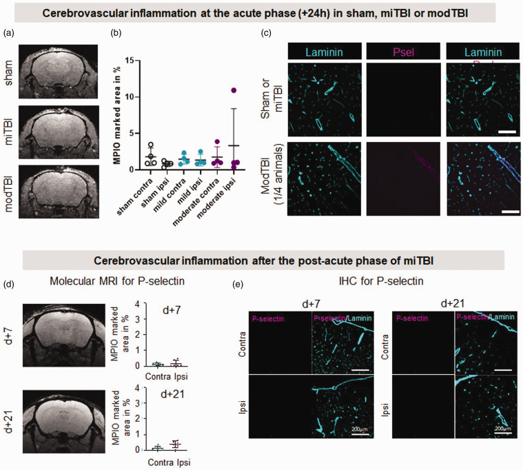
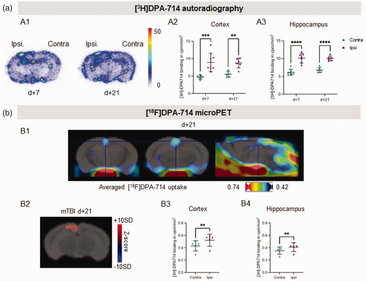
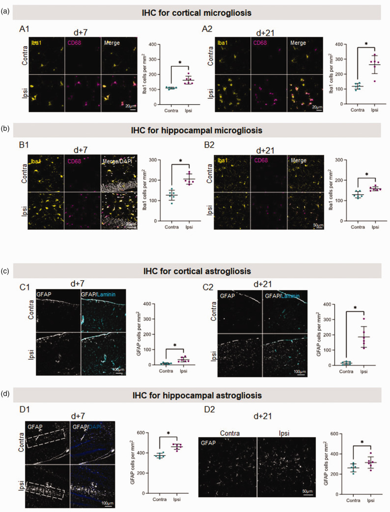
Despite an apparently silent imaging, some patients with mild traumatic brain injury (TBI) experience cognitive dysfunctions, which may persist chronically. Brain changes responsible for these dysfunctions are unclear and commonly overlooked. It is thus crucial to increase our understanding of the mechanisms linking the initial event to the functional deficits, and to provide objective evidence of brain tissue alterations underpinning these deficits. We first set up a murine model of closed-head controlled cortical impact, which provoked persistent cognitive and sensorimotor deficits, despite no evidence of brain contusion or bleeding on MRI, thus recapitulating features of mild TBI. Molecular MRI for P-selectin, a key adhesion molecule, detected no sign of cerebrovascular inflammation after mild TBI, as confirmed by immunostainings. By contrast, in vivo PET imaging with the TSPO ligand [18F]DPA-714 demonstrated persisting signs of neuroinflammation in the ipsilateral cortex and hippocampus after mild TBI. Interestingly, immunohistochemical analyses confirmed these spatio-temporal profiles, showing a robust parenchymal astrogliosis and microgliosis, at least up to 3 weeks post-injury in both the cortex and hippocampus. In conclusion, we show that even one single mild TBI induces long-term behavioural deficits, associated with a persistent neuro-inflammatory status that can be detected by PET imaging.

Keywords: Mild traumatic brain injury; TSPO microPET imaging; molecular MRI; neuroinflammation; persisting behavioural deficits.


Conflict of interest statement

The author(s) declared no potential conflicts of interest with respect to the research, authorship, and/or publication of this article.

Figures

Figure 1.
Figure 1.
Mild TBI is undetectable by conventional MRI but induces long-term behavioural deficits. (a) Experimental design showing that two depths of impact (1.5 mm: mild TBI; 2 mm: moderate TBI) have been compared in the CCI model. (b) Representative T2-weighted (contusion) and T2*-weighted (hemorrhage) MRI acquisitions and corresponding quantifications 24 hours after mild or moderate TBI. n = 7/8 mice/group. *p < 0.05 vs mild TBI; **p < 0.01 vs mild TBI; ***p < 0.001 vs mild TBI, Mann-Whitney test; #p < 0.05 vs d0; ##p < 0.01 vs d0, Wilcoxon paired-test. Red arrows indicate the site of impact. (c) Measures of mouse weights 7 days before TBI, and day 0, 1, 7 and 21 days after TBI. (d) Schematic representation of Y maze behavioural test and quantifications of the percentage of entries in the new arm 7 days before and 7 and 21 days after mild or moderate TBI and (e) Representative sample traces and corresponding quantifications of the total distance moved, velocity, time of movement and time spend in the center on the open field test 7 days before, 7 and 21 days after mild or moderate TBI. n = 10 mice/group, #p < 0.05 vs new arm, Wilcoxon paired-test; ##p < 0.01 vs d-7 Wilcoxon paired-test.
Figure 2.
Figure 2.
Unlike severe TBI, mild TBI does not induce cerebrovascular inflammation. (a, b) Representative T2*-GEFC αP-selectin MPIO acquisitions 24 h after sham surgery, miTBI or modTBI and their respective MPIO-marked area quantifications (n = 4 per condition). (c) Representative photomicrographs of P-selectin immunostaining 24 h after sham surgery, miTBI or modTBI (blood vessels were stained with laminin) (representative of 4 mice/group) (d) Representative T2*-GEFC αP-selectin MPIO acquisitions 7 and 21 days after miTBI and their respective MPIO-marked area quantifications and (e) Representative photomicrographs of P-selectin immunostaining 7 and 21 days after miTBI (blood vessels were stained with laminin) (n = 6 mice/group).
Figure 3.
Figure 3.
[3H]DPA-714 imaging reveals long-lasting neuroinflammation induced by miTBI. (a) Ex vivo autoradiographic experiment. (a1) Representative coronal brain sections of [3H]DPA-714 autoradiography 7 days and 21 days after miTBI. (a2) Quantifications of [3H]DPA-714 binding in cortex and hippocampus. n = 7 mice/group, ***p < 0.001 vs contralateral ROI; 2-way ANOVA and (b) In vivo microPET experiment. (b1) Representative fusion images between a MRI template and images of averaged uptake of [18F]DPA-714 in 2 coronal images (left panels), and one sagittal image (right panel). (b2) Representative coronal brain image of Z-score maps fused with a MRI template showing [18F]DPA-714 uptake. n = 5 mice, p < 0.01 vs contralateral ROI. (b3,4) Quantifications of [18F]DPA-714 uptake in (b3) cortex and (b4) hippocampus 21 days after miTBI. n = 5 mice, p = 0.0015 vs contralateral cortex; p = 0.0022 vs contralateral hippocampus.
Figure 4.
Figure 4.
Immunohistochemistry analyses reveal long-lasting neuroinflammation induced by miTBI. (a and b) Long-lasting microgliosis induced by miTBI using Iba1 and CD68 immunostainings. (a) Cortical microgliosis. Representative photomicrographs of microglia and respective quantifications (a1) 7 days and (a2) 21 days after miTBI. (b) Hippocampal microgliosis. Representative photomicrographs of microglia and respective quantifications (b1) 7 days and (b2) 21 days after miTBI (c,d) Mild TBI provokes a long lasting astrogliosis. (a) GFAP positive astrocytes were found in the cortex 7 days after miTBI. (b) Astrocytes were found abundantly in the cortex 21 days after miTBI. (c) GFAP positive astrocytes were found abundantly in the hippocampus 7 days after miTBI. (d) Astrocytes were found in the hippocampus 21 days after miTBI. n = 6 mice/group, *p < 0.05, **p < 0.01 vs contralateral ROI, Wilcoxon paired-test.

Similar articles

Cited by

References

    1. Maas AIR, Menon DK, Adelson PD, et al.. Traumatic brain injury: integrated approaches to improve prevention, clinical care, and research. Lancet Neurol 2017; 16: 987–1048. - PubMed
    1. Dewan MC, Rattani A, Gupta S, et al.. Estimating the global incidence of traumatic brain injury. J Neurosurg 2019; 130: 1080–1097. - PubMed
    1. Teasdale G, Jennett B. Assessment of coma and impaired consciousness: a practical scale. The Lancet 1974; 304: 81–84. - PubMed
    1. Cruz-Haces M, Tang J, Acosta G, et al.. Pathological correlations between traumatic brain injury and chronic neurodegenerative diseases. Transl Neurodegener 2017; 6: 20. - PMC - PubMed
    1. Gardner RC, Yaffe K. Epidemiology of mild traumatic brain injury and neurodegenerative disease. Mol Cell Neurosci 2015; 66: 75–80. - PMC - PubMed

Publication types

Substances