Giant Prolactinoma Embedded by Pseudoaneurysm of the Cavernous Carotid Artery Treated with a Tailored Therapeutic Scheme
- PMID: 35945997
- PMCID: PMC9357472
- DOI: 10.1055/s-0042-1749662
Giant Prolactinoma Embedded by Pseudoaneurysm of the Cavernous Carotid Artery Treated with a Tailored Therapeutic Scheme
Abstract
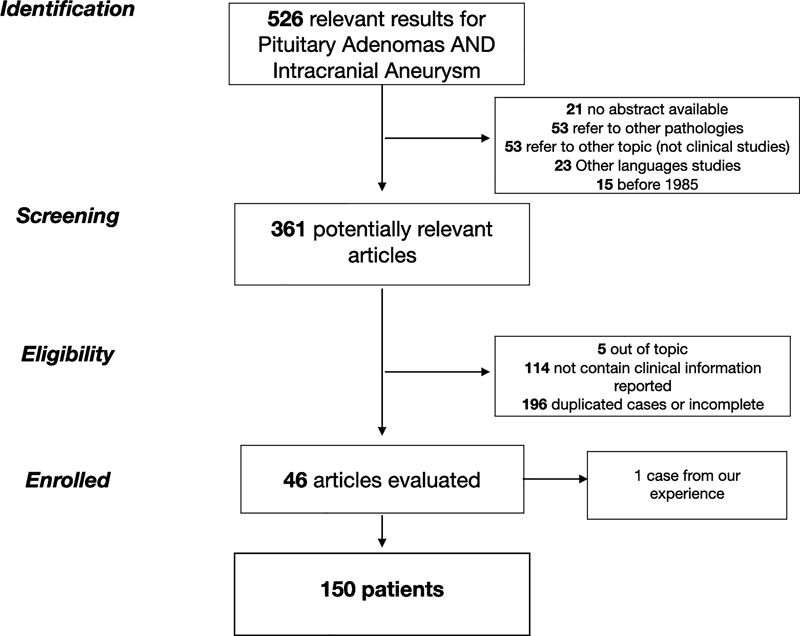
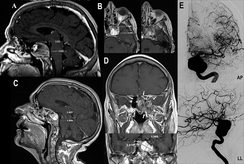
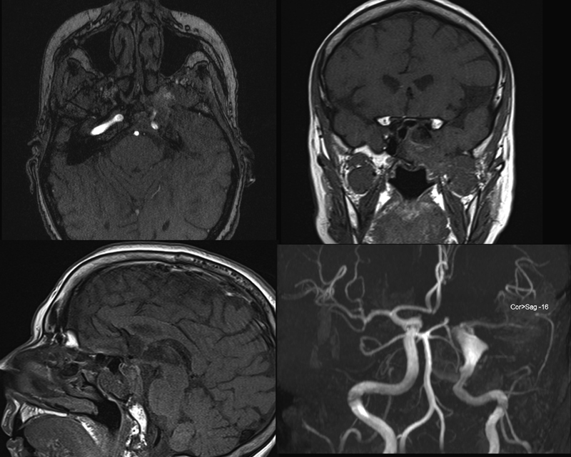
The coexistence of intracranial aneurysm (IA) is generally thought to be highest in patients with pituitary adenomas (PAs). Different mechanisms may play a role in aneurysm formation, but whether the PA contributes to aneurysm formation is still unclear. In the literature, there are numerous reported cases of this association; however, the analyses of the characteristics of PAs, aneurysms, and treatment management are rare and limited to a restricted number of case reports. We report a rare case of an embedded aneurysm in a macroprolactinoma treated with therapeutic management tailored to the clinical, neurological, and radiological characteristics of the patient. To select the best treatment, we reviewed the literature and reported the only cases in which the radiological characteristics of aneurysms, PAs, therapeutic management, and patient outcome are described. We aimed to understand what are the variables that determine the best therapeutic management with the best possible outcome. The presence of a large pseudoaneurysm of the internal carotid artery completely embedded in a giant macroprolactinoma is rare and needs a tailored treatment strategy. The importance of the preoperative knowledge of asymptomatic IA coexisting with PA can avoid accidental rupture of the aneurysm during surgical resection and may lead to planning the best treatment. A high degree of suspicion for an associated aneurysm is needed, and if magnetic resonance imaging shows some atypical features, digital subtraction angiography must be performed prior to contemplating any intervention to avoid iatrogenic aneurysmal rupture. Our multimodal approach with the first-line therapy of low-dose cabergoline to obtain prolactin normalization with minimum risks of aneurysms rupture and subsequent endovascular treatment with flow diverter has not been described elsewhere to our knowledge. In the cases, we suggest adopting a tailored low-dose cabergoline therapy scheme to avoid rupture during cytoreduction and initiate a close neuroradiological follow-up program.
Keywords: cavernous sinus; cerebral aneurysms; flow diverter; internal carotid artery; pituitary adenoma; prolactinoma.
Association for Helping Neurosurgical Sick People. This is an open access article published by Thieme under the terms of the Creative Commons Attribution-NonDerivative-NonCommercial License, permitting copying and reproduction so long as the original work is given appropriate credit. Contents may not be used for commercial purposes, or adapted, remixed, transformed or built upon. ( https://creativecommons.org/licenses/by-nc-nd/4.0/ ).
Conflict of interest statement
Conflict of Interest None declared.
Figures
References
-
- Hu J, Lin Z, Zhang Y. Prevalence of unruptured intracranial aneurysms coexisting with pituitary adenomas. World Neurosurg. 2019;126:e526–e533. - PubMed
-
- Housepian E M, Pool J L. A systematic analysis of intracranial aneurysms from the autopsy file of the Presbyterian Hospital, 1914 to 1956. J Neuropathol Exp Neurol. 1958;17(03):409–423. - PubMed
-
- Wakai S, Fukushima T, Furihata T, Sano K. Association of cerebral aneurysm with pituitary adenoma. Surg Neurol. 1979;12(06):503–507. - PubMed