. 2022 Sep;26(17):4727-4739.
doi: 10.1111/jcmm.17502. Epub 2022 Aug 9.

p53 as a unique target of action of cisplatin in acute leukaemia cells

Affiliations

p53 as a unique target of action of cisplatin in acute leukaemia cells

Sanjay Kumar et al. J Cell Mol Med. 2022 Sep.

Abstract

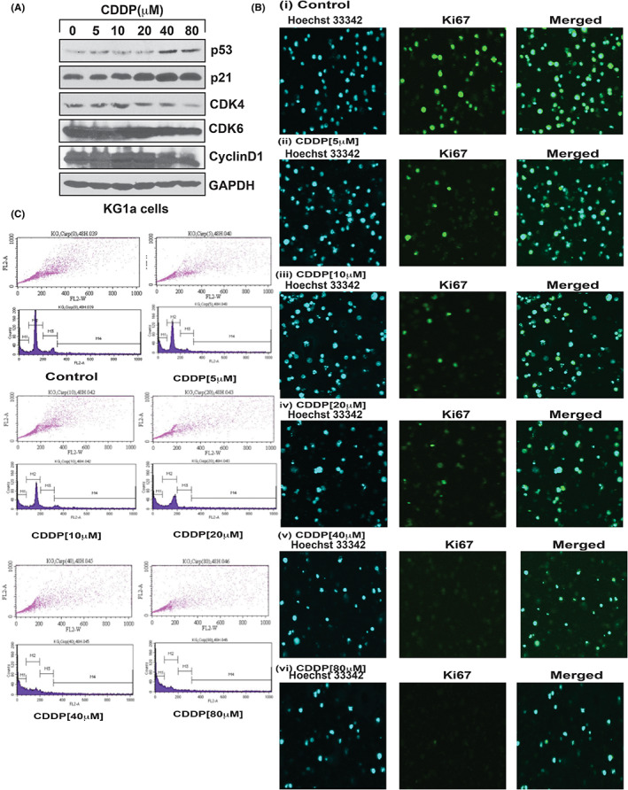
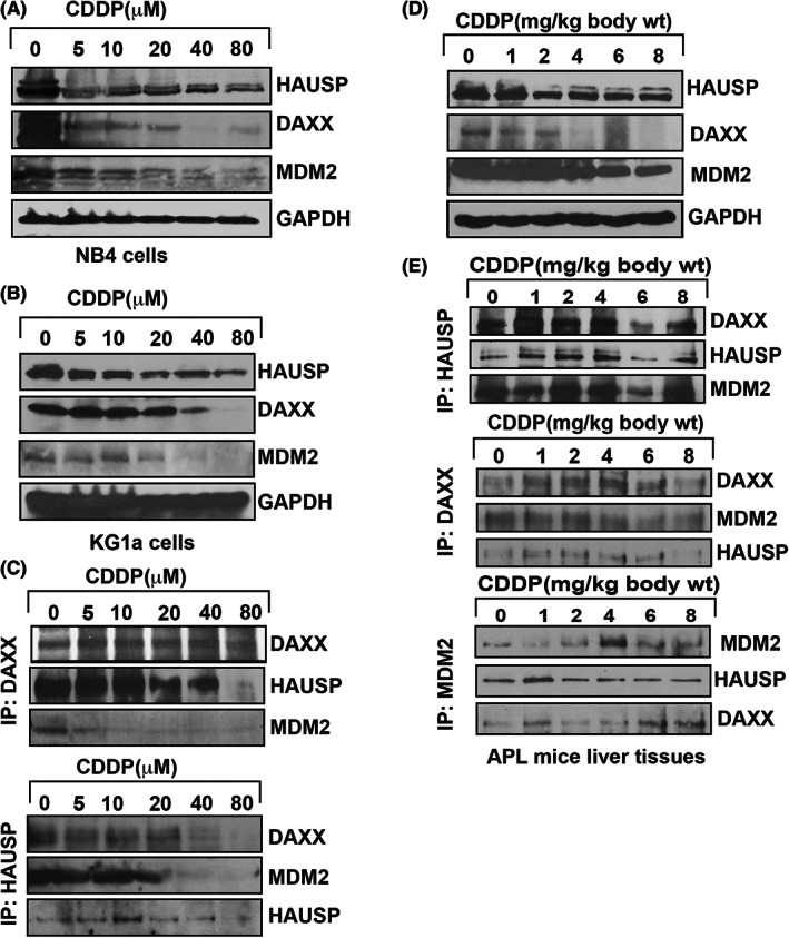
Acute promyelocytic leukaemia (APL) occurs in approximately 10% of acute myeloid leukaemia patients. Arsenic trioxide (ATO) has been for APL chemotherapy, but recently several ATO-resistant cases have been reported worldwide. Cisplatin (CDDP) enhances the toxicity of ATO in ovarian, lung cancer, chronic myelogenous leukaemia, and HL-60 cells. Hence, the goal of this study was to investigate a novel target of CDDP action in APL cells, as an alternate option for the treatment of ATO-resistant APL patients. We applied biochemical, molecular, confocal microscopy and advanced gene editing (CRISPR-Cas9) techniques to elucidate the novel target of CDDP action and its functional mechanism in APL cells. Our main findings revealed that CDDP activated p53 in APL cells through stress signals catalysed by ATM and ATR protein kinases, CHK1 and CHK2 phosphorylation at Ser 345 and Thr68 residues, and downregulation and dissociation of MDM2-DAXX-HAUSP complex. Our functional studies confirmed that CDDP-induced repression of MDM2-DAXX-HAUSP complex was significantly reversed in both nutilin-3-treated KG1a and p53-knockdown NB4 cells. Our findings also showed that CDDP stimulated an increased number of promyelocytes with dense granules, activated p53 expression, and downregulated MDM2 in liver and bone marrow of APL mice. Principal conclusion of our study highlights a novel mode of action of CDDP targeting p53 expression which may provide a basis for designing new anti-leukaemic compounds for treatment of APL patients.

Keywords: CRISPR-Cas9; MDM2-DAXX-HAUSP; acute promyelocytic leukaemia; cisplatin; p53.


Conflict of interest statement

The authors declare that they have no competing interests.

Figures

FIGURE 1
FIGURE 1
CDDP inhibits APL cell proliferation. APL (HL‐60, KG‐1a and NB4) cells were treated with different concentrations (0, 5, 10, 20, 40, and 80 μM) of CDDP for 48 h and further incubated for 4 h in WST/ECS solution. After incubation, absorbance in each well was measured using a microplate reader and the percentages of cell viability were presented with respect to CDDP concentrations. Data represent the means of three independent experiments ± SDs (*p < 0.01) (A), (**p < 0.01) (B) and (#p < 0.01) (C). Highly statistically significant inhibition (p < 0.01) in cell proliferation was observed in NB4 (A), HL‐60 (B), and KG‐1a (C).
FIGURE 2
FIGURE 2
CDDP stimulates p53, inhibits ki67, and causes cell cycle arrest. KG1a cells were treated with different concentrations of CDDP, and the expression levels of p53, p21, cyclins, and cdks proteins were analysed by western blotting. CDDP stimulated p53 and p21 by reduced expression of cyclins (cyclin D1) and cdks (cdk4 and cdk6) in KG1a cells (A). It also reduced the expression of ki67 [B(i–vi)] and induced cell cycle arrest in G1 phase (C) in NB4 cells.
FIGURE 3
FIGURE 3
CDDP causes cell apoptosis. NB4 cells were treated with different concentrations of CDDP for 48 h, and both early and late apoptosis were analysed through expression level of both annexin V and PI dye by confocal imaging. CDDP induced both early and late apoptosis in NB4 cells (i–vi).
FIGURE 4
FIGURE 4
CDDP disrupts MDM2‐DAXX‐HAUSP complex. Both KG1a and NB4 cells were treated with different concentration of CDDP, and complex molecules expression and association were analysed by western blotting and immunoprecipitation (IP). CDDP reduced the expression of complex molecules in both cells (A,B), and molecules were associated each other (C) in KG1a cells. Also, APL mice were treated with different doses of CDDP and complex molecules expression and association were analysed by western blotting and IP. CDDP downregulated the expression of complex molecules (D), and the molecules were associated together (E).
FIGURE 5
FIGURE 5
Functional studies of CDDP role in complex disruption. KG1a cells were treated with different concentrations of CDDP, and both expression and phosphorylation of protein kinases (ATM and ATR) and their downstream targets, CHK1 and CHK2, were analysed by western blotting. CDDP reduced the expression of ATM and ATR by stimulating phosphorylation of CHK1 at S345 residue and CHK2 at Thr68 (A). CDDP treatment did not significantly reduce the expression of complex molecules in p53‐knockout NB4 cells (B). Nutilin almost neutralized CDDP‐induced MDM2 reduced expression in KG1a cells (C). P53 and MDM2 proteins turnover was detected in both CDDP‐treated and untreated cells by the pulse‐chase assay in NB4 cells (D).
FIGURE 6
FIGURE 6
CDDP activates p53 by degradation of MDM2 in mice bone marrow cells. APL mice bone marrow cells were isolated from both control and CDDP‐treated mice, and immunocytochemistry, fluorescence, and confocal imaging were performed to analyse the formation of promyelocytes and the expression levels of MDM2 and p53 proteins. CDDP stimulated the formation of more promyelocytes with dense granules (A) and activated p53 (B), leading to the degradation of MDM2 (C) in bone marrow cells.
FIGURE 7
FIGURE 7
CDDP stimulated p53 by degradation of MDM2 in liver tissue of APL mice. Both control and CDDP‐treated APL mice livers were collected, and both protein lysate and cryosections (5 μM) were made simultaneously. Expression of both p53 and MDM2 in protein lysate was measured by western blotting (A,B) and by immunohistochemistry and fluorescence microscopy of cryosections (C,D).

References

    1. Powell BL. Arsenic trioxide in acute promyelocytic leukemia: potion not poison. Expert Rev Anticancer Ther. 2011;11:1317‐1319. - PubMed
    1. Kumar S, Tchounwou PB. Molecular mechanisms of cisplatin cytotoxicity in acute promyelocytic leukemia cells. Oncotarget. 2015;6:40734‐40746. - PMC - PubMed
    1. Grignani F, Fagioli M, Alcalay M. Acute promyelocytic leukemia: from genetics to treatment. Blood. 1994;83:10‐25. - PubMed
    1. Kumar S, Brown A, Tchounwou PB. Trisenox disrupts MDM2‐ DAXX‐HAUSP complex and activates p53, cell cycle regulation and apoptosis in acute leukemia cells. Oncotarget. 2018;9:33138‐33148. - PMC - PubMed
    1. Lo‐Coco F, Avvisati G, Vignetti M, et al. Retinoic acid and arsenic trioxide for acute promyelocytic leukemia. N Engl J Med. 2013;369:111‐121. - PubMed

Publication types

MeSH terms