

Hexagonal Voronoi pattern detected in the microstructural design of the echinoid skeleton
- PMID: 35946165
- PMCID: PMC9363984
- DOI: 10.1098/rsif.2022.0226
Hexagonal Voronoi pattern detected in the microstructural design of the echinoid skeleton
Abstract
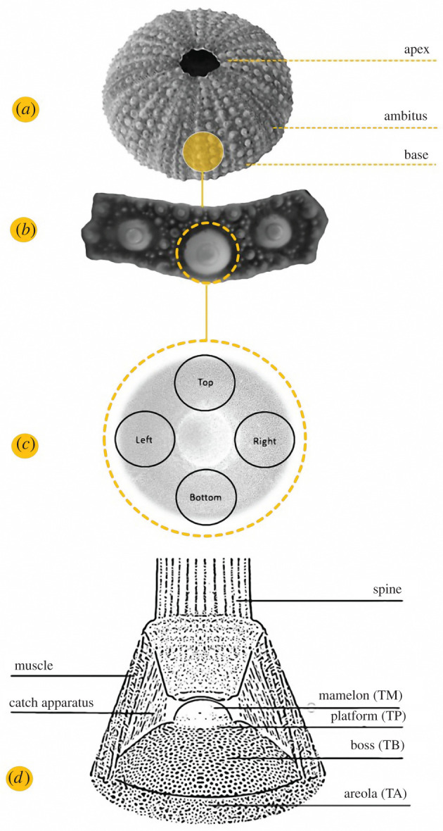
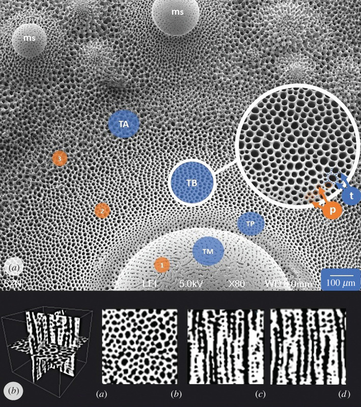
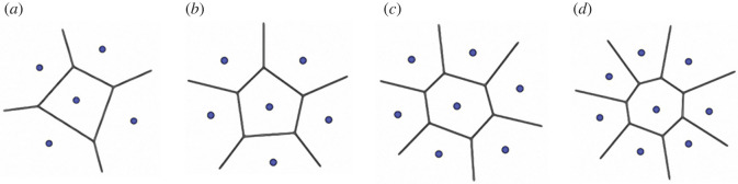
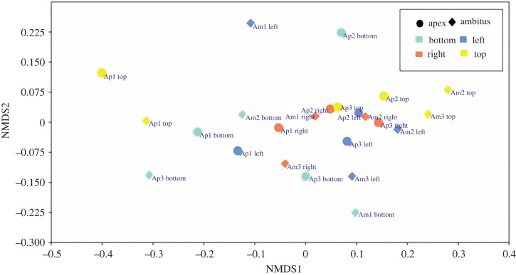
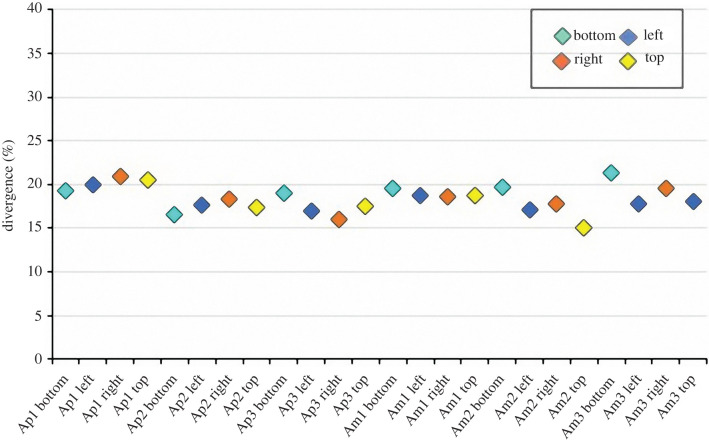
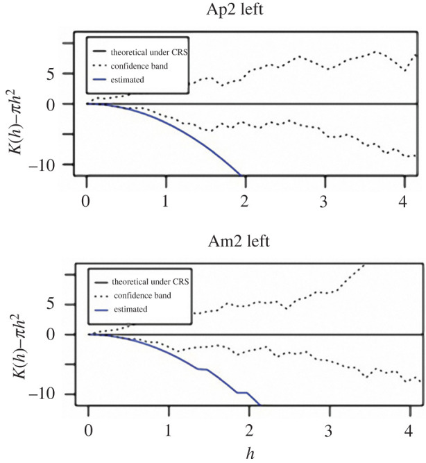
Repeated polygonal patterns are pervasive in natural forms and structures. These patterns provide inherent structural stability while optimizing strength-per-weight and minimizing construction costs. In echinoids (sea urchins), a visible regularity can be found in the endoskeleton, consisting of a lightweight and resistant micro-trabecular meshwork (stereom). This foam-like structure follows an intrinsic geometrical pattern that has never been investigated. This study aims to analyse and describe it by focusing on the boss of tubercles-spine attachment sites subject to strong mechanical stresses-in the common sea urchin Paracentrotus lividus. The boss microstructure was identified as a Voronoi construction characterized by 82% concordance to the computed Voronoi models, a prevalence of hexagonal polygons, and a regularly organized seed distribution. This pattern is interpreted as an evolutionary solution for the construction of the echinoid skeleton using a lightweight microstructural design that optimizes the trabecular arrangement, maximizes the structural strength and minimizes the metabolic costs of secreting calcitic stereom. Hence, this identification is particularly valuable to improve the understanding of the mechanical function of the stereom as well as to effectively model and reconstruct similar structures in view of future applications in biomimetic technologies and designs.
Keywords: Voronoi; echinoids; geometric pattern; stereom; trabecular system.
Figures


Similar articles
-
The microarchitectural variability in the echinoid skeleton: a 3D geometrical and stiffness characterization of Paracentrotus lividus.R Soc Open Sci. 2025 Jun 25;12(6):241439. doi: 10.1098/rsos.241439. eCollection 2025 Jun. R Soc Open Sci. 2025. PMID: 40568548 Free PMC article.
-
Echinoid skeleton: an insight on the species-specific pattern of the Paracentrotus lividus plate and its microstructural variability.J R Soc Interface. 2023 Feb;20(199):20220673. doi: 10.1098/rsif.2022.0673. Epub 2023 Feb 1. J R Soc Interface. 2023. PMID: 36722170 Free PMC article.
-
Constructional design of echinoid endoskeleton: main structural components and their potential for biomimetic applications.Bioinspir Biomim. 2020 Nov 30;16(1). doi: 10.1088/1748-3190/abb86b. Bioinspir Biomim. 2020. PMID: 32927446 Review.
-
Geochemical signatures and nanomechanical properties of echinoid tests from nearshore habitats of Florida: environmental and physiological controls on echinoid biomineralization.PeerJ. 2025 Jan 24;13:e18688. doi: 10.7717/peerj.18688. eCollection 2025. PeerJ. 2025. PMID: 39872031 Free PMC article.
-
²⁶Mg labeling of the sea urchin regenerating spine: Insights into echinoderm biomineralization process.J Struct Biol. 2011 Oct;176(1):119-26. doi: 10.1016/j.jsb.2011.07.008. Epub 2011 Jul 22. J Struct Biol. 2011. PMID: 21803159
Cited by
-
A Devonian crinoid with a diamond microlattice.Proc Biol Sci. 2023 Mar 29;290(1995):20230092. doi: 10.1098/rspb.2023.0092. Epub 2023 Mar 29. Proc Biol Sci. 2023. PMID: 36987636 Free PMC article.
-
Composite material in the sea urchin Cidaris rugosa: ordered and disordered micrometre-scale bicontinuous geometries.J R Soc Interface. 2024 Mar;21(212):20230597. doi: 10.1098/rsif.2023.0597. Epub 2024 Mar 13. J R Soc Interface. 2024. PMID: 38471532 Free PMC article.
-
The microarchitectural variability in the echinoid skeleton: a 3D geometrical and stiffness characterization of Paracentrotus lividus.R Soc Open Sci. 2025 Jun 25;12(6):241439. doi: 10.1098/rsos.241439. eCollection 2025 Jun. R Soc Open Sci. 2025. PMID: 40568548 Free PMC article.
-
Microstructural design of the stalk in the crinoid Seirocrinus supports its pseudoplanktonic lifestyle.Sci Rep. 2025 Aug 26;15(1):31418. doi: 10.1038/s41598-025-16412-8. Sci Rep. 2025. PMID: 40858656 Free PMC article.
-
Echinoid skeleton: an insight on the species-specific pattern of the Paracentrotus lividus plate and its microstructural variability.J R Soc Interface. 2023 Feb;20(199):20220673. doi: 10.1098/rsif.2022.0673. Epub 2023 Feb 1. J R Soc Interface. 2023. PMID: 36722170 Free PMC article.
References
-
- Thompson DAW. 1961. On growth and form, 6th edn. Cambridge, UK: University Press.
-
- Weaire D, Rivier N. 1984. Soap, cells and statistics—random patterns in two dimensions. Contemp. Phys. 25, 59-99. (10.1080/00107518408210979) - DOI
-
- Chiu SN, Stoyan D, Kendall WS, Mecke J. 2013. Stochastic geometry and its applications. New York, NY: John Wiley & Sons.
Publication types
MeSH terms
LinkOut - more resources
Full Text Sources
Miscellaneous
