. 2022 Sep;33(9):777-784.
doi: 10.5152/tjg.2022.21911.

Mitochondrial Homeostasis and Mast Cells in Experimental Hepatic Ischemia-Reperfusion Injury of Rats

Affiliations

Mitochondrial Homeostasis and Mast Cells in Experimental Hepatic Ischemia-Reperfusion Injury of Rats

Suleyman Koc et al. Turk J Gastroenterol. 2022 Sep.

Abstract

Background: Ischemia-reperfusion injury is a histopathological event and is an important cause of morbidity and mortality after hepatobiliary surgery. We aimed to investigate the protective effect of uridine on hepatic ischemia-reperfusion injury in rats.

Methods: The animals were divided into 4 groups (n = 8): group I (control), group II: ischemia-reperfusion (30 minutes ischemia and 120 minutes reperfusion), group III: ischemia-reperfusion+uridine (at the beginning of reperfusion), and group IV: ischemia-reperfusion+uridine (5 minutes before ischemia-reperfusion). Uridine was administered a single dose of 30 mg/kg IV. The 3 elements of the hepatoduodenal ligament (hepatic artery, portal vein, and biliary tract) were obliterated for 30 minutes. Then hepatic reperfusion was achieved for 120 minutes.

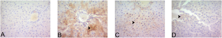
Results: In the ischemia-reperfusion group, both liver tissues and serum chymase activity and high-temperature requirement A2 levels were higher. Severe central vein dilatation and congestion, widening sinusoidal range, diffuse necrotic hepatocytes and dense erythrocyte accumulation in sinusoids, and strongly inducible nitric oxide synthase expression were seen in the ischemia-reperfusion group. A clear improvement was seen in both uridine co-administration and pretreatment groups.

Conclusion: Our results revealed that uridine limits the development of liver damage under conditions of ischemia-reperfusion, thus contributing to an increase in hepatocyte viability.


Figures

Figure 1.
Figure 1.
Representative micrographs showing the liver samples of all experimental groups. Control group (A) shows normal liver architecture. Ischemia-reperfusion (I/R) group (B) shows severe necrotic hepatocytes (*) and congestion (arrowhead) compared to the control group. Section from a rat of group III: I/R + uridine (before reperfusion) (C) shows moderately necrotic hepatocytes (*) and congestion (arrowhead) compared to group II. Group IV: I/R + uridine (5 minutes before I/R) (D) showing mild necrotic hepatocytes (*) and congestion (arrowhead) compared to group II. ×20- hematoxylin–eosin staining
Figure 2.
Figure 2.
Representative micrographs from group I (A), group II (B), group III (C), and group IV (D) shows 8-hydroxy-2’-deoxyguanosine immune negativity. ×20 immunohistochemical staining.
Figure 3.
Figure 3.
Representative micrographs from group I (A), group II (B), group III (C), and group IV (B) shows 8-hydroxy-2’-deoxyguanosine immune negativity. ×20 immunofluorescent staining.
Figure 4.
Figure 4.
Representative micrographs showing iNOS expression for all experimental groups. Control group I (A) showing İNOS immune negativity. Ischemia-reperfusion group II (B) shows severe iNOS expression (arrowhead) compared to the control group. Group III (C) shows moderately iNOS expression (arrowhead) compared to group II. Group IV (D) shows mild iNOS expression (arrowhead) compared to group II. ×20 immunohistochemical staining.
Figure 5.
Figure 5.
Representative micrographs showing iNOS expression for all experimental groups. Control group I (A) showing İNOS immune negativity. Ischemia-reperfusion group II (B) shows severe iNOS expression (arrowhead) compared to the control group. Group III (C) shows moderately iNOS expression (arrowhead) compared to group II. Group IV (D) shows mild iNOS expression (arrowhead) compared to group II. ×20 immunofluorescent staining.

References

    1. Aldi S, Marino A, Tomita K.et al. E-NTPDase1/CD39 modulates renin release from heart mast cells during ischemia/reperfusion: a novel cardioprotective role. FASEB J. 2015;29(1):61 69. 10.1096/fj.14-261867) - DOI - PMC - PubMed
    1. Atiakshin D, Buchwalow I, Tiemann M. Mast cell chymase: morphofunctional characteristics. Histochem Cell Biol. 2019;152(4):253 269. 10.1007/s00418-019-01803-6) - DOI - PubMed
    1. Barrier A, Olaya N, Chiappini F.et al. Ischemic preconditioning modulates the expression of several genes, leading to the overproduction of IL-1RA, iNOS, and Bcl-2 in a human model of liver ischemia-reperfusion. FASEB J. 2005;19(12):1617 1626. 10.1096/fj.04-3445com) - DOI - PubMed
    1. Bertoni S, Ballabeni V, Barocelli E, Tognolini M. Mesenteric ischemia-reperfusion: an overview of preclinical drug strategies. Drug Discov Today. 2018;23(7):1416 1425. 10.1016/j.drudis.2018.05.034) - DOI - PubMed
    1. Bulion VV, Selina EN, Krylova IB. Zashchitnoe deĭstvie uridina na metabolicheskie protsessy v miokarde krys pri ego reperfuzionnom povrezhdenii [Protective effect of uridine on metabolic processes in rat myocardum during its ischemia/reperfusion damage]. Biomed Khim. 2019;65(5):398 402. 10.18097/PBMC20196505398) - DOI - PubMed

LinkOut - more resources