

Multiple tumorous lesions of the pituitary gland
- PMID: 35947342
- PMCID: PMC9712358
- DOI: 10.1007/s42000-022-00392-9
Multiple tumorous lesions of the pituitary gland
Abstract
Purpose/objective: Multiple tumorous lesions in one pituitary gland are rare and mostly described in case reports. Their incidences and combinations are defined in larger collectives. Therefore, we analyzed our large collection for double tumors and combinations of tumors, cysts, and inflammation.
Methods: The German Registry of Pituitary Tumors, including cases from 1990 to 2018, served as the database. Our collection comprises a total of 16,283 cases up until the end of 2018. Of these cases, 12,673 originated from surgical and 3,610 from autopsy material. All specimens were fixed in formalin and embedded in paraffin. The sections were stained with hematoxylin-eosin and PAS. Monoclonal (prolactin, TSH, FSH, LH, and α subunit) or polyclonal (GH and ACTH) antibodies were used to detect pituitary hormones in the lesions. Since 2017, antibodies against the transcription factors Pit-1, T-Pit, and SF-1 have been used in difficult cases. The criteria of the 2017 WHO classification have been basic principles for classification since 2018 (Osamura et al. 2017). For differentiation of other sellar tumors, such as meningiomas, chordomas, or metastases, the use of additional antibodies was necessary. For these cases, it was possible to use a broad antibody spectrum. Autopsy pituitaries were generally studied by H&E and PAS sections. If any lesions were demonstrated in these specimens, additional immunostaining was performed.
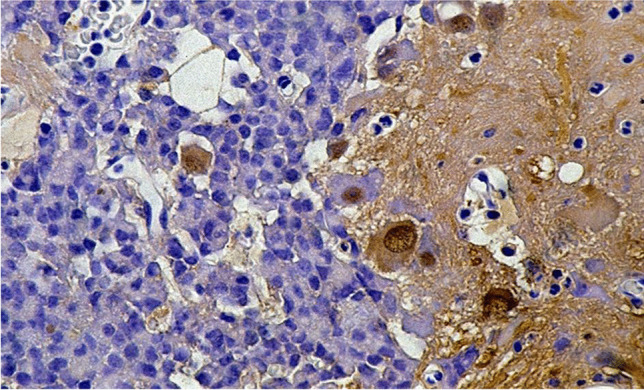
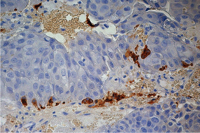
Results: Multiple tumorous lesions with more than one pituitary neuroendocrine tumor (PitNET) respectively adenoma make up 1.4% (232 cases) in our collection. Within the selected cases, synchronous multiple pituitary neuroendocrine tumors (PitNETs) account for 17.3%, PANCH cases (pituitary adenoma with neuronal choristoma) for 14.7%, PitNETs and posterior lobe tumors for 2.2%, PitNETs and metastases for 5.2%, PitNETs and mesenchymal tumors for 2.6%, PitNETs and cysts for 52.2%, and PitNETs and primary inflammation for 6.0%. The mean patient age was 53.8 years, with a standard deviation of 18.5 years. A total of 55.3% of the patients were female and 44.7% were male. From 1990 to 2018, there was a continuous increase in the number of multiple tumorous lesions.
Conclusion: From our studies, we conclude that considering possible tumorous double lesions during surgeries and in preoperative X-ray analyses is recommended.
Keywords: Adenoma; Double adenomas; Gangliocytoma; Granular cell tumor; Metastasis; Multiple tumorous lesions; PANCH; PitNETs; Pituicytoma; Pituitary; Pituitary cysts; Rathke’s cleft cyst; Spindle cell oncocytoma.
© 2022. The Author(s).
Conflict of interest statement
The authors declare no competing interests.
Figures
References
-
- Osamura RY, Lopes MBS, Grossmann A, Kontogeorgos G, Trouillas J. WHO classification of tumours of the pituitary. In: Lloyd RV, Osamura, R. Y., Klöppel, G., Rosai, J., editor. WHO classification of tumours of endocrine organs. 4. Lyon: International Agency for Research on Cancer; 2017. p. 11-63
MeSH terms
LinkOut - more resources
Full Text Sources
Medical
