. 2022 Oct 1;33(12):ar111.
doi: 10.1091/mbc.E22-07-0262. Epub 2022 Aug 10.

Chaperone requirements for de novo folding of Saccharomyces cerevisiae septins

Affiliations

Chaperone requirements for de novo folding of Saccharomyces cerevisiae septins

Daniel Hassell et al. Mol Biol Cell. .

Abstract

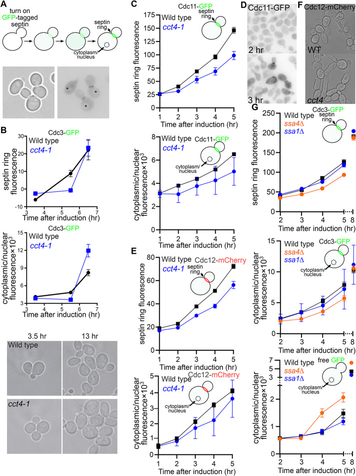
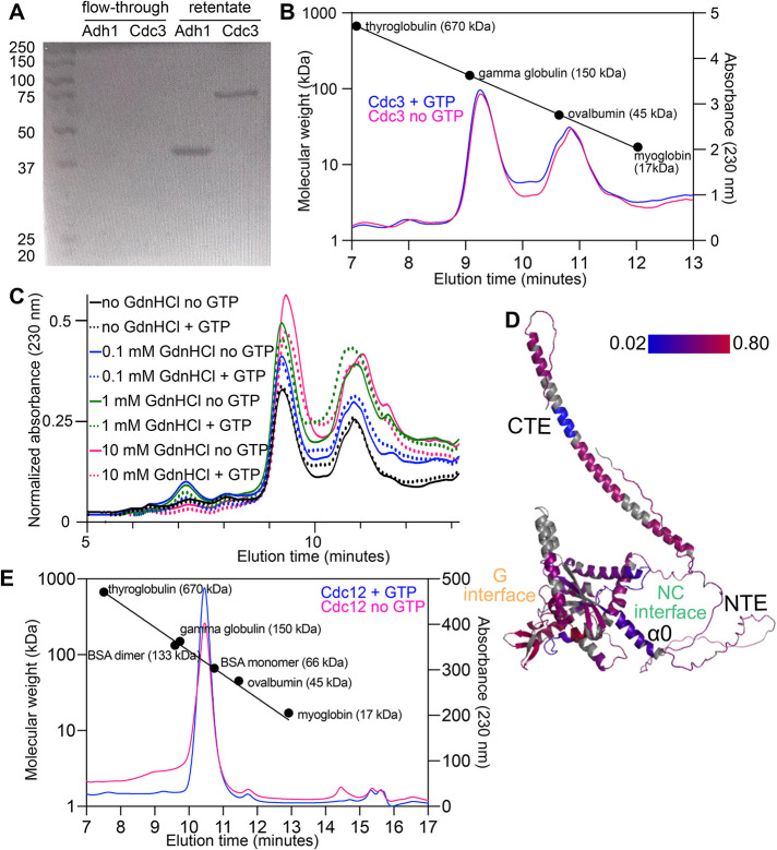
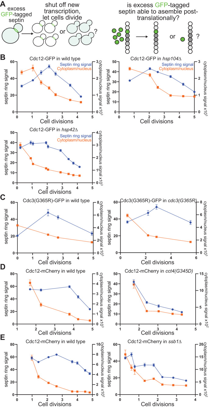
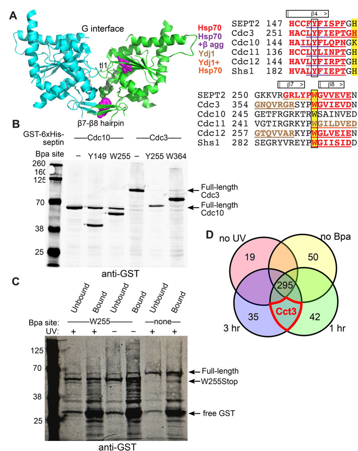
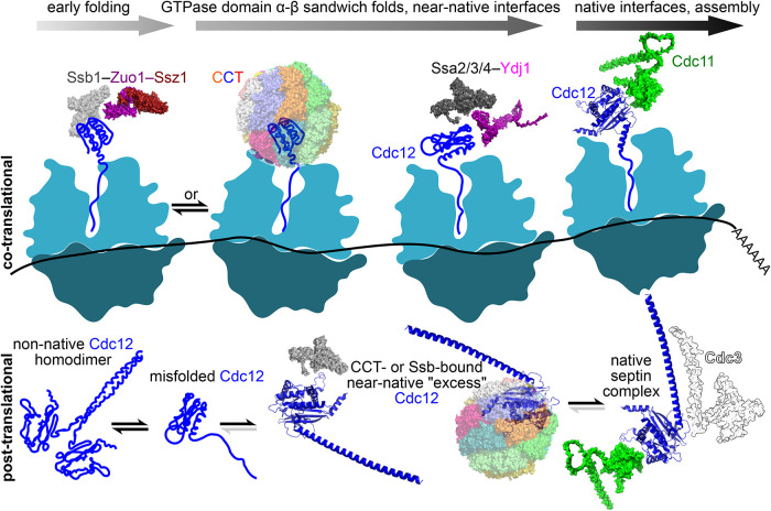
Polymers of septin protein complexes play cytoskeletal roles in eukaryotic cells. The specific subunit composition within complexes controls functions and higher-order structural properties. All septins have globular GTPase domains. The other eukaryotic cytoskeletal NTPases strictly require assistance from molecular chaperones of the cytosol, particularly the cage-like chaperonins, to fold into oligomerization-competent conformations. We previously identified cytosolic chaperones that bind septins and influence the oligomerization ability of septins carrying mutations linked to human disease, but it was unknown to what extent wild-type septins require chaperone assistance for their native folding. Here we use a combination of in vivo and in vitro approaches to demonstrate chaperone requirements for de novo folding and complex assembly by budding yeast septins. Individually purified septins adopted nonnative conformations and formed nonnative homodimers. In chaperonin- or Hsp70-deficient cells, septins folded slower and were unable to assemble posttranslationally into native complexes. One septin, Cdc12, was so dependent on cotranslational chaperonin assistance that translation failed without it. Our findings point to distinct translation elongation rates for different septins as a possible mechanism to direct a stepwise, cotranslational assembly pathway in which general cytosolic chaperones act as key intermediaries.


Figures

FIGURE 1:
FIGURE 1:
Chaperone requirements for efficient de novo septin folding. (A) Schematic illustration of experimental approach. Localization to septin filaments in septin rings at bud necks or in the cytoplasm/nucleus is monitored at time points following the induction of new synthesis of a GFP-tagged septin under control of the galactose-inducible GAL1/10 promoter. Signal is quantified at the bud neck or in a circular region of the cytoplasm/nucleus, avoiding the vacuole. Micrographs are representative cells from a middle time point in an actual experiment. Left panel, transmitted light; right panel, fluorescent signal shown as dark pixels on a light background. Asterisks are centered in presumptive vacuoles. (B) Wild-type cells (BY4741) or cct4 mutant cells (CBY11211) carrying plasmid pMVB1 were grown in synthetic medium with 2% raffinose and loaded into a microfluidic chamber. Medium containing 1.95% raffinose and 0.05% galactose was introduced, and images were taken at the indicated time points thereafter. At least 46 cells were analyzed per genotype per time point. Points show means, error bars are SEM. The micrographs show the same fields of cells at two time points, demonstrating similar rates of cell division for the wild-type and mutant cells. (C, E) As in B but with Cdc11-GFP plasmid pMVB3 or Cdc12-mCherry plasmid pGF-IVL-470. Cells were grown in culture tubes, and aliquots were removed for imaging at the indicated time points. At least 22 cells were analyzed per genotype per time point. (D) Fluorescence micrographs for representative wild-type cells from the Cdc11-GFP experiment in C imaged after the indicated time postinduction. (F) Cells (“WT,” BY4741; “cct4,” CBY11211) with Cdc12-mCherry plasmid pGF-IVL-470 were cultured in selective medium containing 1.95% raffinose and 0.05% galactose for 24 h. At 11 h, cells were diluted to prevent culture saturation. (G) As in B but with strains H00504 (“ssa1∆”) and H00507 (“ssa4∆”) and, as indicated, Cdc3-GFP plasmid pMVB1 or GFP alone plasmid pTS395. At least 30 cells were analyzed per genotype per time point.
FIGURE 2:
FIGURE 2:
Nonnative homodimerization by purified yeast septins. (A) Purified tetrameric Adh1 (native molecular weight ∼150 kDa) and purified 6xHis-Cdc3 (monomeric molecular weight ∼60 kDa) were centrifuged through a 100-kDa-cutoff filtration device. Portions of the flow-through and retentate were separated by SDS–PAGE and stained with Coomassie. Leftmost lane contains molecular weight ladder (Thermo Fisher Scientific #SM0331). (B) Samples of molecular weight standards or 6xHis-Cdc3 were analyzed by size exclusion chromatography, and the 230-nm absorbance profiles, indicative of protein concentration, were plotted as a function of elution time. Colored lines show 6xHis-Cdc3 with or without added GTP. Circles show peak elution times for the indicated standards. (C) As in B, but only with samples of 6xHis-Cdc3 containing or lacking GTP (2 mM) and/or GdnHCl at the indicated concentrations. GdnHCl was also included in the running buffer at the same concentrations. (D) Hydrogen-deuterium exchange ratios after 7200 s incubation of 6xHis-Cdc3 in deuterated buffer were mapped onto the predicted structure of Cdc3. Color indicates exchange ratio; regions in gray were not detected by MS. Locations of the CTE, NTE, α0 helix, and G and NC interfaces are for illustration purposes only. (E) As in B, but with purified Cdc12.
FIGURE 3:
FIGURE 3:
Chaperone requirements for posttranslational septin assembly. (A) Schematic illustration of experimental approach. An excess of a single, fluorescently tagged septin is generated by transient overexpression from the galactose-inducible, glucose-repressible GAL1/10 promoter, and the fluorescent signal in septin filaments at the bud neck or in the cytoplasm/nucleus is quantified via microscopy at various time points after cessation of new expression (addition of glucose). If the excess septin is capable of assembling with newly made partner septins long after it was synthesized, then the bud neck signal should increase initially and decrease slowly thereafter via a mix of incorporation into septin filaments and dilution through cell divisions. If, on the other hand, the excess septin is unable to assemble with other septins, bud neck signal should decrease exclusively via dilution, similar to the more rapid and approximately exponential kinetics expected for the cytoplasmic/nuclear signal. (B) Wild-type diploid cells (BY4743), hsp104∆/hsp104∆ mutant cells (strain 31514), or hsp42∆hsp42∆ mutant cells (diploid made by mating strains H00481 and H00419) carrying plasmid pMVB2 were grown in synthetic medium with 0.1% galactose and 1.9% raffinose. Following the addition of glucose to final 2%, aliquots were taken at time points and imaged (50 cells per genotype per time point). Cell concentration in the culture was also monitored at each time point to calculate the number of cell divisions following glucose addition. Points show means, error bars are SEM. (C) As in B but with haploid strain BY4741 (“wild type”) or CBY07236 (“cdc3(G365R)”) carrying plasmid pPgal-Cdc3(G365R)-GFP, and with 40 cells per genotype per time point. (D) As in C but with plasmid pGF-IVL-470 and cct4 strain CBY11211. At least 33 cells were imaged per genotype per time point. (E) As in B but with plasmid pGF-IVL-470 and ssb1∆ mutant haploid cells (strain H00421). Forty cells per genotype per time point.
FIGURE 4:
FIGURE 4:
In vivo cross-linking between the chaperonin CCT and the yeast septin Cdc10. (A) Predicted chaperone binding sites. At left, ribbon structure of human septin-2 G homodimer (PDB 2QNR). Residues shown as spheres were chosen for replacement by the photocrosslinker Bpa and are boxed in the sequence alignment at right. The β7-β8 hairpin and trans loop 1 (“tl1”) make contacts across the G interface. Predicted sites are underlined and in bold and color-coded for β-aggregation, Hsp70 and/or Ydj1. Hsp70 binding sites have LIMBO score >11.08 (Van Durme et al., 2009). Ydj1-binding sites match the consensus GX[LMQ]{P}X{P}{CIMPVW} (Kota et al., 2009). β-Aggregation–prone sequences are according to TANGO (Fernandez-Escamilla et al., 2004). (B) Yeast cells (strain YRP2838) carrying plasmid pSNRtRNA-pBPA-RS and a plasmid encoding a GST-6xHis–tagged septin (Cdc3 or Cdc10) with or without sites of incorporation of the photocrosslinker amino acid Bpa were grown in the presence of Bpa and, to induce tagged septin expression, 2% galactose. Total protein was extracted via alkaline lysis and TCA precipitation and separated by SDS–PAGE before immunoblotting with anti-GST antibodies. (C) As in B but just for Cdc10 and cells were exposed to UV to induce cross-linking (or not, as indicated). Following protein extraction in denaturing conditions 6xHis-tagged proteins were purified by Ni-NTA affinity. (D) Venn diagram of proteins identified by mass spectrometry in samples prepared as in C. Circles are not drawn to scale. Numbers indicate numbers of unique proteins identified; there were two independent biological replicates of all samples except the no-UV sample. For this diagram, the replicates were pooled together. Cct3 was the only protein identified in all experimental samples and none of the controls. “No Bpa” means wild-type Cdc10, with no UAG codon for Bpa incorporation.
FIGURE 5:
FIGURE 5:
In vivo cross-linking between the E. coli chaperonin GroEL and the yeast septin Cdc10. (A) E. coli cells (strain BL21(DE3)) carrying plasmid pEVOL-pBpF and a Cdc10-6xHis plasmid with the indicated site of Bpa incorporation were grown in the presence of Bpa. IPTG was added to induce tagged Cdc10 expression and cells were exposed to UV and then lysed in denaturing conditions. Proteins bound to Ni-NTA were separated by SDS–PAGE and stained with Coomassie. Boxes indicate regions of the gel that were excised for mass spectrometry. (B, C) Samples as in A were separated by SDS–PAGE and then immunoblotted for DnaK (B) or GroEL (C). The leftmost lanes in all gels contained molecular weight ladder (Li-Cor Biosciences # 928-60000).
FIGURE 6:
FIGURE 6:
Chaperonin requirement for translation of the septin Cdc12 by prokaryotic ribosomes. (A) Coomassie-stained SDS–PAGE of proteins purified via Ni-NTA from wild-type or groEL-mutant E. coli cells carrying plasmids encoding the indicated septins, plus other plasmids as necessary for induction or viability. Strains were B002 (“WT”), B003 (“∆clpB”), B004 (“∆dnaJ”), B005 (“∆dnaK”), B006 (“∆htpG”), and 461 (“groEL(E461K)”). Septin-encoding plasmids were pMVB128, pMVB133, and pMAM54. Leftmost lane contains a molecular weight ladder (Li-Cor Biosciences #928-60000). (B) Immunoblot of bound proteins prepared as in A, plus a sample from wild-type cells (B002) carrying no septin-encoding plasmid (“No Septins”), using a cocktail of antibodies recognizing Cdc3, Cdc11, and the 6xHis tag. Magenta asterisk indicates an ∼70 kDa protein weakly reactive with the Cdc3 and/or Cdc11 antibody. (C) As in B but for urea-solubilized pellets (cell debris and insoluble material) obtained following cell lysis. The blue asterisk indicates the band corresponding to Cdc12. (D) DNA agarose gel electrophoresis and ethidium bromide staining of products of RT-PCR for Cdc3 or Cdc12 mRNA from total RNA extracted from groEL-mutant strain 461 carrying plasmid pMAM54 and induced with IPTG at the indicated temperatures. The presence or absence of reverse transcriptase (“RT”) in the reactions is indicated. Leftmost lane contains molecular weight ladder (Thermo Fisher Scientific #SM0331). (E) Published Ribo-seq/ribosome profiling data (Sen et al., 2015) showing the positions of ribosomes along the Cdc12 mRNA as numbers of reads of ribosome-protected mRNA fragments. Below, the Cdc12 ORF is illustrated with Met codons in green, Pro codons in blue, and the stop codon in red. The first 14 amino acids of Cdc12 are indicated with the same color scheme. (F) As in B but including samples from groEL-mutant cells carrying the plasmid pMAM88, which encodes Cdc3 and 6xHis-tagged Cdc12 with the four Pro residues highlighted in E mutated to Ala (“(AVAAA)Cdc12”). Samples from cells with pMAM54, encoding wild-type Cdc12, are indicated by “(PVPPP)Cdc12.” The blue asterisk indicates the band corresponding to Cdc12. (G) Chaperone-free in vitro transcription/translation reactions lacking release factors (PURExpress ∆RF123) and containing fluorescent puromycin to label translation products were resolved by SDS–PAGE. The template plasmid pMVB128 encodes Cdc10 and 6xHis-Cdc12. Purified GroEL/ES chaperonin was added to one reaction, as indicated. Leftmost lane contains a molecular weight ladder (Bio-Rad #1610395). (H) As in G but gel was run longer, the order of lanes is reversed, and brightness and contrast were adjusted in FIJI.
FIGURE 7:
FIGURE 7:
Model for cotranslational chaperone-mediated septin folding in the context of septin complex assembly. In this illustration, folding and translation proceed from left to right. CCT and Ssb (Ssb1 or Ssb2) initially interact with the same exposed surface composed of strands β1 and β3 from the α-β sandwich, until α is synthesized and buries that surface. Not shown: CCT may also bind later at the β4 and/or β7-β8 hairpin, as evidenced by our in vivo photocrosslinking. Ssa-family chaperones (but not Ssa1) interact with nascent Cdc12 to promote native folding until Cdc12 has achieved a conformation capable of stably interacting with partner septins. Ydj1 is shown as the presumptive Hsp40 for Ssa. If folded Cdc11 is available during Cdc12 translation, a Cdc12–Cdc11 heterodimer assembles cotranslationally (“co-translational”). Alternatively, if Cdc12 is in excess to other septins, it completes translation without having interacted with another septin (“post-translational”). Here, Cdc12 is susceptible to “off-pathway” nonnative conformations prone to nonnative homodimerization. CCT and Ssb continue to interact with full-length Cdc12 (as illustrated), inhibiting off-pathway conformations until stable septin complexes assemble. Not illustrated: their dysfunction during translation produces full-length Cdc12 that is irreversibly misfolded and has no path to stable septin assembly. Other septins presumably require similar chaperone assistance for de novo folding but Cdc12 is the slowest to be translated and thus a potential “platform” for cotranslational assembly. Protein structures are AlphaFold predictions (the NTE of Cdc3 is hidden, for clarity) or cryo-EM structures (human CCT; PDB 7LUM [Knowlton et al., 2021]; Zuo1–Ssz1, PDB 7 × 3K [Chen et al., 2022]).

Similar articles

Cited by

References

    1. Abbey M, Hakim C, Anand R, Lafera J, Schambach A, Kispert A, Taft MH, Kaever V, Kotlyarov A, Gaestel M, et al. (2016). GTPase domain driven dimerization of SEPT7 is dispensable for the critical role of septins in fibroblast cytokinesis. Sci Rep 6, 20007. - PMC - PubMed
    1. Ahmed N, Friedrich UA, Sormanni P, Ciryam P, Altman NS, Bukau B, Kramer G, O’Brien EP (2020). Pairs of amino acids at the P- and A-sites of the ribosome predictably and causally modulate translation-elongation rates. J Mol Biol 432, 166696. - PMC - PubMed
    1. Alamgir M, Erukova V, Jessulat M, Azizi A, Golshani A (2010). Chemical-genetic profile analysis of five inhibitory compounds in yeast. BMC Chem Biol 10, 6. - PMC - PubMed
    1. Albanèse V, Yam AY-W, Baughman J, Parnot C, Frydman J (2006). Systems analyses reveal two chaperone networks with distinct functions in eukaryotic cells. Cell 124, 75–88. - PubMed
    1. Amberg D, Burke D, Strathern J (2005). Methods in Yeast Genetics: A Cold Spring Harbor Laboratory Course Manual, 2005 ed, Cold Spring Harbor, NY: Cold Spring Harbor Laboratory Press.

Publication types

MeSH terms

LinkOut - more resources