. 2022 Aug 10;17(8):e0272776.
doi: 10.1371/journal.pone.0272776. eCollection 2022.

Bee venom-loaded EGFR-targeting peptide-coupled chitosan nanoparticles for effective therapy of hepatocellular carcinoma by inhibiting EGFR-mediated MEK/ERK pathway

Affiliations

Bee venom-loaded EGFR-targeting peptide-coupled chitosan nanoparticles for effective therapy of hepatocellular carcinoma by inhibiting EGFR-mediated MEK/ERK pathway

Shaymaa Abdulmalek et al. PLoS One. .

Retraction in

Abstract

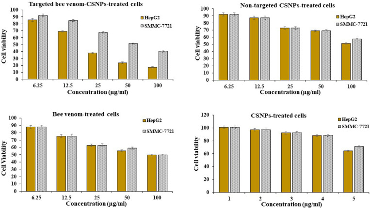
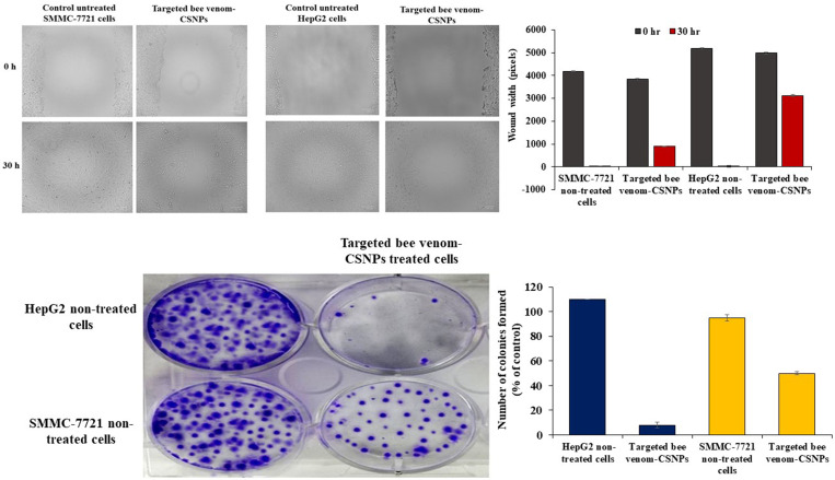
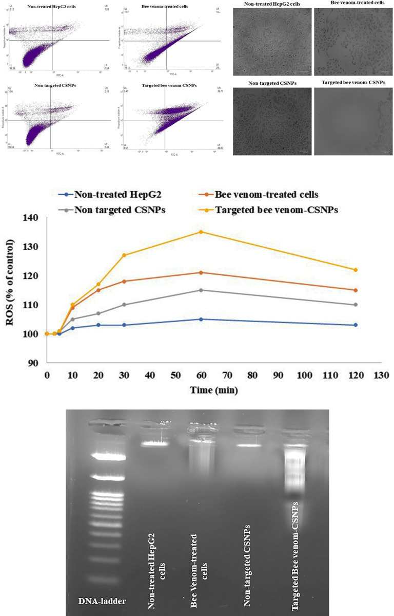
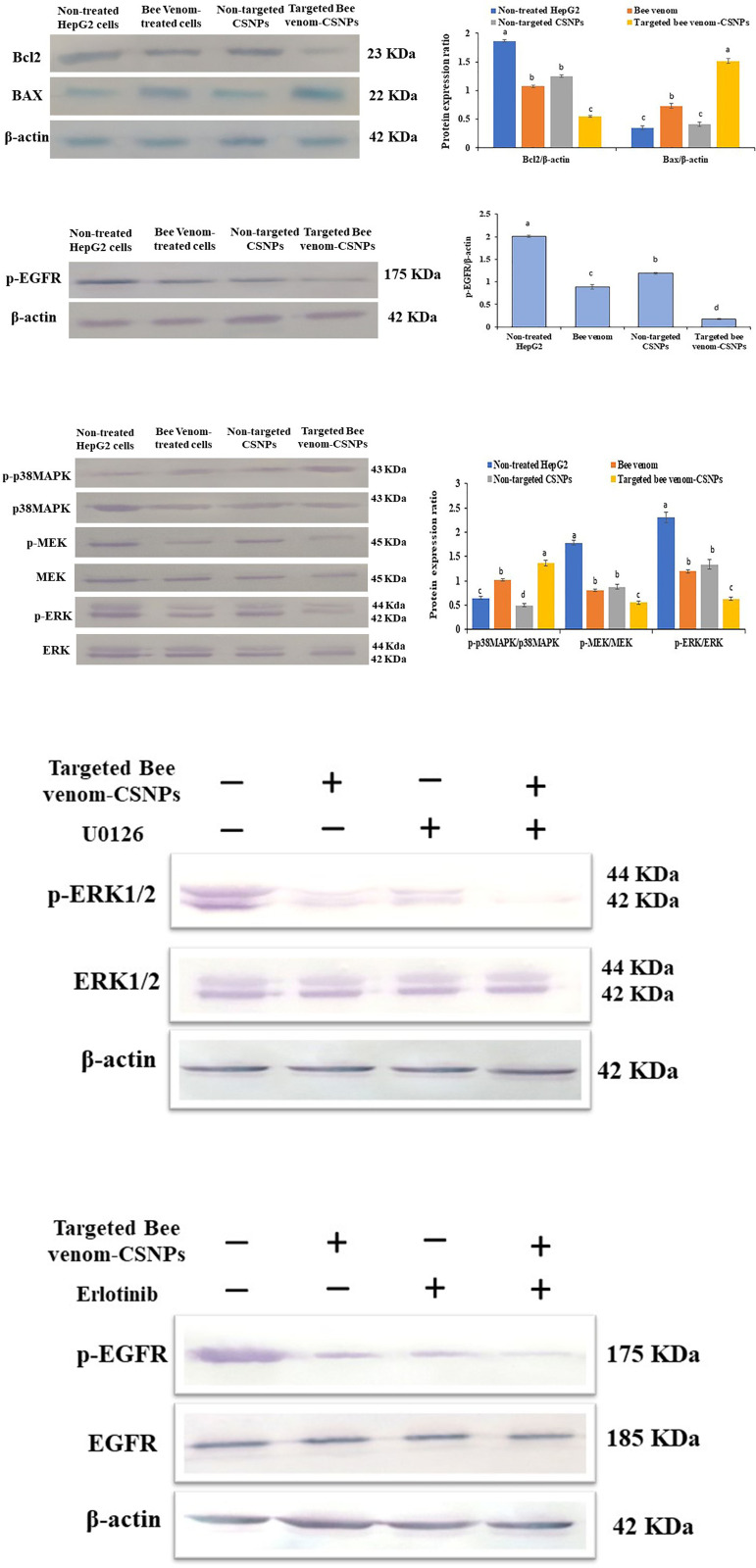
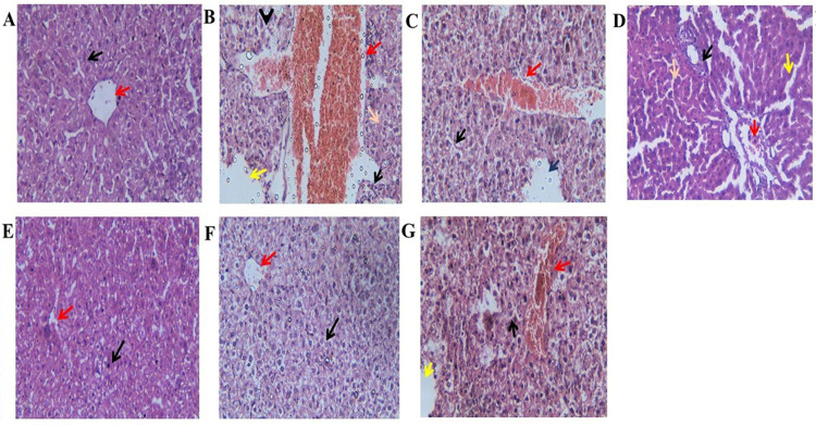
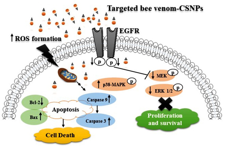
Hepatocellular carcinoma (HCC) is one of the world's most risky diseases due to the lack of clear and cost-effective therapeutic targets. Currently, the toxicity of conventional chemotherapeutic medications and the development of multidrug resistance is driving research into targeted therapies. The nano-biomedical field's potential for developing an effective therapeutic nano-sized drug delivery system is viewed as a significant pharmaceutical trend for the encapsulation and release of numerous anticancer therapies. In this regard, current research is centered on the creation of biodegradable chitosan nanoparticles (CSNPs) for the selective and sustained release of bee venom into liver cancer cells. Furthermore, surface modification with polyethylene glycol (PEG) and GE11 peptide-conjugated bee venom-CSNPs allows for the targeting of EGFR-overexpressed liver cancer cells. A series of in vitro and in vivo cellular analyses were used to investigate the antitumor effects and mechanisms of targeted bee venom-CSNPs. Targeted bee venom-CSNPs, in particular, were found to have higher cytotoxicity against HepG2 cells than SMMC-7721 cells, as well as stronger cellular uptake and a substantial reduction in cell migration, leading to improved cancer suppression. It also promotes cancer cell death in EGFR overexpressed HepG2 cells by boosting reactive oxygen species, activating mitochondria-dependent pathways, inhibiting EGFR-stimulated MEK/ERK pathway, and elevating p38-MAPK in comparison to native bee venom. In hepatocellular carcinoma (HCC)-induced mice, it has anti-cancer properties against tumor tissue. It also improved liver function and architecture without causing any noticeable toxic side effects, as well as inhibiting tumor growth by activating the apoptotic pathway. The design of this cancer-targeted nanoparticle establishes GE11-bee venom-CSNPs as a potential chemotherapeutic treatment for EGFR over-expressed malignancies. Finally, our work elucidates the molecular mechanism underlying the anticancer selectivity of targeted bee venom-CSNPs and outlines therapeutic strategies to target liver cancer.


Conflict of interest statement

The authors have declared that no competing interests exist.

Figures

Fig 1
Fig 1. Chromatogram of bee venom.
(A) Standard mixture; and (B) Sample mixture.
Fig 2
Fig 2. Characterization of prepared nanoparticles.
(A) Transmission electron microscope image; (B) particle size; (C) Zeta potential; (D) Release profile of bee venom from CSNPs relative to time.
Fig 3
Fig 3
TEM micrographs of (A) fresh prepared targeted bee venom-CSNPs and (B) targeted bee venom-CSNPs after 30 days incubation in a buffer.
Fig 4
Fig 4. Cell viability of HepG2 and SMMC-7721.
Relationship between targeted bee venom-CSNPs, non-targeted CSNPs, native bee venom, and CSNPs concentrations and viability percentage on HepG2 and SMMC-7721 cells.
Fig 5
Fig 5. Cellular uptake of bee venom-loaded CSNPs.
(A) Cellular uptake of coumarin-6-loaded targeted bee venom nanoparticles in HepG2 and SMMC-7721 cells; (B) coumarin-6-loaded non-targeted nanoparticles in HepG2 and SMMC-7721 cells, n = 3.
Fig 6
Fig 6. EGFR expression and selective cellular uptake of targeted bee venom-CSNPs.
(A) EGFR expression; (B) selective cellular uptake of coumarin-6 loaded targeted bee venom-CSNPs; (C) and anti-EGFR antibody blocking in HepG2 and SMMC-7721 cells.
Fig 7
Fig 7. Migration ability of HepG2 and SMMC-7721 cells.
(A) wound-healing assays; (B) colony formation in HepG2 and SMMC-7721 cells treated with targeted bee venom-CSNPs.
Fig 8
Fig 8. Studying the effects of targeted bee venom-CSNPs, non-targeted CSNPs, or native bee venom on induction of cell apoptosis.
(A) Flow cytometry analysis; (B) ROS; and (C) DNA fragmentation in HepG2 treated cells.
Fig 9
Fig 9. Activation of mitochondria-dependent apoptosis pathway and regulation of EGFR-mediated p38-MAPK/MEK/ERK pathway by targeted bee venom-CSNPs.
(A) Western blot analysis for the expression of Bcl-2 and Bax in HepG2 cells; (B) Western blot analysis for the expression of p-EGFR; (C) Western blot analysis for the expression of p38MAPK, p-p38MAPK, MEK, p-MEK, ERK, and p-ERK; (D) HepG2 cells were treated with targeted bee venom-CSNPs and/or U0126 for 24 h, and the protein levels of p-ERK1/2 and total ERK1/2 were detected; (E) HepG2 cells were treated with targeted bee venom-CSNPs and/or Erlotinib for 24 h, and the protein levels of p-EGFR and total EGFR were detected. β-Actin was used as a loading control. Data are expressed as mean ± SEM (n = 3), means for the same parameter with different letters in each bar are significantly different (p < 0.01), where the highest data value takes the letter (a).
Fig 10
Fig 10. Histopathological examination of liver tissue sections in each group.
(A) Control healthy showing normal hepatocytic cords with normal hepatic cells (Black arrows) and with clear central vein (Red arrows) (H&E, X400); (B) HCC-induced showing complete hepatocellular carcinoma, the hepatic tissue is necrotic and occupied by mononuclear cells infiltration (Black arrows) and pyknotic nucleus (orange arrows) and congested dilated portal tracts along with hemorrhage (Red arrows), and dilated sinusoids in between (yellow arrows) (H&E, X400); (C) HCC-Bee venom (1 mg/kg) showing focal area of hepatic necrosis occupied by pyknotic nucleus (Green arrows) and congested dilated portal tracts along with hemorrhage (Red arrows), and dilated sinusoids in between (Blue arrows), and all hepatocytes suffer from vacuolar and hydropic degeneration (Black arrows) (H&E, X400); (D) HCC-Bee venom (2 mg/kg) showing hepatic tissue is necrotic and occupied by mononuclear cells infiltration (Black arrows) and pyknotic nucleus (orange arrows) and congested dilated portal tracts along with hemorrhage (Red arrows), and dilated sinusoids in between (yellow arrows) (H&E, X400); (E) HCC-Targeted bee venom-CSNPs (0.5 mg/kg) showing clear central vein (Red arrows) and almost normal hepatocyte with some hepatocytes suffer from vacuolar and hydropic degeneration (Black arrows) (H&E, X400); (F) HCC-Targeted bee venom-CSNPs (1 mg/kg) showing central vein with some hemorrhage (Red arrows) and almost normal hepatocyte with some hepatocytes suffer from vacuolar and hydropic degeneration (Black arrows) (H&E, X400); (G) HCC-Non-targeted CSNPs (2 mg/kg) showing focal area of hepatic necrosis occupied by pyknotic nucleus (Black arrows) and larger degenerated area occupied by centrilobular congestion and congested dilated portal tracts along with hemorrhage (Red arrows), and dilated sinusoids in between (yellow arrows).
Fig 11
Fig 11. Schematic representation of the different pathways for targeted bee venom-CSNPs inside cells.

Similar articles

Cited by

References

    1. Llovet JM, Zucman-Rossi J, Pikarsky E, Sangro B, Schwartz M, Sherman M, et al.. Hepatocellular carcinoma. Nat Rev Dis Primers. 2016; 2:16018.‏ doi: 10.1038/nrdp.2016.18 - DOI - PubMed
    1. Berasain C, Perugorria MJ, Latasa MU, Castillo J, Goni S, Santamaria M, et al.. The epidermal growth factor receptor: a link between inflammation and liver cancer. Exp Biol Med. 2009;234:713–725.‏ doi: 10.3181/0901-MR-12 - DOI - PubMed
    1. Jura N, Zhang X, Endres NF, Seeliger MA, Schindler T, Kuriyan J. Catalytic control in the EGF receptor and its connection to general kinase regulatory mechanisms. Mol cell. 2001;42:9–22.‏ - PMC - PubMed
    1. Llovet JM, Bruix J. Molecular targeted therapies in hepatocellular carcinoma. Hepatology. 2008;48:1312–1327.‏ doi: 10.1002/hep.22506 - DOI - PMC - PubMed
    1. Kaufman NE, Dhingra S, Jois SD, Vicente MDGH. Molecular Targeting of Epidermal Growth Factor Receptor (EGFR) and Vascular Endothelial Growth Factor Receptor (VEGFR). Molecules. 2021;26:1076.‏ doi: 10.3390/molecules26041076 - DOI - PMC - PubMed

Publication types