. 2022 Aug 10;14(657):eabl9605.
doi: 10.1126/scitranslmed.abl9605. Epub 2022 Aug 10.

Broadly neutralizing antibodies to SARS-related viruses can be readily induced in rhesus macaques

Affiliations

Broadly neutralizing antibodies to SARS-related viruses can be readily induced in rhesus macaques

Wan-Ting He et al. Sci Transl Med. .

Abstract

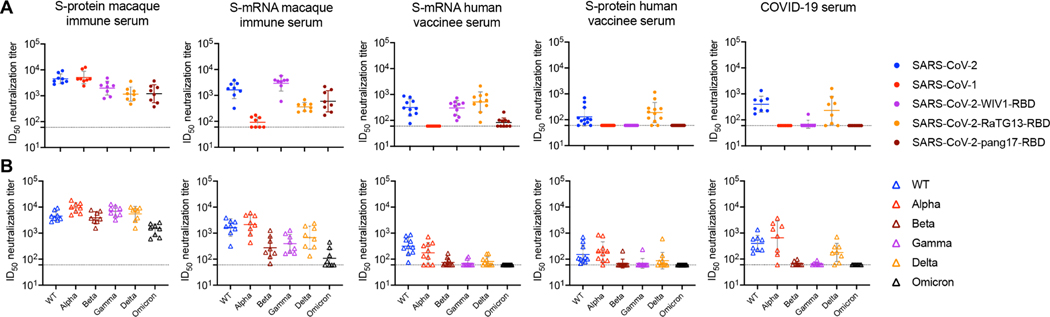
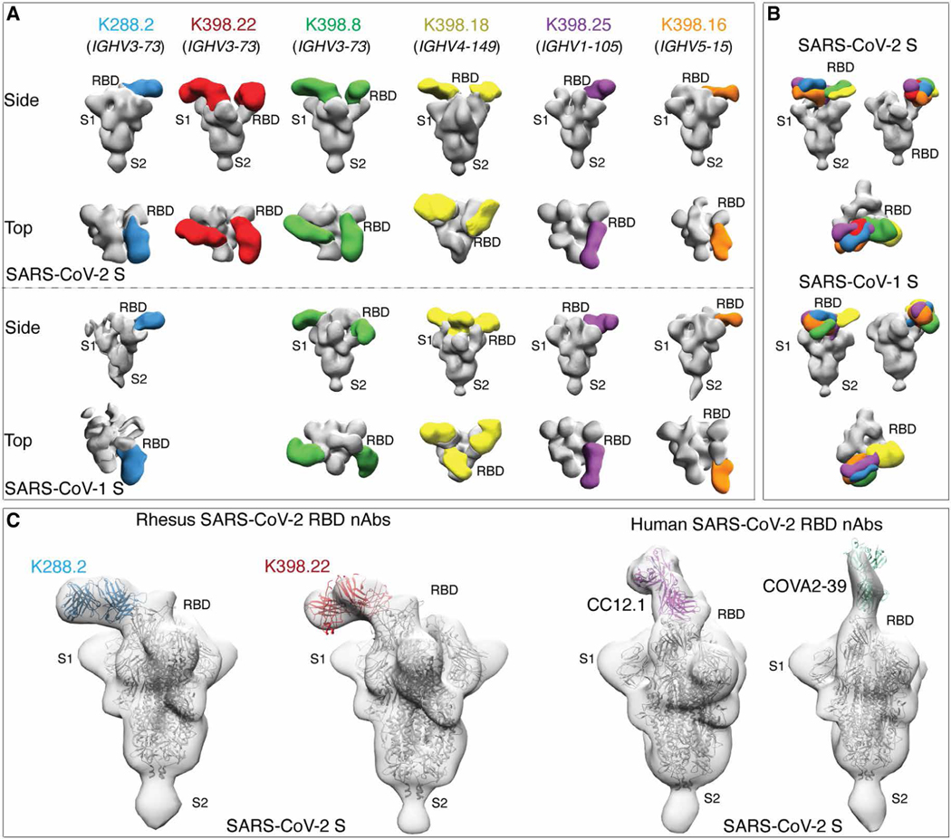
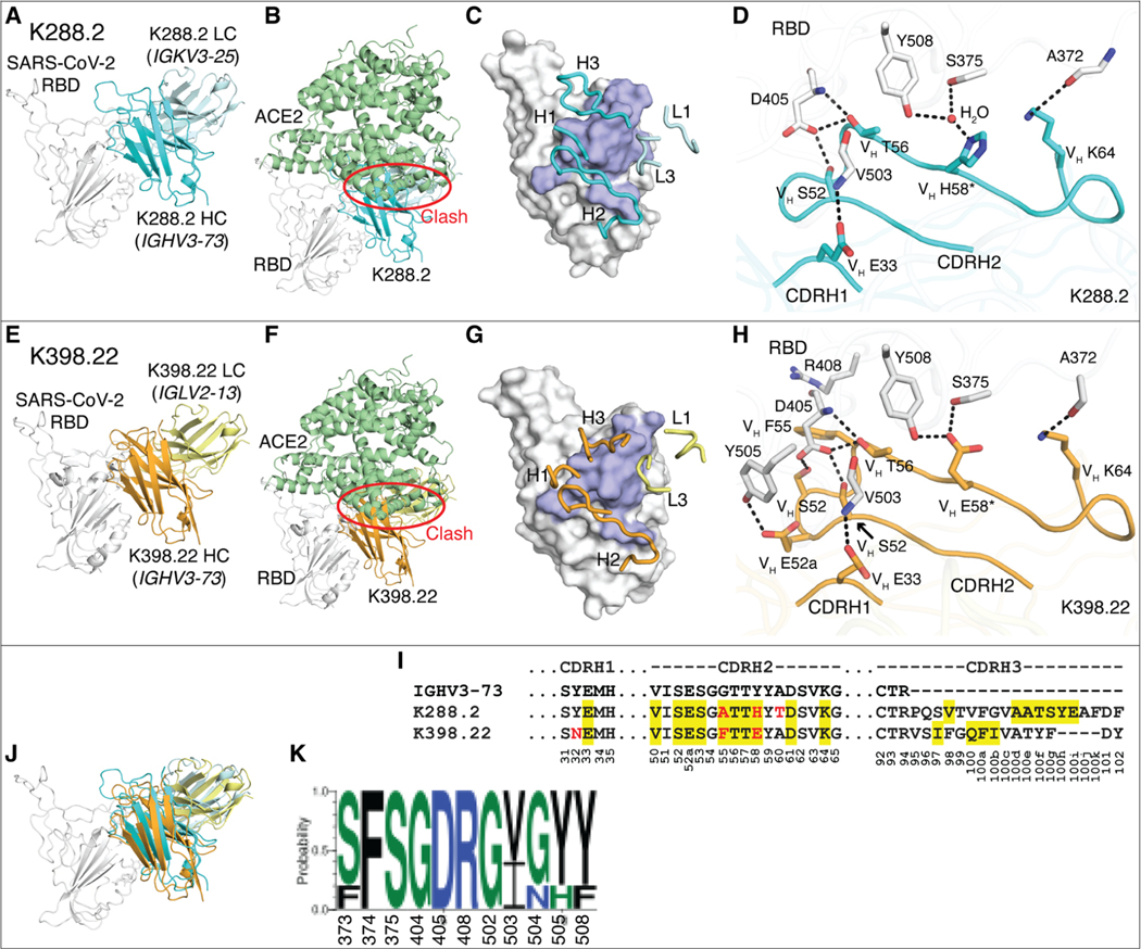
To prepare for future coronavirus (CoV) pandemics, it is desirable to generate vaccines capable of eliciting broadly neutralizing antibody responses to CoVs. Here, we show that immunization of macaques with SARS-CoV-2 spike (S) protein with a two-shot protocol generated potent serum receptor binding domain cross-neutralizing antibody responses to both SARS-CoV-2 and SARS-CoV-1. Furthermore, responses were equally effective against most SARS-CoV-2 variants of concern (VOCs) and some were highly effective against Omicron. This result contrasts with human infection or many two-shot vaccination protocols where responses were typically more SARS-CoV-2 specific and where VOCs were less well neutralized. Structural studies showed that cloned macaque neutralizing antibodies, particularly using a given heavy chain germline gene, recognized a relatively conserved region proximal to the angiotensin converting enzyme 2 receptor binding site (RBS), whereas many frequently elicited human neutralizing antibodies targeted more variable epitopes overlapping the RBS. B cell repertoire differences between humans and macaques appeared to influence the vaccine response. The macaque neutralizing antibodies identified a pan-SARS-related virus epitope region less well targeted by human antibodies that could be exploited in rational vaccine design.


Figures

Fig. 1.
Fig. 1.. SARS-CoV-2 and SARS-CoV-1 cross-reactive antibody binding and neutralizing responses were observed in SARS-CoV-2 S-protein–immunized rhesus macaques.
(A) The SARS-CoV-2 S-protein prime-boost immunization in rhesus macaques and sampling schedule is shown. Animals were primed at week 0 with 100 μg of SARS-CoV-2 S-protein along with adjuvant SMNP, by bolus (group 1: n = 4) or escalating dose (ED) (group 2: n = 4) immunization, and both groups further boosted at week 10 with 100 μg of the S-protein immunogen. Serum and peripheral blood mononuclear cells (PBMCs) were collected every 2 weeks for immune response analysis. (B) The heatmap shows EC50 ELISA binding of SARS-CoV-2 S-protein–immunized NHP serum from bolus and ED schemes against different S-proteins. EC50 binding responses for prebleed (PB), post-prime (WK 4 and WK 10), and post-boost (WK 14) sample time points are shown. (C) SARS-CoV-2– and SARS-CoV-1–specific serum ID50 neutralizing antibody titers were measured in SARS-CoV-2 S-protein–immunized rhesus macaques. ID50 neutralizing antibody responses are shown for PB, post-prime (WK 4 and WK 10), and post-boost (WK 14) sample time points. (D) NHP immune serum binding to SARS-CoV-2 or SARS-CoV-1 S-proteins correlated modestly with neutralization against the corresponding virus. SARS-CoV-2 or SARS-CoV-1 EC50 serum antibody binding titers and ID50 nAb titers were compared by nonparametric Spearman correlation two-tailed test with 95% confidence interval. The Spearman correlation coefficient r and the P value are indicated. (E) A comparison of cross-neutralizing activities is shown for SARS-CoV-2 S-protein–immunized NHP immune serum (week 14, after two immunizations at weeks 0 and 10), serum from S-mRNA–vaccinated NHPs, serum from S-mRNA–vaccinated humans, serum from S-protein–vaccinated human, serum from COVID-19–recovered humans, and serum from S-protein–immunized mice. Horizontal bars indicate geometric mean. Dashed horizontal lines indicate limit of detection. Statistical comparisons between groups were performed using a Mann-Whitney two-tailed test. **P < 0.01; ***P < 0.001; ****P < 0.0001; not significant (ns), P > 0.05. (F) Neutralization of SARS-CoV-2 by post-prime (WK 10) and post-boost (WK 14) macaque immune serum and the corresponding anti-RBD antibody–depleted serum (RBD-depleted) is shown. Anti-RBD polyclonal antibodies in the immune serum were depleted by adsorption on recombinant monomeric RBD, and the depleted serum was tested for SARS-CoV-2 neutralization. The SARS-CoV-2 ID50 neutralizing antibody titers for untreated and RBD-depleted serum were compared by Mann-Whitney two-tailed test; **P < 0.01 and ****P < 0.0001.
Fig. 2.
Fig. 2.. SARS-CoV-2 S-protein–immunized macaque serum samples show neutralization with sarbecovirus chimeric SARS-CoV-2 pseudotyped viruses and SARS-CoV-2 variants.
(A) The dot plots show ID50 neutralizing antibody titers of SARS-CoV-2 S-protein–immunized week 14 macaque immune serum, S-mRNA macaque immune serum, S-mRNA human vaccinee serum samples, S-protein human vaccinee serum samples, and COVID-19 convalescent serum samples with SARS-CoV-2, SARS-CoV-1, and RBD-swapped WIV1, RaTG13, and pang17 chimeric SARS-CoV-2 pseudoviruses. (B) The dot plots show ID50 neutralizing antibody titers of the same serum samples from (A) with SARS-CoV-2 WT (Wuhan strain) and VOCs that have circulated in humans (Alpha, Beta, Gamma, Delta, and Omicron). For (A) and (B), ID50 neutralization titers less than 60 are shown as 60, and the dashed lines at 60 indicate the lower limit of detection. Horizontal bars indicate geometric mean values.
Fig. 3.
Fig. 3.. Isolation and characterization of cross-neutralizing monoclonal antibodies from two SARS-CoV-2 S-protein–immunized rhesus macaques.
(A) The flow cytometry plots show the sorting strategy used to isolate single isotype-switched B cells that specifically bind to fluorescently labeled SARS-CoV-1 and SARS-CoV-2 S-protein probes. The class-switched memory B cells were gated as single, scattered, live (SSL), CD3, CD4, CD8, CD14, CD19+/CD20+, IgM, and IgG+. The antigen-specific IgG+ B cells selected, as indicated by a red arrow, were used to obtain paired heavy and light chain immunoglobulin sequences for mAb expression and characterization. mAbs in this study were only isolated from S-protein–vaccinated rhesus macaques. (B) The heatmap shows the BLI binding responses of isolated monoclonal antibodies from rhesus macaques (K398 and K288) against SARS-CoV-2 S-protein, SARS-CoV-2 S-protein–derived domains and subdomains (NTD, RBD, RBD-SD1, and RBD-SD1–2), SARS-CoV-1 S-protein, and SARS-CoV-1-RBD (indicated with a green color scale). The IC50 neutralizing titer against SARS-CoV-1 and SARS-CoV-2 pseudoviruses is shown in the right columns with a red color scale. (C) Neutralization of SARS-CoV-2 RBD–swapped pseudoviruses by macaque cross-neutralizing mAbs are shown. IC50 values are color-coded, and an IC50 greater than 5 μg/ml is indicated in gray. (D) IC50 values are shown for macaque cross-neutralizing mAbs against pseudoviruses with single amino acid mutations in the SARS-CoV-2 S-protein and variants of concern (Alpha, Beta, Gamma, Delta, and Omicron). IC50 values greater than 5000 ng/ml are shown in gray. A dengue virus–specific antibody, DEN3, was used as a negative control in (B) to (D). (E) Neutralization IC50 fold changes of mAbs (n = 15) with mutants compared with the WT SARS-CoV-2 pseudovirus are shown in the dot plots with geometric mean labeled by the bar in gray. The gain or loss of neutralizing potency is indicated by the arrows on the right.
Fig. 4.
Fig. 4.. Immunogenetic and epitope properties of sarbecovirus cross-neutralizing macaque antibodies.
(A) Phylogenetic trees derived from heavy chain (VH)–gene sequences of S-protein–specific antibodies isolated from K398 and K288 macaques are shown. Gene assignments used the rhesus macaque (M. mulatta) germline database described in (59). The IGHV gene usage for each mAb depicted in the tree is shown by different colors indicated in the bottom of the color scheme. Neutralizing and nonneutralizing mAbs are shown in red and blue, respectively. (B) IGHV gene usage for neutralizing and nonneutralizing mAbs in K288 and K398 macaques are shown. (C) CDRH3 length in amino acids (aa) and number of SHMs were quantified for neutralizing and nonneutralizing mAbs. *P < 0.1 and ****P < 0.0001. (D) The heatmap shows BLI competition–based epitope binning of macaque cross-neutralizing mAbs with human RBD-specific nAbs CC12.3, CC12.19, CR3022, and S309. The gene usage, geometric mean neutralization potency, and breadth [calculated from neutralization with five viruses in Fig. 3 (B and C)] for each nAb are indicated. The BLI competition experiment was performed with SARS-CoV-2 RBD, and the degree of competition is indicated as red (strong), orange (moderate), light blue (weak), and gray (very weak competition).
Fig. 5.
Fig. 5.. Electron microscopy structures of sarbecovirus cross-neutralizing macaque mAbs with SARS-CoV-2 and SARS-CoV-1 S-proteins.
(A) Electron microscopy 3D reconstructions of rhesus macaque nAb Fabs with SARS-CoV-2 and SARS-CoV-1 S-proteins are shown. Fabs of six rhesus macaque nAbs, K288.2 (blue), K398.22 (red), K398.8 (green), (K398.18 (yellow), K398.25 (purple), and K398.16 (orange) were complexed with SARS-CoV-2 and SARS-CoV-1 S-proteins, and 3D reconstructions were generated from 2D class averages. The heavy chain germline gene usage is indicated for each macaque nAb. The S-protein S1 and S2 subunits and the RBD sites are labeled. (B) Side and top views showing 3D reconstructions of all six RBD-directed macaque cross-neutralizing Abs bound to SARS-CoV-2 (top) and SARS-CoV-1 (bottom) S-proteins. (C) S-protein binding angles of approach were compared between macaque IGHV3–73–encoded RBD cross nAbs, K288.2 and K398.22, and human IGHV3–53–encoded RBD nAbs, CC12.1 and COVA2–39, isolated from COVID-19 donors. The crystal structures of the respective nAbs were docked into EM density maps. The RBD, S1, and S2 subunits of SARS-CoV-2 S-protein are labeled. The macaque nAbs approach S-protein RBD from the side, and the two human nAbs approach at a more perpendicular angle close to the S-protein threefold axis.
Fig. 6.
Fig. 6.. Crystal structures of two rhesus macaque IGHV3–73–encoded neutralizing antibodies bound to SARS-CoV-2 RBD.
The SARS-CoV-2 RBD is shown in white throughout the figure and human ACE2 (hACE2) is shown in pale green. The heavy (HC) and light chains (LC) of K288.2 are in cyan and light cyan, and those of K398.22 are in orange and yellow. For clarity, only variable domains of the antibodies are shown. (A to H) Structures of SARS-CoV-2 RBD in complex with K288.2 antibody (A to D) and in complex with K398.22 antibody (E to H) are shown. (A and E) Germline genes encoding K288.2 (A) and K398.22 (D) macaque antibodies are labeled. (B and F) Structures of SARS-CoV-2 RBD in complex with K288.2 (B) or K398.22 (F) superimposed onto an RBD-hACE2 complex structure (PDB 6M0J) (97) show that the K288.2/K398.22 antibody would clash with the hACE2 receptor (indicated with a red circle). (C and G) The epitopes of K288.2 (C) and K398.22 (G) are shown in purple. CDR loops that interact with the RBD are shown in blue and labeled. (D and H) Detailed molecular interactions of K288.2 (D) or K398.22 (H) CDRs H1 and H2 with SARS-CoV-2 RBD are shown. Somatically mutated residues are indicated with *. The CDRH2 of IGHV3–73 antibodies interacts extensively with the RBD. (I) The IGHV3–73 germline sequence and the sequences of K288.2 and K398.22 are shown. The IGHV3–73 sequence is shown in black. Only CDR sequences are shown for clarity. Lengths of CDR H3 can vary. Yellow shades indicate paratope regions in antibody heavy chain defined as buried surface area (BSA > 0 Å2). Red letters indicate somatic mutations. Bottom numbers indicate Kabat numbering. Y58 is mutated to E/H. Our structures show germline Y58 would clash, whereas E/H is able to interact with the RBD. (J) The Fab-RBD complex structures of the two antibodies are shown superimposed on the RBD. (K) Sequence conservation of the epitope residues in SARS-related strains recognized by IGHV3–73–encoded paratopes is shown (cutoff = 4 Å). SARS-CoV-2, SARS-CoV-1, WIV-1, RaTG13, and pang17 were used for the conservation analysis.

References

    1. Dai L, Gao GF, Viral targets for vaccines against COVID-19. Nat. Rev. Immunol. 21, 73–82 (2020). - PMC - PubMed
    1. Amanat F, Krammer F, SARS-CoV-2 vaccines: Status report. Immunity 52, 583–589 (2020). - PMC - PubMed
    1. Krammer F, SARS-CoV-2 vaccines in development. Nature 586, 516–527 (2020). - PubMed
    1. Ahmed SF, Quadeer AA, McKay MR, Preliminary identification of potential vaccine targets for the COVID-19 coronavirus (SARS-CoV-2) based on SARS-CoV immunological studies. Viruses 12, 254 (2020). - PMC - PubMed
    1. Klasse PJ, Nixon DF, Moore JP, Immunogenicity of clinically relevant SARS-CoV-2 vaccines in nonhuman primates and humans. Sci. Adv. 7, eabe8065 (2021). - PMC - PubMed

Publication types

Supplementary concepts