. 2023 Jan 12;44(1):70-116.
doi: 10.1210/endrev/bnac019.

History of Adrenal Research: From Ancient Anatomy to Contemporary Molecular Biology

Affiliations

History of Adrenal Research: From Ancient Anatomy to Contemporary Molecular Biology

Walter L Miller et al. Endocr Rev. .

Abstract

The adrenal is a small, anatomically unimposing structure that escaped scientific notice until 1564 and whose existence was doubted by many until the 18th century. Adrenal functions were inferred from the adrenal insufficiency syndrome described by Addison and from the obesity and virilization that accompanied many adrenal malignancies, but early physiologists sometimes confused the roles of the cortex and medulla. Medullary epinephrine was the first hormone to be isolated (in 1901), and numerous cortical steroids were isolated between 1930 and 1949. The treatment of arthritis, Addison's disease, and congenital adrenal hyperplasia (CAH) with cortisone in the 1950s revolutionized clinical endocrinology and steroid research. Cases of CAH had been reported in the 19th century, but a defect in 21-hydroxylation in CAH was not identified until 1957. Other forms of CAH, including deficiencies of 3β-hydroxysteroid dehydrogenase, 11β-hydroxylase, and 17α-hydroxylase were defined hormonally in the 1960s. Cytochrome P450 enzymes were described in 1962-1964, and steroid 21-hydroxylation was the first biosynthetic activity associated with a P450. Understanding of the genetic and biochemical bases of these disorders advanced rapidly from 1984 to 2004. The cloning of genes for steroidogenic enzymes and related factors revealed many mutations causing known diseases and facilitated the discovery of new disorders. Genetics and cell biology have replaced steroid chemistry as the key disciplines for understanding and teaching steroidogenesis and its disorders.

Keywords: Addison disease; adrenal hyperplasia; cortisone; cytochrome P450; genetic disease; steroid.


Figures

Graphical Abstract
Graphical Abstract
Figure 1.
Figure 1.
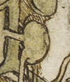
A sketch by DaVinci ca. 1485-1490 of the kidney with an apparent adrenal in the correct anatomic location (RCIN 912597, in the public domain at https://commons.wikimedia.org/wiki/File:Leonardo_da_Vinci_-_RCIN_912597,_The_major_organs_and_vessels_c.1485-90.jpg).
Figure 2.
Figure 2.
Left, Plate 2 from Eustacchio’s “Opuscola Anatomica,” as reproduced by JM Lancisi in 1714 as “Tabulae Anatomicae” (image from page 64 of Lancisi’s volume, in the public domain at https://search.lib.virginia.edu/sources/uva_library/items/u3606395?idx=0&page=64. This image was also published as Fireu. 2 in Endocrine Reviews 41(1):1-46, 2020). Right, Faceplate of Lancisi’s book (image captured online at https://en.wikipedia.org/wiki/Bartolomeo_Eustachi).
Figure 3.
Figure 3.
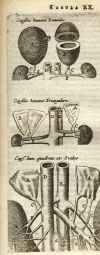
Left, Giulio Cesare Casseri, from the frontispiece of his book Tabulae Anatomica (Deuchinus, 1627) (in the public domain at https://en.wikipedia.org/wiki/Giulio_Cesare_Casseri#/media/File:Casserius.png). Right, Casseri’s illustration of the kidneys, featuring the adrenals, indicated by “G” and labeled “corpuscula reni incumbentia sive Renes succenturiati” (renal corpuscles lying on or above the kidney) (images from page 174 of Casseri’s volume in the public domain at https://archive.org/details/tabulaeanatomica00cass/page/174/mode/2up).
Figure 4.
Figure 4.
Thomas Bartholin’s varied adrenal anatomy. The top figure shows hollow, ovoid adrenals, supporting the notion that the adrenals were filled with “black bile”; a variant of this illustration was also published by Johann Vesling (1647) [from (14)] (in the public domain at http://mateo.uni-mannheim.de/camenaref/bartholin/bartholin1/jpg/s123.html).
Figure 5.
Figure 5.
Left, Thomas Addison (photographer unknown); in the public domain at (https://commons.wikimedia.org/w/index.php?curid=35152865). Right, Reproduction of Plate 11, showing a postmortem “sketch” of “Mr. S” from Thomas Addison’s 1855 monograph (30) (https://wellcomecollection.org/works/xsmzqpdw). Most of the patients described by Addison had tuberculous adrenalitis, but the patchy vitiligo and scant pubic hair illustrated here suggests autoimmune polyglandular syndrome type 2f.
Figure 6.
Figure 6.
Autopsy of genitourinary structures of a virilized female patient with congenital adrenal hyperplasia in 1833 (44). The larger illustration, labeled “Fig. 1” is accompanied by a listing of 15 numbered structures; notable structures include (1) the urethra immediately inferior to the glans penis; (6 and 7) bladder and ureters, respectively; (8) vagina; (10) uterus; (11,12) ovaries; (11) 1 of the Fallopian tubes. The smaller illustration, labeled “Fig. 2” is accompanied by a listing of 7 structures labeled a though g, showing the urethra, Cowper’s glands, corpora cavernosa, and accompanying musculature (in the public domain at https://www.sciencedirect.com/science/article/abs/pii/S0140673602937700).
Figure 7.
Figure 7.
Plate 3 from De Crecchio’s paper (45), containing 3 drawings. Fig. 1: longitudinal view of the pelvic contents. Some relevant structures are: 5. Urethra (opened). 10. Vagina. 14. Uterus (opened). 15. Cervix. 17. Right broad ligament, containing the right ovary and Fallopian tube 19. Left Fallopian tube and fimbriae. 20. Left ovary. 23. Right corpus cavernosum (cut). 24. Urethral meatus. Fig. 2: connection of the vagina to the prostatic urethra; numbers as in Fig. 1; for details see (46). Fig. 3: drawing of a Graafian follicle from Marzo, as seen microscopically at 620× (in the public domain; from the University of Michigan Library, http://hdl.handle.net/2027/mdp.39015013772457).
Figure 8.
Figure 8.
10½-month-old girl with “adrenogenital syndrome” and “adrenocortical obesity” caused by a 3 cm diameter, 12 g right adrenal adenoma (51) (in the public domain at https://jamanetwork.com/journals/jamapediatrics/fullarticle/1178993).
Figure 9.
Figure 9.
Structures proposed for cholesterol. Left, structure according to Heinrich Wieland (111) (in the public domain at https://www.nobelprize.org/uploads/2018/06/wieland-lecture.pdf). Center, structure as proposed by Adolf Windaus (112) (in the public domain at https://www.nobelprize.org/uploads/2018/06/windaus-lecture.pdf). Right, structure as proposed by Rosenheim and King (114).
Figure 10.
Figure 10.
Structures of the 28 steroids that were chemically characterized by Reichstein between 1938 and 1949 (123) (in the public domain at https://www.nobelprize.org/uploads/2018/06/reichstein-lecture.pdf).
Figure 11.
Figure 11.
Left, Tadeus Reichstein (1897-1996) as a well-dressed graduate student at the Swiss Federal Institute of Technology in Zürich, 1923. From (118); reproduced courtesy of Schweitzerische Stiftung für die Photographie. Right, The Kendall lab, 1948, with the principal investigators of the use of cortisone for rheumatoid arthritis. Left to right: Charles H. Slocumb, Howard F. Polley, Edward C. Kendall, and Philip S. Hench [from (126), reprinted with permission].
Figure 12.
Figure 12.
Patient with congenital adrenal hyperplasia reported by Wilkins et al (159), with scrotal enlargement suggesting the presence of testicular adrenal rest tumors.
Figure 13.
Figure 13.
The physiology of congenital adrenal hyperplasia, described by Bartter et al in 1951 (178) (in the public domain at https://www.jci.org/articles/view/102438/pdf). In these diagrams, “S” does not refer to Reichstein’s compound S (11-deoxycortisol) but to the then-unknown steroid active in sugar metabolism (now known to be cortisol); “N” refers to the then-unknown adrenal androgen that influences nitrogen retention (now known to be 11-keto-testosterone). In the right-hand panel, “E” refers to Kendall’s compound E (cortisone).
Figure 14.
Figure 14.
The first steroidogenic pathway, as proposed by Hechter and Pincus in 1954 (193); reprinted with permission. The pathway is largely correct, as acetate is converted to cholesterol through a complex, 30-step pathway, represented by “x.” However, none of the intermediates in that pathway is converted to progesterone or other steroids without first being converted to cholesterol.
Figure 15.
Figure 15.
Bongiovanni’s steroidogenic pathway from 1963 (199). Cortisol is (5) and corticosterone is (7). The various enzymes, A (3 β-hydroxysteroid dehydrogenase), B (17-hydroxylase), C (21-hydroxylase), and D (11-hydroxylase) are illustrated, and their sites of action are shown by the encircled heavy type. The most common form of congenital adrenal hyperplasia is characterized by a lack of 21-hydroxylase (C) and hence an accumulation of 17-hydroxyprogesterone (3) with the excretion of large quantities of the latter’s reduced metabolite, pregnanetriol (III) (reprinted with permission; ©1963 Massachusetts Medical Society. All rights reserved).
Figure 16.
Figure 16.
Left, Spectrum for the light-mediated reversal of the inhibition of bovine adrenal microsomal 21-hydroxylase by carbon monoxide [from (224)]. Right, Diagram of home-made apparatus built by Cooper et al to measure the reversal of CO-induced P450 spectrum and 21-hydroxylase activity of bovine adrenal microsomes [from (225); reprinted with permission].
Figure 17.
Figure 17.
Cooper lab, 1964. Left to right: Levine, Slade, Narasimhulu, Omura, Foroff, Rosenthal, Cooper, and their homemade apparatus for measuring the photochemical action spectra of adrenal microsomal cytochrome P450 [from (230); reprinted with permission].
Figure 18.
Figure 18.
Unequal crossing-over between CYP11B1 and CYP11B2. CYP11B2 is on the left and pairs with the 95% similar CYP11B1. The direction of transcription is from left to right. A and C and B and D are homologous regions 5’ and 3’, respectively, of the CYP11B genes. A crossover between these paired genes during meiosis results in 1 gamete with duplicated genetic material, containing a hybrid gene with 5’ sequences from CYP11B1 and 3’ sequences from CYP11B2. The presence of a single copy of such an allele is sufficient to explain the features of glucocorticoid-remediable aldosteronism. The second gamete is deficient, containing only the complementary hybrid gene [from (292); figure provided by Prof. Perrin C. White].
Figure 19.
Figure 19.
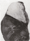
A sectioned adrenal gland from a patient with congenital lipoid adrenal hyperplasia, termed “lipidosis of the adrenal.” [from Sandison (380); reprinted with permission].
Figure 20.
Figure 20.
Newborn SF-1 (NR5A1) null mice lack adrenal glands and gonads. Abbreviations: A, SF-1 null female. B, wild-type female. k, kidney; a, adrenal; o, ovary; od, oviduct [from (480) reprinted with permission].

Similar articles

Cited by

References

    1. Shumacker HB. The early history of the adrenal glands: with particular reference to theories of function. Bull Inst History Med 1936;4(1):39-56. https://www.jstor.org/stable/44433654.
    1. Lenard A. The history of research on the adrenals; 1563-1900. J Hist Med Allied Sci. 1951;6(4):496-505. doi:10.1093/jhmas/vi.autumn.496. - DOI - PubMed
    1. Carmichael SW. The history of the adrenal medulla. Rev Neurosci. 1989;2(2):83-100. doi:10.1515/REVNEURO.1989.2.2.83. - DOI - PubMed
    1. Leoutsakos B, Leoutsakos A. The adrenal glands: a brief historical perspective. Hormones (Athens) 2008;7(4):334-336. doi:10.14310/horm.2002.1216. - DOI - PubMed
    1. Miller WL. A brief history of adrenal research: Steroidogenesis - the soul of the adrenal. Mol Cell Endocrinol. 2013;371(1-2):5-14. doi:10.1016/j.mce.2012.10.023. - DOI - PubMed

Publication types