. 2022 Sep 9;50(16):9470-9489.
doi: 10.1093/nar/gkac671.

Biochemical and structural characterization of hepatitis A virus 2C reveals an unusual ribonuclease activity on single-stranded RNA

Affiliations

Biochemical and structural characterization of hepatitis A virus 2C reveals an unusual ribonuclease activity on single-stranded RNA

Pu Chen et al. Nucleic Acids Res. .

Abstract

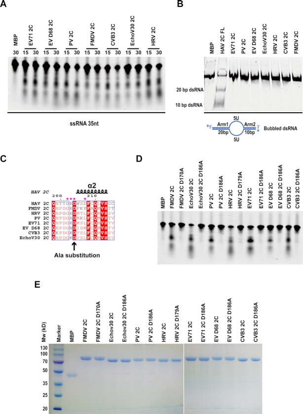
The HAV nonstructural protein 2C is essential for virus replication; however, its precise function remains elusive. Although HAV 2C shares 24-27% sequence identity with other 2Cs, key motifs are conserved. Here, we demonstrate that HAV 2C is an ATPase but lacking helicase activity. We identified an ATPase-independent nuclease activity of HAV 2C with a preference for polyuridylic single-stranded RNAs. We determined the crystal structure of an HAV 2C fragment to 2.2 Å resolution, containing an ATPase domain, a region equivalent to enterovirus 2C zinc-finger (ZFER) and a C-terminal amphipathic helix (PBD). The PBD of HAV 2C occupies a hydrophobic pocket (Pocket) in the adjacent 2C, and we show the PBD-Pocket interaction is vital for 2C functions. We identified acidic residues that are essential for the ribonuclease activity and demonstrated mutations at these sites abrogate virus replication. We built a hexameric-ring model of HAV 2C, revealing the ribonuclease-essential residues clustering around the central pore of the ring, whereas the ATPase active sites line up at the gaps between adjacent 2Cs. Finally, we show the ribonuclease activity is shared by other picornavirus 2Cs. Our findings identified a previously unfound activity of picornavirus 2C, providing novel insights into the mechanisms of virus replication.


Figures

Figure 1.
Figure 1.
Overall structural feature of HAV 2C-ΔN. (A) Expression and purification of WT HAV 2C and HAV 2C-ΔN. Left, expression of WT HAV 2C in bacteria yielded insoluble proteins. Mw: molecular weight standards. The molecular weight of the standard is indicated on the left in kDa. Lane 1, insoluble fraction of the bacteria overexpressing WT HAV 2C; lane 2, supernatant of the bacteria lysate. Middle, production of soluble WT HAV 2C in the presence of DDM. Lane 1, supernatant of sf-21 cell lysate overexpressing WT HAV 2C; lane 2, elution from Ni-NTA resin. Right, production of HAV 2C-ΔN. Mw: molecular weight standards; lane 1, pellet of bacteria lysate; lane 2, supernatant of bacteria lysate; lane 3, flow through of Ni-NTA resin; lane 4, wash of unbound materials; lane 5, sample eluted from Ni-NTA resin; lane 6, Ni-NTA elution digested by TEV protease; lane 7, flow through of the sample from lane 6 reloaded on Ni-NTA. (B) Left, the rate of ATP hydrolysis by WT HAV 2C is plotted as the function of ATP concentration; the data was fitted with nonlinear regression using Michaelis–Menten equation to calculate kinetic parameters; Km, Vmax, kcat and R2 of the fitting are indicated. ATPase activity of WT HAV 2C, three ATPase-dead mutants harboring mutation K149A, D194A and N241A and HAV 2C-ΔN. (C) Top, diagram of domain/motif organization of HAV 2C protein; key features are indicated. Bottom, ribbon model of HAV 2C-ΔN structure colored by secondary structural elements with annotation; α-helix colored cyan and β-sheet colored magenta. (D) Structural superimposition of HAV 2C ZFER (orange ribbon) with the zinc-fingers of PV 2C (blue ribbon) and EV71 2C (green ribbon). The zinc-coordinating residues of two enterovirus 2C proteins are shown with the stick model, demonstrating they are completely shortcut in HAV 2C ZFER. (E) Structural superimposition of the C-terminus (α6) of HAV 2C, PV 2C and EV71 2C. The C-terminus helix of HAV 2C breaks down in to two helices (α6A and α6B) that are nearly perpendicular to each other.
Figure 2.
Figure 2.
HAV 2C undergoes C-terminus helix dependent self-oligomerization. (A) Crystal packing analysis of HAV 2C-ΔN crystals reveals all 2C copies in the lattice are connected via a specific interaction involving two contact sits, indicated as ‘1’ and ‘2′. Site 1 is between α6A and α1’–β2 loop of the adject 2C, and site 2 is between α6B (termed PBD) and a hydrophobic pocket (termed Pocket) on the adject 2C molecule. (B) Magnified view of contact site 1; residues implicated in the interaction are labelled and shown with a stick model. (C) Magnified view of contact site 2; residues implicated in the interaction are labelled and shown with a stick model. (D) Size-exclusion chromatography profiles of HAV 2C-ΔN and a selection of mutants/truncation harboring mutations at the contact sites. Retention volume from Superdex 200 10/300 GL column, calculated molecular mass and oligomeric state of the proteins are indicated.
Figure 3.
Figure 3.
Comparison of the PBD–Pocket interaction in the crystal structures of HAV 2C, EV71 2C and PV 2C. (A, C and E) Ribbon model of HAV 2C dimer, EV71 2C dimer (PDB id: 5GQ1) and PV 2C dimer (PDB id: 5Z3Q) found in crystal lattice. The adjacent 2C molecules are colored differently. (B, D and F) Magnified views at the 2C-2C juncture illustrating the PBD–Pocket interaction observed in the structures of HAV 2C, EV71 2C and PV 2C. The C-terminus helix is colored gold, and residues occupying the hydrophobic Pocket (highlighted with magenta surface) on are shown in a blue stick model.
Figure 4.
Figure 4.
Impact of mutations on HAV 2C ATPase activity and viral RNA replication. (A) ATP activity of WT HAV 2C and a selection of 2C mutants. The ATPase activity is expressed as ‘μmol of ATP hydrolyzed per μmol of enzyme per minute’. Images below are thin-layer chromatography of the ATP hydrolysis reaction visualized by Typhoon Trio Variable Mode Imager. (B–E). RNA replication efficiency of WT HAV and a selection of HAV 2C mutants. Huh7-Lunet cells were electroporated with subgenomic reporter replicons representing WT HAV or containing the indicated mutations in 2C region. At the respective timepoints, replication was determined by firefly luciferase assay in the lysates of transfected cells, representing replication efficiency. Data are normalized to the value obtained 4 hours after electroporation to correct for transfection efficiency. Mean values with SD from technical replicates of a representative experiment (n = 4 repetitions with comparable outcome). ATPase mutants and irrelevant mutants (refer to mutations outside of the conserved motifs critical to HAV 2C activities, E169A, S206A, K267A and H276A) are shown in panel B; Pocket (the hydrophobic pocket formed between ATPase domain and ZEFR) mutants are shown in panel C; ZFER (Zinc-finger equivalent region) mutants are shown in panel D; PBD (Pocket-Binding Domain) mutants are shown in panel E.
Figure 5.
Figure 5.
HAV 2C is a ribonuclease that specifically digests U-rich ssRNA regions. (A) Purified NSP13 of MERS-CoV, WT HAV 2C or 2C-ΔN were incubated with a dsRNA substrate containing a 18 bp duplex with 10U 5′ssRNA overhang (RNA1 & RNA4). The top strand of the duplex was labelled with Cy3, marked by an asterisk in the scheme below. The reaction mixtures were analysed by native-PAGE. Heat denatured substrate was used as a positive control for mimicking helicase activity. (B) HAV 2C-ΔN was incubated with two dsRNA substrates containing 18 bp duplex with 5′ (RNA1 & RNA4) or 3′ (RNA1 & RNA3) ssRNA overhangs. The top strand of the duplex was labelled with Cy3, marked by asterisks. The reaction mixtures were analysed by native-PAGE. 18bp dsRNA (RNA1 and RNA2) was loaded as a size marker. (C) Digestions of a Cy3 labelled 35 nt ssRNA by HAV 2C WT (FL) and 2C-ΔN. The incubation time is indicated above each lane and reaction mixtures were analyzed by urea–PAGE. (D) The same 35 nt ssRNA with 2′-O-Me modification at uridine residues (RNA6, left panel), a uridine-rich ssRNA (RNA7) and an adenosine-rich ssRNA (RNA8) were incubated with HAV 2C-ΔN and analysed as in (C) (middle panel). A dsRNA (RNA5 & RNA9) with an internal U-rich mismatch region flanked by 20 bp and 10 bp arms was used as a substrate and analyzed by Native-PAGE. (E) Digestion of a 35 nt ssRNA (RNA5) by HAV 2C-ΔN and a set of mutants harboring alanine substitution of the indicated acidic residues. The resulting mixtures were resolved by Urea-PAGE. Mutants lacking ribonuclease activity are marked by asterisk.
Figure 6.
Figure 6.
Multiple picornavirus 2C proteins exhibit ribonuclease activity. MBP-tagged full-length purified 2C proteins from Enterovirus 71 (EV71 2C), Enterovirus D68 (EV-D68 2C), Poliovirus (PV 2C), Foot-and-mouth disease virus (FMDV 2C), Coxsackievirus B3 (CVB3 2C), Echovirus 30 (EchoV30 2C) and Human Rhinovirus (HRV 2C), either WT or mutant, subjected to RNase assays using different substrates. (A) Picornaviral 2C proteins were incubated with a 35-nt single-stranded RNA substrate (RNA5) and the products were analyzed by 15% urea-PAGE. The incubation time (in minutes) is indicated on the top of the gel. (B) A bubble RNA substrate was prepared by annealing RNA5 with RNA9, and the substrate was incubated with the indicated 2C proteins at 37°C for 30 min. HAV 2C FL was the non-tagged WT protein. The products of the RNase assays were analyzed by 15% native PAGE. ‘MBP’ is the control of the MBP-tag alone. (C) An acidic residue D204 essential to HAV 2C RNase activity is invariant among various picornaviral 2C proteins (indicated by an arrow). (D) Various MBP-tagged 2C proteins and their mutants harboring alanine substitution of the invariant aspartic acid shown in panel C were tested for RNase activity on a 35-nt single-stranded RNA substrate (RNA5); the reaction mixtures were incubated at 37°C for 30 min before analysing by a 15% urea-PAGE. (E) SDS-PAGE analysis of various 2C proteins and their mutants used in the RNase assay shown in panel D.
Figure 7.
Figure 7.
Impact of mutations in the RNase domain of HAV 2C and EV71 on replication. (A) Huh7-Lunet cells were electroporated with subgenomic HAV luciferase reporter replicon, wild-type (WT), replication dead (3D mut) or with mutations relevant to nuclease activity. At the indicated time points luciferase activity was determined, normalized to the 4-h value, and depicted in logarithmic scale. Graph depicts mean and standard deviation of two biological replicates performed in technical duplicates. RLU = relative light units. (B) Bar graph focusing on specific data of panel A. * P < 0.05, ** P < 0.01, *** P < 0.005. (C) An infectious cDNA clone of EV71 was mutated at position D186A in the RNase domain (corresponding to D204A in HAV). A lethal mutation in the ATPase domain K135A (22) was used as a control. In vitro transcripts from WT and the respective mutants were transfected in Vero cells. Serial dilutions of freeze-thaw lysates obtained at 72 h after transfection were titrated on RD cells to quantify infectivity (TCID50/ml). DL: detection limit.
Figure 8.
Figure 8.
Hexameric model of HAV 2C. (A) Left, hexameric model of HAV 2C-ΔN was built by superimposing six copies of HAV 2C-ΔN structure to each subunit of JCV LTag hexamer (PDB: 5J4Y); the C-terminus α6B helix was merged with α6A helix, so that the PBD could be readily fitted into the Pocket on the adjacent 2C. The missing N-terminal portion of HAV 2C (Chain F) harboring membrane binding motif is indicated with the dashed line. Right, hexameric model of full-length HAV 2C. The monomeric model of full-length HAV 2C was predicted using program AlphaFold2. Six copies of full-length HAV 2C model were then superimposed to each subunit of the hexameric model of HAV 2C-ΔN. (B) Front, side and back views of surface electrostatic potential plot of the hexameric model of HAV 2C-ΔN. The dimensions of the hexameric model are marked. (C) Left, magnified view of the central pore on cytoplasm side; acidic residues important to ribonuclease activity (black stick model) are gathered around the pore on the cytoplasm side. Residues essential to nuclease activity of HAV 2C are indicated with asterisk. (D) Acidic residues important to ribonuclease activity are highlighted with stick model and colored magenta, Walker A motif is colored red, Walker B motif is colored blue. Residues essential to nuclease activity of HAV 2C are indicated with an asterisk.

Similar articles

Cited by

References

    1. International Committee on Taxonomy of Viruses Virus Taxonomy: Classification and Nomenclature of Viruses: Sixth Report of the International Committee on Taxonomy of Viruses. 1995; NY: Springer-Verlag Wien Austria.
    1. Lemon S.M., Ott J.J., Van Damme P., Shouval D.. Type a viral hepatitis: a summary and update on the molecular virology, epidemiology, pathogenesis and prevention. J. Hepatol. 2017; 68:167–184. - PubMed
    1. Averhoff F.M., Khudyakov Y., Nelson N.P.. Plotkin S.A., Orenstein W.A., Offit P.A., Edwards K.M.. 24 - Hepatitis A Vaccines. Plotkin's Vaccines. 7th edn. 2018; Elsevier; 319–341.
    1. Foster M.A., Hofmeister M.G., Kupronis B.A., Lin Y., Xia G.L., Yin S., Teshale E.. Increase in hepatitis a virus infections - United States, 2013-2018. MMWR Morb. Mortal. Wkly. Rep. 2019; 68:413–415. - PMC - PubMed
    1. Hu X., Collier M.G., Xu F.. Hepatitis a outbreaks in developed countries: detection, control, and prevention. Foodborne Pathog Dis. 2020; 17:166–171. - PubMed

Publication types