. 2022 Aug 19;17(8):2344-2354.
doi: 10.1021/acschembio.2c00469. Epub 2022 Aug 10.

Subtype-Selective Positive Modulation of KCa2.3 Channels Increases Cilia Length

Affiliations

Subtype-Selective Positive Modulation of KCa2.3 Channels Increases Cilia Length

Young-Woo Nam et al. ACS Chem Biol. .

Abstract

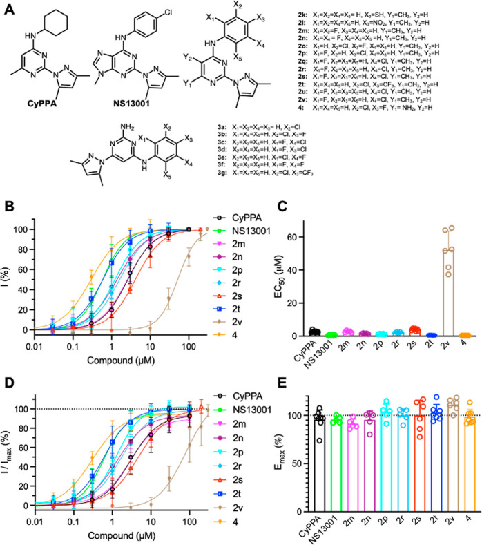
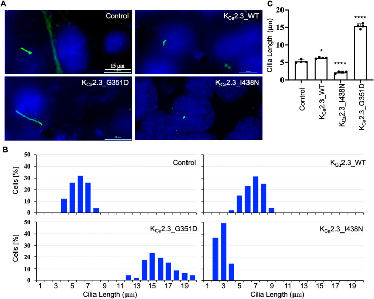
Small-conductance Ca2+-activated potassium (KCa2.x) channels are gated exclusively by intracellular Ca2+. The activation of KCa2.3 channels induces hyperpolarization, which augments Ca2+ signaling in endothelial cells. Cilia are specialized Ca2+ signaling compartments. Here, we identified compound 4 that potentiates human KCa2.3 channels selectively. The subtype selectivity of compound 4 for human KCa2.3 over rat KCa2.2a channels relies on an isoleucine residue in the HA/HB helices. Positive modulation of KCa2.3 channels by compound 4 increased flow-induced Ca2+ signaling and cilia length, while negative modulation by AP14145 reduced flow-induced Ca2+ signaling and cilia length. These findings were corroborated by the increased cilia length due to the expression of Ca2+-hypersensitive KCa2.3_G351D mutant channels and the reduced cilia length resulting from the expression of Ca2+-hyposensitive KCa2.3_I438N channels. Collectively, we were able to associate functions of KCa2.3 channels and cilia, two crucial components in the flow-induced Ca2+ signaling of endothelial cells, with potential implications in vasodilation and ciliopathic hypertension.


Conflict of interest statement

The authors declare no competing financial interest.

Figures

Figure 1
Figure 1
Positive modulation of human KCa2.3 channels by compounds. (A) Chemical structures of compounds 2k–2v, 3a–3g, and 4, compared with those of CyPPA and NS13001. (B) Concentration-dependent potentiation of KCa2.3 channels by compounds. (C) EC50 values to compounds of KCa2.3 channels. (D) Responses to compounds of KCa2.3 channels were normalized to the maximal currents induced by 10 μM Ca2+. (E) Emax to compounds of KCa2.3 channels. The numbers of independent recordings are shown in parentheses for CyPPA (8), NS13001 (5), 2m (5), 2n (5), 2p (5), 2r (5), 2s (6), 2t (7), 2v (6), and 4 (7). Data are presented as mean ± SD.
Figure 2
Figure 2
Subtype selectivity of compound 4 relies on the HA/HB helices. (A) Amino acid sequence alignment of rat KCa2.2a [GenBank: NP_062187.1], human KCa2.3 [GenBank: NP_002240.3], and human KCa3.1 [GenBank: NP_002241.1] channels at the proximal C terminus. HA and HB helices are highlighted in green. I568 in KCa2.3 channels and their equivalent residues are shown in bold. (B) Potentiation by compound 4 of the WT and mutant human KCa2.3 channels. (C) EC50 values for potentiation by compound 4. ***P < 0.001 compared with human KCa2.3_WT. (D) Responses to compound 4 were normalized to the maximal currents induced by 10 μM Ca2+. (E) Emax to compound 4 of the WT and mutant KCa2.3 channels. The numbers of independent recordings are shown in parentheses for KCa2.3_WT (7) and KCa2.3_I568V (6). Data are presented as mean ± SD.
Figure 3
Figure 3
The effects of KCa2.3 channel potentiation by compound 4 on cilia length in ET cells. (A) Cells were stained with the antibody of the ciliary marker acetylated α-tubulin (green) and the nuclear marker (DAPI; blue). (B) Cilia length was grouped in a discreet range, and percent distribution was tabulated. (C) Cilia length is significantly longer in cells treated with the positive modulator, compound 4 (20 μM). N = 50–70 for each slide preparation, and a total of four independent slides were used in each group. Data are presented as mean ± SD. *p < 0.05 compared to the control.
Figure 4
Figure 4
Mutant mouse KCa2.3 channels with altered Ca2+ sensitivity. Mutations channels were expressed in ET cells and their apparent Ca2+ sensitivity was evaluated using inside-out patch clamp recordings. (A) Representative KCa2.3_WT channel currents in response to Ca2+. (B) Concentration-dependent activation by Ca2+ of the mutant and WT KCa2.3 channels. (C) EC50 values to Ca2+ of the mutant and WT KCa2.3 channels. The numbers of independent recordings are shown in parentheses for KCa2.3_WT (6), KCa2.3_G351D (7), and KCa2.3_I438N (5). Data are presented as mean ± SD. ***P < 0.001 compared with KCa2.3_WT.
Figure 5
Figure 5
Expression of mouse KCa2.3 channels changes the primary cilia length in ET cells. (A) Cells were stained with the antibody of the ciliary marker acetylated α-tubulin (green) and the nuclear marker DAPI (blue). (B) Cilia length was grouped in a discreet range, and percent distribution was tabulated. (C) Cilia length is significantly longer in cells expressing KCa2.3_WT and KCa2.3_G351D but shorter in cells expressing KCa2.3_I438N channels. N = 50–70 for each slide preparation, and a total of four independent slides were used in each group. Data are presented as mean ± SD. *p < 0.05 and ****p < 0.0001 compared to the control.
Figure 6
Figure 6
Positive and negative modulation of KCa2.3 channels affected flow-induced cytosolic Ca2+ signaling. Fluorescence Ca2+ measurements of ET cells treated with (A) solvent control, (B) negative modulator AP14145 (20 μM), and (C) positive modulator compound 4 (20 μM). (D) Peak Ca2+ values are significantly increased by compound 4 but reduced by AP14145. The numbers of independent measurements are shown in parentheses for the control (5), AP14145 (5), and compound 4 (5). Data are presented as mean ± SD. *p < 0.05 and ****p < 0.0001 compared with the control.

Similar articles

Cited by

References

    1. Adelman J. P.; Maylie J.; Sah P. Small-conductance Ca2+-activated K+ channels: form and function. Annu. Rev. Physiol. 2012, 74, 245–269. 10.1146/annurev-physiol-020911-153336. - DOI - PubMed
    1. Brown B. M.; Shim H.; Christophersen P.; Wulff H. Pharmacology of small- and intermediate-conductance calcium-activated potassium channels. Annu. Rev. Pharmacol. Toxicol. 2020, 60, 219–240. 10.1146/annurev-pharmtox-010919-023420. - DOI - PMC - PubMed
    1. Liu Y.; Xie A.; Singh A. K.; Ehsan A.; Choudhary G.; Dudley S.; Sellke F. W.; Feng J. Inactivation of Endothelial Small/Intermediate Conductance of Calcium-Activated Potassium Channels Contributes to Coronary Arteriolar Dysfunction in Diabetic Patients. J. Am. Heart Assoc. 2015, 4, e00206210.1161/JAHA.115.002062. - DOI - PMC - PubMed
    1. Köhler R.; Brakemeier S.; Kühn M.; Behrens C.; Real R.; Degenhardt C.; Orzechowski H. D.; Pries A. R.; Paul M.; Hoyer J. Impaired hyperpolarization in regenerated endothelium after balloon catheter injury. Circ. Res. 2001, 89, 174.10.1161/hh1401.093460. - DOI - PubMed
    1. Köhler R.; Degenhardt C.; Kühn M.; Runkel N.; Paul M.; Hoyer J. Expression and function of endothelial Ca(2+)-activated K(+) channels in human mesenteric artery: A single-cell reverse transcriptase-polymerase chain reaction and electrophysiological study in situ. Circ. Res. 2000, 87, 496–503. 10.1161/01.res.87.6.496. - DOI - PubMed

Publication types

MeSH terms

Substances