Review
. 2022 Nov 14;107(5):1177-1204.
doi: 10.1093/biolre/ioac160.

Advanced bioengineering of female germ cells to preserve fertility

Affiliations
Review

Advanced bioengineering of female germ cells to preserve fertility

Sadegh Ghorbani et al. Biol Reprod. .

Abstract

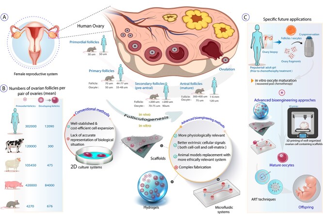
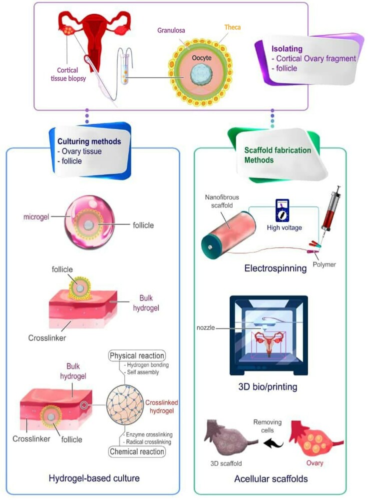
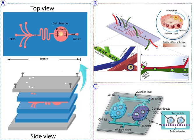
Oogenesis and folliculogenesis are considered as complex and species-specific cellular differentiation processes, which depend on the in vivo ovarian follicular environment and endocrine cues. Considerable efforts have been devoted to driving the differentiation of female primordial germ cells toward mature oocytes outside of the body. The recent experimental attempts have laid stress on offering a suitable microenvironment to assist the in vitro folliculogenesis and oogenesis. Despite developing a variety of bioengineering techniques and generating functional mature gametes through in vitro oogenesis in earlier studies, we still lack knowledge of appropriate microenvironment conditions for building biomimetic culture systems for female fertility preservation. Therefore, this review paper can provide a source for a large body of scientists developing cutting-edge in vitro culture systems for female germ cells or setting up the next generation of reproductive medicine as feasible options for female infertility treatment. The focal point of this review outlines advanced bioengineering technologies such as 3D biofabricated hydrogels/scaffolds and microfluidic systems utilized with female germlines for fertility preservation through in vitro folliculogenesis and oogenesis.

Keywords: advanced tissue engineering; biomaterials; female germ cells; female reproductive system; in vitro folliculogenesis; in vitro oogenesis; stem cells.


Conflict of interest statement

The authors have declared that no conflict of interest exists.

Figures

Graphical Abstract
Graphical Abstract
Figure 1
Figure 1
Schematic representation of applicable state-of-art technologies in female fertility preservation and in vitro folliculogenesis through utilizing various hydrogels and bioscaffolds. Bioscaffolds in forms of hydrogels, porous or fibrous structures can be fabricated via different approaches like acellularization of tissues, electrospinning, 3D printers, and so on using natural (collagen, alginate, agar, decellularized testis tissue-driven ECM, etc.) or synthetic (poly-L-lactide (PLLA), Poly(vinyl alcohol) (PVA), PCL, etc.) materials. The ovarian cortical fragments or isolated ovarian follicles could be cultured in vitro through the fabricated scaffolds to accomplish their goal for female fertility preservation.
Figure 2
Figure 2
Several hydrogels applied to provide a suitable environment for in vitro folliculogenesis/oogenesis. (A) Survival of primary follicle cultured with ascorbic acid on days 0, 6, 10, and 16 (i–iv). Confocal images illustrate the growth of the follicle cultured in the presence of ascorbic acid after 6 days (v) and the control secondary follicle (vi) (staining represents red for laminin, green for f-actin, and blue for nucleus). It was observed that the survival rate increased by ascorbic acid, and primary follicles reached a diameter of around 250 μm. The scale bars: 100 μm for i–iv and 10 μm for iv. Reprinted with permission from Tagler et al. (52). (B) Morphological changes of rat follicle in the 2D (i–iv) and 3D (v–viii) in vitro growth (IVG). In both cultures at day 0, immature oocytes were observed at the center (i, v); however, the difference is distinguishable later as the follicles started to collapse in 2D (ii) compared with well-maintained follicles in 3D alginate bead (vi). During 10 days of culture, there were a few antrum-like cavities in 2D-cultured follicles (ii, iv), although in 3D condition, follicles were observed larger forming antrum-like cavities (vii, viii). (C) The IVG of rat oocytes in 2D (i–iv) and the 3D (v–viii) culture conditions. The release of COCs from follicles is shown by arrows (i, v), 20 h after exposure to hCG and EGF to induce ovulation. Germinal vesicles were detected in both of the cultures (ii, vi) and oocyte transition from a Germinal Vesicle (GV) to Germinal vesicle breakdown (GVBD) (iii, vii). Meiosis also continued as the mature oocytes released the first polar body (PB) (iv–viii). Reprinted with permission from Zhang et al. (58). (D) The ECM proteins and distribution visualized by a fluorescent microscope in ovarian tissue and antral follicles. (a) Staining represents blue for fibronectin and yellow for collagen I, throughout the stroma and within ovarian follicles, while laminin (in red) is only observed in the perifollicular space. Scale bars = 100 μm. (b) Staining represents green for fibronectin and yellow for collagen I, the granulosa cell layers of antral follicles isolated from ovarian tissue, but laminin (in red) and perlecan (in magenta) are detected in theca layers. Scale bars = 50 μm and 100 μm (inset). Reprinted with permission from Tomaszewski et al. (65).
Figure 3
Figure 3
3D scaffolds used in supporting folliculogenesis and oogenesis. (A) In vitro culture of the primary follicle after 9 days on a decellularized scaffold. The control culture was in base medium, IAM on intact human amniotic membrane and DAM group represents the ones cultured on decellularized human amniotic membrane. Star represents hyperproliferation that occurred for granulosa cells in primary follicles cultured on IAM. Scale bar =50 μm. Reprinted with permission from Motamed et al. (103). (B) The histological sections of autotransplanted ovarian grafts in mice after 28 days (arrows show follicles). (i) Control group (mice without ovariectomy or grafting), (ii) autografted group which is detected by fewer follicles, (iii) autografted + platelet-rich fibrin bioscaffold group, which exhibited a significantly higher number of follicles. Scale bar: 100 μm. M: Muscle. Reprinted with permission from Shojafar et al. (110). (C) Porcine follicles seeded on electrospun PCL fibers, PCL/gel, and control (PET membrane) illustrated by fluorescence microscope (actin in red and nucleus in blue) and SEM images 10 days post-seeding. SEM micrographs with higher magnifications represented interaction between follicles and scaffolds, as no adhesion spots were observed for the control. Reprinted with permission from Liverani et al. (118). (D) i, The schematic represents the fabricated 3D glass scaffold and 2D culture methods. ii, Hoechst 33342 staining showed the total number of cells, and TUNEL assay exhibited apoptotic cells (the arrows point to TUNEL-positive nuclei). Reprinted with permission from Shen et al. (119).
Figure 4
Figure 4
Schematic representation of specific microfluidic systems applications in female fertility preservation and in vitro folliculogenesis. (A) Schematic representation of a microfluidic device containing five layers of which the middle one was used for follicle culture (reproduced with permission (146) ). (B) A schematic for a microfluidic chip including several inlets and outlets to create shelled hydrogel microtissue (reproduced with permission (20)). (C) A multiwell microfluidic device used for oocyte culture, cumulus cell removal, and contraceptive agents screening by (reproduced with permission (150)).

References

    1. Mahajan N. Fertility preservation in female cancer patients: an overview. J Hum Reprod Sci 2015; 8(1):3. - PMC - PubMed
    1. Goswami D, Conway GS. Premature ovarian failure. Hum Reprod Update 2005; 11(4):391–410. - PubMed
    1. Salama M, Woodruff TK. New advances in ovarian autotransplantation to restore fertility in cancer patients. Cancer Metastasis Rev 2015; 34(4):807–22. - PMC - PubMed
    1. Woodruff TK, Shea LD. The role of the extracellular matrix in ovarian follicle development. Reprod Sci 2007; 14(8_suppl):6–10. - PMC - PubMed
    1. Tingen C, Kim A, Woodruff TK. The primordial pool of follicles and nest breakdown in mammalian ovaries. Mol Hum Reprod 2009; 15(12):795–803. - PMC - PubMed

Publication types