. 2022 Sep 15;29(9):1381-1395.e13.
doi: 10.1016/j.chembiol.2022.07.003. Epub 2022 Aug 10.

Discovery of compounds that reactivate p53 mutants in vitro and in vivo

Affiliations

Discovery of compounds that reactivate p53 mutants in vitro and in vivo

Geetha Durairaj et al. Cell Chem Biol. .

Abstract

The tumor suppressor p53 is the most frequently mutated protein in human cancer. The majority of these mutations are missense mutations in the DNA binding domain of p53. Restoring p53 tumor suppressor function could have a major impact on the therapy for a wide range of cancers. Here we report a virtual screening approach that identified several small molecules with p53 reactivation activities. The UCI-LC0023 compound series was studied in detail and was shown to bind p53, induce a conformational change in mutant p53, restore the ability of p53 hotspot mutants to associate with chromatin, reestablish sequence-specific DNA binding of a p53 mutant in a reconstituted in vitro system, induce p53-dependent transcription programs, and prevent progression of tumors carrying mutant p53, but not p53null or p53WT alleles. Our study demonstrates feasibility of a computation-guided approach to identify small molecule corrector drugs for p53 hotspot mutations.

Keywords: cryptic pocket; ensemble based virtual screening; molecular dynamics simulations; mutant p53; p53 reactivation; small molecule p53 corrector drugs.


Conflict of interest statement

Declaration of interests Several authors of this manuscript are listed as inventors on the patent listed below. The patent describes compounds reported in this manuscript. Amaro, R. E., Baronio, R., Demir, O., Kaiser, P., Lathrop, R. H., Salehi-Amiri, S.-F., Wassman, C. Small molecules to enhance p53 activity. US patent US20160193214 A1. Approved March 2017.

Figures
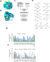
Figure 1:
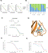
Figure 1:. Fourteen potential p53 cancer mutant reactivation compounds.
A) Closed conformation of the L1/S3 pocket in p53 WT crystal structure with pdbID:1TSR, chain B where Cysteine 124 is occluded. B) Open conformation of the L1/S3 pocket in a molecular-dynamics-generated frame for the R273H mutant. The black arrow indicates Cysteine 124. The protein surface in panels A and B is colored according to atom types; N: blue, O: red, S: yellow, C: cyan. Secondary structure of the site is depicted in ribbons with beta sheets in yellow, alpha helices in red and loops in white. C) A flowchart depicting the virtual screen targeting the L1/S3 pocket of p53. D) Saos-2 cells lacking p53 (p53null) or expressing cancer mutant p53R175H were cultured in the presence of different concentrations of computationally predicted reactivation compounds for 3 days. Data is presented as standard error of mean (n= 3). Stictic acid (37μM); 35ZWF, 25KKL, 22LSV, 28NZ6 and 27TGR (100μM); 32CTM (40μM); 26RQZ and 32LDE (10μM); 33AG6 (200μM), 33BAZ (60μM), 27VFS (20μM), 35LVZ, 36EB5 (50μM), and 27UDP (25μM). Data are presented as standard error of mean (n= 3). PRIMA-1 and Stictic acid were previously identified (Lambert et al., 2009; Wassman et al., 2013). E) Structures of fourteen computationally predicted p53 reactivation compounds that showed activity in panel D. F) Effect of the fourteen predicted compounds on Saos-2 cells (p53null) and 4 major p53 cancer mutants (p53R175H p53G245S, p53R273H, p53R248W). Data is presented as standard error of mean (n= 4).
Figure 2:
Figure 2:. Activity of UCI-LC0023 series of compounds in reactivating p53.
A) Effect of UCI-LC0023 on different p53 cancer mutants. Saos-2 cells lacking p53 (p53null) or expressing different p53 mutations (p53R175H, p53G245S, p53Y220C, p53R273H, p53R248W, p53R282W), breast (MCF7) and ovarian cancer (TOV112D) cell lines harboring p53WT and p53R175H, respectively, were cultured in the presence of different concentrations of UCI-LC0023 for 3 days. Cell numbers were measured using the CellTiter-Glo® reagent. Data is presented as standard deviation of mean (n= 3). B) Structural modifications to UCI-LC0023. C) Structures of UCI-LC0023 derivatives and their corresponding IC50 values in Saos-2 cells lacking p53 (p53null), expressing p53G245S and TOV112D (p53R175H) cells.
Figure 3:
Figure 3:. LC0023 directly binds to mutant p53.
UCI-A) TOV112D cells were cultured in the presence of different concentrations of UCI-LC0023, UCI-LC0019 or APR-246 for 24, 48 and 72 hours respectively. Cell numbers were measured using the CellTiter-Glo® reagent. Data is presented as standard deviation of mean (n= 3). B) Flow cytometry analysis of TOV112D cells stained with Annexin V and propidium iodide. TOV112D cells were treated with DMSO, 30μM UCI-LC0023, 30μM UCI-LC0019 and 40μM APR-246 for 3, 12 and 24 hours. Data is presented as standard error of the mean (n=3). C) Effect of photo-cross linkable diazirine analog UCI-LC0045 and UCI-LC0023 on TOV112D. Ovarian cancer (TOV112D) cell lines harboring p53R175H were cultured in the presence of different concentrations of UCI-LC0045 and UCI-LC0023 for 3 days. Cell numbers were measured using the CellTiter-Glo® reagent. Data is presented as standard error of mean (n= 3). D) Predicted binding pose of UCI-LC0045 in the L1/S3 pocket of p53R175H by docking guided by MS/crosslinking analyses. E) Effect of UCI-LC0023 on Saos-2 cells expressing p53R175H and p53C124A_R175H mutants. Cells were cultured in the presence of different concentrations of UCI-LC0023 for 3 days. Cell numbers were measured using the CellTiter-Glo® reagent. Data is presented as standard deviation of mean (n= 3).
Figure 4:
Figure 4:. UCI-LC0023 induces conformational changes in p53R175H.
A), B) and C) TOV112D cells were treated with vehicle, 40μM and 60 μM APR-246, 25μM and 30μM UCI-LC0019 and 25μM and 30μM UCI-LC0023 in the presence or absence of 5mM NAC for 3 days. Cell numbers were measured using the CellTiter-Glo® reagent. Data is presented as standard deviation of mean (n=3). D) UCI-LC0023 induced conformational changes of p53R175H in ovarian cancer cell line TOV112D, as observed by immunofluorescent staining of cells with p53-conformation selective antibodies PAB240 (mutant conformation) and PAB1620 (WT conformation). All scale bars represent a size of 10μM. Quantification of PAB240 and PAB1620 staining is shown. Results from two independent experiments with 16 blinded quantified cells are shown in the graph. The error bars represent mean ± S.E.M. ** p < 0.05 and *** p < 0.005. E) Immunoprecipitation of mutant p53 in TOV112D (p53R175H) cells using PAB240 antibodies and detection by p53 (FL393) and GAPDH antibodies.
Figure 5:
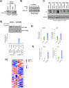
Figure 5:. UCI-LC0023 restores DNA binding of p53R175H in vivo and in vitro and induces p53 target gene expression.
A) and B) Cells treated with compound or vehicle were fractionated into cytosolic and nuclear soluble fractions as well as chromatin fractions as indicated by histone H3. UCI-LC0023 restores chromatin binding of p53R175H. C) Effect of UCI-LC0023 on chromatin binding of Saos-2 cells expressing different p53 mutations (p53R175H p53G245S, p53Y220C, p53R273H, p53R248W, p53R282W). D) Restoration of site-specific DNA binding of mutant p53 in a purified reconstituted system. The recombinant purified DNA binding domains of p53WT and p53R175H were incubated with biotinylated DNA fragments representing the promoter of the p53-dependent GADD45A gene or a p53 independent (E2F) promoter along with 100 μM UCI-LC0023 or DMSO (vehicle). Protein bound to the DNA fragments was analyzed by immunoblotting with antibodies against p53. Drug induced binding of p53 to p53 consensus and E2F non-consensus sequences is represented as a fold change of UCI-LC0023 treated samples over their respective vehicle controls. E) Chromatin immunoprecipitation (ChIP) analysis demonstrates induction of site-specific promoter binding of p53R175H by UCI-LC0023 in TOV112D cells. Eluted DNA was examined by quantitative real time PCR using primers that target the p53 binding site in the promoter regions of p21 and PUMA. Data is represented as standard error of mean (n=3). F) q-RTPCR of Noxa and p21 in TOV112D (p53R175H) cells treated with 30μM UCI-LC0023 or DMSO (vehicle) for 3 hours. Data is represented as standard deviation of mean (n=3). G) p53 target gene expression induced by treatment with 30μM UCI-LC0023 or DMSO (vehicle) in TOV112D (p53R175H) cells for 3 hours on a custom p53 nanostring panel. 3 independent experiments are shown as log2 values of the mean.
Figure 6:
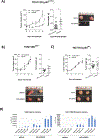
Figure 6:. In-vivo evidence of UCI-LC0019 mediated p53 reactivation.
A), B), and C) Xenograft tumors were generated from TOV112D (p53R175H), TOVII2D p53−/− and HCT116 (p53 +/+). Tumors were allowed to grow to 50mm3 prior to daily intraperitoneal injections of UCI-LC0019 (10mg/kg). Tumor dimensions were measured every other day and their volumes were calculated by length (L) and width (W) by using the formula: volume = L × W2 × 0.523. The tumor volumes of treated and untreated tumors for TOV112D (p53R175H), TOVII2D p53−/− and HCT116 (p53 +/+) are represented in figures A, B and C. Tumor weight and images of treated and untreated tumors of TOV112D (p53R175H), TOVII2D p53−/− and HCT116 (p53 +/+) are also represented in figures A, B and C respectively. The error bars represent the mean ± SEM * p<0.05 by Turkey’s multiple comparison test compared to vehicle control. D) Mice carrying xenograft tumors (TOV112D [p53R175H]) were intraperitoneal injected with vehicle or 10mg/kg UCI-LC0019. Tumors were harvested 1h and 2h after compound injection and intratumor GSH and UCI-LC0019 levels measured by LC-MS.

References

    1. Aggarwal M, Saxena R, Sinclair E, Fu Y, Jacobs A, Dyba M, Wang X, Cruz I, Berry D, Kallakury B, et al. (2016). Reactivation of mutant p53 by a dietary-related compound phenethyl isothiocyanate inhibits tumor growth. Cell Death Differ. - PMC - PubMed
    1. Bargonetti J, and Prives C (2019). Gain-of-function mutant p53: History and speculation. J. Mol. Cell Biol. - PMC - PubMed
    1. Baronio R, Danziger SA, Hall LV, Salmon K, Hatfield GW, Lathrop RH, and Kaiser P (2010). All-codon scanning identifies p53 cancer rescue mutations. Nucleic Acids Res 38, 7079–7088. - PMC - PubMed
    1. Beraza N, and Trautwein C (2007). Restoration of p53 function: a new therapeutic strategy to induce tumor regression? Hepatology 45, 1578–1579. - PubMed
    1. Blanden AR, Yu X, Loh SN, Levine AJ, and Carpizo DR (2015). Reactivating mutant p53 using small molecules as zinc metallochaperones: Awakening a sleeping giant in cancer. Drug Discov. Today. - PMC - PubMed

Publication types